D.C. Heath and Co. – Publishers
Original copyright 1909
such a manner as not only to confer knowledge which is
useful in itself, but to serve the purpose of a training in
accurate observation, and in the methods of reasoning of
physical science.”—Huxley.
Preface
The aim in the preparation of this treatise on the human body has
been, first, to set forth in a teachable manner the actual science of physiology; and
second, to present the facts of hygiene largely as applied physiology. The view is held that “right living”
consists in the harmonious adjustment of one’s habits to the nature and
plan of the body, and that the best preparation for such living is a
correct understanding of the physical self. It is further held that the
emphasizing of physiology augments in no small degree the educative
value of the subject, greater opportunity being thus afforded for
exercise of the reasoning powers and for drill in the modus operandi of natural forces. In the
study of physiology the facts of anatomy have a place, but in an
elementary course these should be restricted to such as are necessary
for revealing the general structure of the body.
Although no effort has been spared to bring this work within the
comprehension of the pupil, its success in the classroom will depend
largely upon the method of handling the subject by the teacher. It is
recommended, therefore, that the relations which the different organs and processes sustain
to each other, and to the body as a whole, be given special prominence.
The pupil should be impressed with the essential unity of the body and
should see in the diversity of its activities the serving of a common
purpose. In creating such an impression the introductory paragraphs at
the beginning of many of the chapters and the summaries throughout the
book, as well as the general arrangement of the subject-matter, will be
found helpful.
Since the custom largely prevails of teaching physiology in advance
of the sciences upon which it rests—biology, physics, and chemistry—care
should be exercised to develop correct ideas of the principles and
processes derived from these sciences. Too much latitude has been taken
in the past in the use of comparisons and illustrations drawn from
“everyday life.” To teach that the body is a “house,” “machine,” or
“city”; that the nerves carry “messages”; that the purpose of oxygen is
to “burn up waste”; that breathing is to “purify the blood,” etc., may
give the pupil phrases which he can readily repeat, but teaching of this
kind does not give him correct ideas of his body.
The method of teaching, however, that uses the pupil’s experience as
a basis upon which to build has a value not to be overlooked. The fact
that such expressions as those quoted above are so easily remembered
proves the value of connecting new knowledge with the pupil’s
experience. But the inadequacy of this
experience must be recognized and taken into account. The concepts
of the average pupil are entirely too indefinite and limited to supply
the necessary foundation for a
science such as physiology. Herein lies the great value of
experiments and observations. They supplement the pupil’s experience,
and increase both the number and definiteness of his concepts. No degree
of success can be attained if this phase of the study is omitted.
The best results in physiology teaching are of course attained where
laboratory work is carried on by the pupils, but where this cannot be
arranged, class experiments and observations must suffice. The Practical
Work described at the close of most of the chapters is mainly for class
purposes. While these serve a necessary part in the development of the
subject, it is not essential that all of the experiments and
observations be made, the intention being to provide for some choice on
the part of the teacher. A note-book should be kept by the pupil.
To adapt the book to as wide a range of usefulness as possible, more
subject-matter is introduced than is usually included in an elementary
course. Such portions, however, as are unessential to a proper
understanding of the body by the pupil are set in small type, to be used
at the discretion of the teacher.
The use of books of reference is earnestly recommended. For this
purpose the usual high school texts may be employed to good advantage. A
few more advanced works should, however, be frequently consulted. For
this purpose Martin’s Human Body
(Advanced Course), Rettger’s Advanced
Lessons in Physiology, Thornton’s Human Physiology, Huxley’s Lessons in Elementary Physiology, Howell’s A Text-book of Physiology, Hough and
Sedgwick’s Hygiene and Sanitation,
and Pyle’s Personal Hygiene will be
found serviceable.
In the preparation of this work valuable assistance has been rendered
by Dr. C.N. McAllister, Department of Psychology, and by Professor B.M.
Stigall, Department of Biology, along the lines of their respective
specialties, and in a more general way by President W.J. Hawkins and
others of the Warrensburg, Missouri, State Normal School. Expert advice
from Professor S.D. Magers, Instructor in Physiology and Bacteriology,
State Normal School, Ypsilanti, Michigan, has been especially helpful,
and many practical suggestions from the high school teachers of
physiology of Kansas City, Missouri, Professor C.H. Nowlin, Central
High School, Dr. John W. Scott, Westport High
School, and Professor A.E. Shirling, Manual Training High School, all of
whom read both manuscript and proofs, have been incorporated.
Considerable material for the Practical Work, including the respiration
experiment (page 101) and the reaction time experiment (page 323), were
contributed by Dr. Scott. Professor Nowlin’s suggestions on
subject-matter and methods of presentation deserve special mention. To
these and many others the author makes grateful acknowledgment.
F.M.W.
Missouri State Normal
School,
Second District, May 1,
1909.
Contents
- Preface
- Contents
- PART I: THE VITAL PROCESSES
- CHAPTER I – INTRODUCTION
- CHAPTER II – GENERAL VIEW OF THE BODY
- CHAPTER III – THE BODY ORGANIZATION
- CHAPTER IV – THE BLOOD
- CHAPTER V – THE CIRCULATION
- CHAPTER VI – THE LYMPH AND ITS MOVEMENT THROUGH THE BODY
- CHAPTER VII – RESPIRATION
- CHAPTER VIII – PASSAGE OF OXYGEN THROUGH THE BODY
- CHAPTER IX – FOODS AND THE THEORY OF DIGESTION
- CHAPTER X – ORGANS AND PROCESSES OF DIGESTION
- CHAPTER XI – ABSORPTION, STORAGE, AND ASSIMILATION
- CHAPTER XII – ENERGY SUPPLY OF THE BODY
- CHAPTER XIII – GLANDS AND THE WORK OF EXCRETION
- PART II: MOTION, COORDINATION, AND SENSATION
- CHAPTER XIV – THE SKELETON
- CHAPTER XV – THE MUSCULAR SYSTEM
- CHAPTER XVI – THE SKIN
- CHAPTER XVII – STRUCTURE OF THE NERVOUS SYSTEM
- CHAPTER XVIII – PHYSIOLOGY OF THE NERVOUS SYSTEM
- CHAPTER XIX – HYGIENE OF THE NERVOUS SYSTEM
- CHAPTER XX – PRODUCTION OF SENSATIONS
- CHAPTER XXI – THE LARYNX AND THE EAR
- CHAPTER XXII – THE EYE
- CHAPTER XXIII – THE GENERAL PROBLEM OF KEEPING WELL
- APPENDIX
- INDEX
PART I: THE VITAL PROCESSES
CHAPTER I – INTRODUCTION
To derive strength equal to the daily task; to experience the
advantages of health and avoid the pain, inconvenience, and danger of
disease; to live out contentedly and usefully the natural span of life:
these are problems that concern all people. They are, however, but
different phases of one great problem—the problem of properly managing
or caring for the body. To supply knowledge necessary to the solution of
this problem is the chief reason why the body is studied in our public
schools.
Divisions of the Subject.—The body
is studied from three standpoints: structure, use of parts, and care or
management. This causes the main subject to be considered under three
heads, known as anatomy, physiology, and hygiene.
Anatomy treats of the construction
of the body—the parts which compose it, what they are like, and where
located. Its main divisions are known as gross anatomy and histology.
Gross anatomy treats of the larger
structures of the body, while histology treats of the minute structures of which these
are composed—parts too small to be seen with the naked eye and which
have to be studied with the aid of the microscope.
[pg 002]Physiology treats of the function, or use, of the different
parts of the body—the work which the parts do and how they do it—and of
their relations to one another and to the body as a whole.
Hygiene treats of the proper care
or management of the body. In a somewhat narrower sense it treats of the
“laws of health.” Hygiene is said to be personal, when applied by the individual to his own body;
domestic, when applied to a small
group of people, as the family; and public, or general, when applied to the community as a whole or to the
race.
The General Aim of Hygiene.—There
are many so-called laws of health, and for these laws it is essential in
the management of the body to find a common basis. This basic law,
suggested by the nature of the body and conditions that affect its
well-being, may be termed the Law of
Harmony: The mode of living must harmonize with the plan of the
body. To live properly one must supply the conditions which his
body, on account of its nature and plan, requires. On the other hand, he
must avoid those things and conditions which are injurious, i.e., out of harmony with the body plan.
To secure these results, it is necessary to determine what is and what
is not in harmony with the plan of the body, and to find the means of
applying this knowledge to the everyday problems of living. Such is the
general aim of hygiene. Stated in other words: Hygiene has for its
general aim the bringing about of an essential harmony between the body
and the things and conditions that affect it.1
[pg 003]Relation of Anatomy and Physiology to the Study of
Hygiene.—If the chief object in studying the body is that of
learning how to manage or care for it, and hygiene supplies this
information, why must we also study anatomy and physiology? The answer
to this question has already been in part suggested. In order to
determine what things and conditions are in harmony with the plan of the
body, we must know what that plan is. This knowledge is obtained through
a study of anatomy and physiology. The knowledge gained through these
subjects also renders the study of hygiene more interesting and
valuable. One is enabled to see why
and how obedience to hygienic laws
benefits, and disobedience to them injures, the body. This causes the
teachings of hygiene to be taken more seriously and renders them more
practical. In short, anatomy and physiology supply a necessary basis for
the study of hygiene.
Advantages of Properly Managing the
Body.—One result following the mismanagement of the body is loss of
health. But attending the loss of health are other results which are
equally serious and far-reaching. Without good health, people fail to
accomplish their aims and ambitions in life; they miss the joy of
living; they lose their ability to work and become burdens on their
friends or society. The proper management of the body means health, and
it also means the capacity for work and for enjoyment. Not only should
one seek to preserve his health from day to day, but he should so manage
his body as to use his powers to the best advantage and prolong as far
as possible the period during which he may be a capable and useful
citizen.
[pg 004]
CHAPTER II – GENERAL VIEW OF THE BODY
External Divisions.—Examined from
the outside, the body presents certain parts, or divisions, familiar to
all. The main, or central, portion is known as the trunk, and to this are attached the head, the upper
extremities, and the lower
extremities. These in turn present smaller divisions which are also
familiar. The upper part of the trunk is known as the thorax, or chest, and the lower part as
the abdomen. The portions of the
trunk to which the arms are attached are the shoulders, and those to which the legs are joined are the
hips, while the central rear portion
between the neck and the hips is the back. The fingers, the hand, the wrist, the forearm, the
elbow, and the upper arm are the main divisions of each of the upper
extremities. The toes, the foot, the ankle, the lower leg, the knee, and
the thigh are the chief divisions of each of the lower extremities. The
head, which is joined to the trunk by the neck, has such interesting
parts as the eyes, the ears, the nose, the jaws, the cheeks, and the
mouth. The entire body is inclosed in a double covering, called the skin, which protects it in various
ways.
The Tissues.—After examining the
external features of the body, we naturally inquire about its internal
structures. These are not so easily investigated, and much which is of
interest to advanced students must be omitted from an elementary course.
We may, however, as a first step in this study, determine what kinds of
materials enter into [pg 005]the
construction of the body. For this purpose the body of some small animal
should be dissected and studied. (See observation at close of chapter.)
The different materials found by such a dissection correspond closely to
the substances, called tissues, which
make up the human body. The main tissues of the body, as ordinarily
named, are the muscular tissue, the
osseous tissue, the connective tissue, the nervous tissue, the adipose tissue, the cartilaginous tissue, and the epithelial and glandular
tissue. Most of these present different varieties, making all together
some fifteen different kinds of tissues that enter into the construction
of the body.2
General Purposes of the
Tissues.—The tissues, first of all, form the body. As a house is constructed of wood, stone,
plaster, iron, and other building materials, so is the body made up of
its various tissues. For this reason the tissues have been called the
building materials of the body.
In addition to forming the body, the tissues supply the means through
which its work is carried on. They are thus the working materials of the body. In serving this purpose the
tissues play an active rôle. All of them must perform the activities of
growth and repair, and certain ones (the so-called active tissues) must
do work which benefits the body as a whole.
Purposes of the Different
Tissues.—In the construction of the body and also in the work which
it carries on, the different tissues are made to serve different
purposes. The osseous tissue is the chief substance in the bony
framework, or skeleton, while the muscular tissue produces the different
movements of the body. The connective[pg 006] tissue, which is everywhere abundant, serves the general
purpose of connecting the different parts together. Cartilaginous tissue
forms smooth coverings over the ends of the bones and, in addition to
this, supplies the necessary stiffness in organs like the larynx and the
ear. The nervous tissue controls the body and brings it into proper
relations with its surroundings, while the epithelial tissue (found upon
the body surfaces and in the glands) supplies it with protective
coverings and secretes liquids. The adipose tissue (fat) prevents the
too rapid escape of heat from the body, supplies it with nourishment in
time of need, and forms soft pads for delicate organs like the
eyeball.
Properties of the Tissues.—If we
inquire how the tissues are able to serve such widely different
purposes, we find this answer. The tissues differ from one another both
in composition and in structure and, on this account, differ in their
properties.3 Their different properties enable them
to serve different purposes in the body. Somewhat as glass is adapted by
its transparency, hardness, and toughness to the use made of it in
windows, the special properties of the tissues adapt them to the kinds
of service which they perform. Properties that adapt tissues to their
work in the body are called essential
properties. The most important of these essential properties are as
follows:
1. Of osseous tissue, hardness, stiffness, and toughness. 2. Of
muscular tissue, contractility and irritability. 3. Of nervous tissue,
irritability and conductivity. 4. Of cartilaginous tissue, stiffness and
elasticity. 5. Of connective tissue, toughness and pliability. 6. Of
epithelial tissue, ability to resist the action of external forces and
power to secrete.

connective tissues in the organ for grasping.
Tissue Groups.—In the construction
of the body the tissues are grouped together to form its various
divisions or parts. A group of tissues which serves some special purpose
is known as an organ. The hand, for
example, is an organ for grasping (Fig. 1). While the different organs
of the body do not always contain the same tissues, and never contain
them in the same proportions, they do contain such tissues as their work
requires and these have a special arrangement—one adapted to the work
which the organs perform.
In addition to forming the organs, the tissues are also grouped in
such a manner as to provide supports for organs and to form cavities in
which organs are placed. The various cavities of the body are of
particular interest and importance. The three largest ones are the cranial cavity, containing the brain; the
thoracic cavity, containing the heart
and the lungs; and the abdominal
cavity, containing the stomach, the liver, the intestines, and other
important organs (Fig. 2). Smaller cavities serving different purposes
are also found.

large cavities and the organs which they contain.
Organs and Systems.—The work of the
body is carried on by its various organs. Many, in fact the majority, of
these organs serve more than one purpose. The tongue[pg 009]
is used in talking, in
masticating the food, and in swallowing. The nose serves at least three
distinct purposes. The mouth, the arms, the hands, the feet, the legs,
the liver, the lungs, and the stomach are also organs that serve more
than one purpose. This introduces the principle of economy into the
construction of the body and diminishes the number of organs that would
otherwise be required.
The various organs also combine
with one another in carrying on the work of the body. An illustration of
this is seen in the digestion of the food—a process which requires the
combined action of the mouth, stomach, liver, intestines, and other
organs. A number of organs working together for the same purpose form a
system. The chief systems of the body
are the digestive system, the circulatory system, the respiratory
system, the muscular system, and the nervous system.
The Organ and its Work.—A most
interesting question relating to the work of the organ is this: Does the
organ work for its own benefit or for the benefit of the body as a
whole? Does the hand, for example, grasp for itself or in order that the
entire body may come into possession? Only slight study is sufficient to
reveal the fact that each organ performs a work which benefits the body
as a whole. In other words, just as the organ itself is a part of the
body, the work which it does is a part of the necessary work which the
body has to do.
But in working for the general good, or for the body as a whole, each
organ becomes a sharer in the benefits of the work done by every other
organ. While the hand receives only a little of the nourishment
contained in the food which it places in the mouth or of the heat from,
fuel which it places on the fire, it is aided and supported by the work
of all the other organs of the body—eyes, [pg 010] feet, brain, heart, etc. The hand does not and cannot work
independently of the other organs. It is one of the partners in a very
close combination where, by doing a particular work, it, shares in the
profits of all. What is true of the hand is true of every other organ of
the body.
An Organization.—The relations
which the different organs sustain to each other and to the body as a
whole suggest the possibility of classifying the body as an
organization. This term is broadly applied to a variety of combinations.
An organization is properly defined as any
group of individuals which, in working together for a common purpose,
practices the division of labor. This definition will be better
understood by considering a few familiar examples.
A baseball team is an organization. The team is made up of individual
players. These work together for the common purpose of winning games.
They practice the division of labor in that the different players do
different things—one catching, another pitching, and so on. A
manufacturing establishment which employs several workmen may also be an
organization. The article manufactured provides the common purpose
toward which all strive; and, in the assignment of different kinds of
work to the individual workmen, the principle of division of labor is
carried out. For the same reason a school, a railway system, an army,
and a political party are organizations.
An organization of a lower order of individuals than these human
organizations is to be found in a hive of bees. This is made up of the
individual bees, and these, in carrying on the general work of the hive,
are known to practice the division of labor.
Is the Body an Organization?—If the
body is an organization, it must fulfill the conditions of the
definition. It[pg 011] must be made up of
separate or individual parts. These must work together for the same
general purpose, and, in the accomplishment of this purpose, must
practice the division of labor. That the body practices the division of
labor is seen in the related work of the different organs. That it is
made up of minute, but individual, parts will be shown in the chapter
following. That it carries on a general
work which is accomplished through the combined action of its
individual parts is revealed through an extended study of its various
activities. The body is an
organization. Moreover, it is one of the most complex and, at the
same time, most perfect of the organizations of which we have
knowledge.
Summary.—Viewed from the outside,
the body is seen to be made up of divisions which are more or less
familiar. Viewed internally, it is found to consist of different kinds
of materials, called tissues. The tissues are adapted, by their
properties, to different purposes both in the construction of the body
and in carrying on its work. The working parts of the body are called
organs and these in their work combine to form systems. The entire body,
on account of the method of its construction and the character of its
work, may be classed as an organization.
Exercises.—1. Name and locate the
chief external divisions of the body.
2. What tissues may be found by dissecting the leg of a chicken?
3. Name the most important properties and the most important uses of
muscular tissue, osseous tissue, and connective tissue.
4. Define an organ. Define a system. Name examples of each.
5. Name the chief cavities of the body and the organs which they
contain.
6. What tissues are present in the hand? How does each of these aid
in the work of the hand?
[pg 012]7. Define an organization. Show
that a railway system, an army, and a school are organizations.
8. What is meant by the phrase “division of labor”? In what manner is
the division of labor practiced in a shoe or watch factory? What are the
advantages?
9. What are the proofs that the body is an organization?
PRACTICAL WORK
Observation on the Tissues.—Examine
with care the structures in the entire leg of a chicken, squirrel,
rabbit, or other small animal used for food. Observe, first of all, the
external covering, consisting of cuticle and hair, claws, scales, or
feathers, according to the specimen. These are similar in structure, and
they form the epidermis, which is one kind of epithelial tissue. With a sharp knife lay open the skin and
observe that it is attached to the parts underneath by thin, but tough,
threads and sheaths. These represent a variety of connective tissue. The reddish material which forms the
greater portion of the specimen is a variety of muscular tissue, and its divisions are called muscles. With
a blunt instrument, separate the muscles, by tearing apart the
connective tissue binding them together, and find the glistening white
strips of connective tissue (tendons) which attach them to the bones.
Find near the central part of the leg a soft, white cord (a nerve) which
represents one variety of nervous
tissue. The bones, which may now be examined, form the osseous tissue. At the ends of the bones
will be found a layer of smooth, white material which represents one
kind of cartilaginous tissue. The adipose, or fatty, tissue, which is found
under the skin and between the other tissues, is easily recognized.
Relation of the Tissues to the
Organs.—Observe in the specimen just studied the relation of the
different tissues to the organ as a whole (regarding the leg as an
organ), i.e., show how each of the
tissues aids in the work which the organ accomplishes. Show in
particular how the muscles supply the foot with motion, by tracing out
the tendons that connect them with the toes. Pull on the different
tendons, noting the effect upon the different parts of the foot.
[pg 013]
CHAPTER III – THE BODY ORGANIZATION
What is the nature of the body organization? What are the individual
parts, or units, that make it up? What general work do these carry on
and upon what basis do they practice the division of labor? The answers
to these questions will suggest the main problems in the study of the
body.

showing the relation of the cells and the intercellular material. C. Cells. I. Intercellular material.
Complex Nature of the Tissues.—To
the unaided eye the tissues have the appearance of simple structures.
The microscope, however, shows just the reverse to be true. When any one
of the tissues is suitably prepared and carefully examined with this
instrument, at least two classes of materials can be made out. One of
these consists of minute particles, called cells; the other is a substance lying between the cells,
known as the intercellular material
(Fig. 3). The cells and the intercellular material, though varying in
their relative proportions, are present in all the tissues.
The Body a Cell Group.—The
biologist has found that the bodies of all living things, plants as well
as animals, consist either of single cells or of groups of cells. The
single cells live independently of one another, but the cells that form
groups are attached to, and are more or less dependent upon, one
another. In the first condition are [pg 014]
found the very lowest forms of life. In the second, life reaches its
greatest development. The body of man, which represents the highest type
of life, is recognized as a group of cells. In this group each cell is
usually separate and distinct from the others, but is attached to them,
and is held in place by the intercellular material.
Protoplasm, the Cell Substance.—The
cell is properly regarded as an organized bit of a peculiar material, called protoplasm. This is a semi-liquid and
somewhat granular substance which resembles in appearance the white of a
raw egg. Its true nature and composition are unknown, because any
attempt to analyze it kills it, and dead protoplasm is essentially
different from living protoplasm. It is known, however, to be a highly
complex substance and to undergo chemical change readily. It appears to
be the only kind of matter with which life is ever associated, and for
this reason protoplasm is called the physical basis of life. Its organization into separate
bits, or cells, is necessary to the life activities that take place
within it.
Structure of the Cell.—Though all
portions of the cell are formed from the protoplasm, this essential
substance differs both in structure and in function at different places
in the cell. For this reason the cell is looked upon as a complex body
having several distinct parts. At or near the center is a clear, rounded
body, called the nucleus. This plays
some part in the nourishment of the cell and also in the formation of
new cells. If it be absent, as is sometimes the case, the cell is
short-lived and unable to reproduce itself. The variety of protoplasm
contained in the nucleus is called the nucleoplasm.
. 1. Main body. 2. Nucleus. 3. Attraction sphere. 4. Food particles and waste. 5. Cell-wall. 6. Masses of active material found in certain cells, called plastids.")
typical cell (after Wilson). 1. Main body. 2. Nucleus. 3.
Attraction sphere. 4. Food particles and waste. 5. Cell-wall. 6. Masses
of active material found in certain cells, called plastids.
Surrounding the nucleus is the main
body of the cell, sometimes referred to as the “protoplasm.” Since
the[pg 015] protoplasm forms all parts of
the cell, this substance is more properly called the cytoplasm, or cell plasm. Surrounding and
inclosing the cytoplasm, in many cells, is a thin outer layer, or
membrane, which affords more or less protection to the contents of the
cell. This is usually referred to as the cell-wall. A fourth part of the cell is also described,
being called the attraction sphere.
This is a small body lying near the nucleus and coöperating with that
body in the formation of new cells. Food particles, wastes, and other
substances may also be present in the cytoplasm. The parts of a typical
cell are shown in Fig. 4.
Importance of the Cells.—The cells
must be regarded as the living, working parts of the body. They are the
active agents in all of the tissues, enabling them to serve their
various purposes. Working through the tissues, they build up the body
and carry on its different activities. They are recognized on this
account as the units of structure and of
function, and are the “individuals” in the body organization. Among
the most important and interesting of the activities of the cells are
those by which they build up the body, or cause it to grow.
[pg 016]How the Cells enable the Body to
Grow.—Every cell is able to take new material into itself and to
add this to the protoplasm. This tends to increase the amount of the
protoplasm, thereby causing the cells to increase in size. A general
increase in the size of the cells has the effect of increasing the size
of the entire body, and this is one way by which they cause it to grow.
There is, however, a fixed limit, varying with different cells, to the
size which they attain, and this is quite low. (The largest cells are
scarcely visible to the naked eye.) Any marked increase in the size of
the body must, therefore, be brought about by other means. Such a means
is found in the formation of new cells, or cell reproduction. The new cells are always formed by and from the old cells, the essential process being known as
cell-division.
. Note that the process begins with the division of the attraction sphere, then involves the nucleus, and finally separates the main body.")
cell-division (after Wilson). Note that the process begins with the
division of the attraction sphere, then involves the nucleus, and
finally separates the main body.
Cell-Division.—By dividing, a
single cell will, on attaining its growth, separate into two or more new
cells. The process is quite complex and is imperfectly understood. It is
known, however, that the act of separation is preceded by a series of
changes in which the attraction sphere[pg 017] and the nucleus actively
participate, and that, as a result of these changes, the contents of the
old cell are rearranged to form the new cells. Some of the different
stages in the process, as they have been studied under the microscope,
are indicated in Fig. 5.
Gradually, through the formation of new cells and by the growth of
these cells after they have been formed, the body attains its full size.
When growth is complete, cell reproduction is supposed to cease except
where the tissues are injured, as in the breaking of a bone, or where
cells, like those at the surface of the skin, are subject to wear. Then
new material continues to be added to the protoplasm throughout life,
but in amount only sufficient to replace that lost from the protoplasm
as waste.

with water, suggesting the relations of the cells to the lymph.
Cell Surroundings.—All cells are
said to be aquatic. This means simply
that they require water for carrying on their various activities. The
cells, in order to live, must take in and give out materials, and water
is necessary to both processes. It is also an essential part of the
protoplasm. Deprived of water, cells become inactive and usually die.
Aquatic surroundings are provided for the cells of the body through a
liquid known as the lymph, which is
distributed throughout the intercellular material (Fig. 6). This
consists of water containing oxygen and food substances in solution.
Besides supplying these to the cells, the lymph also receives their
wastes. Through the lymph the necessary conditions for cell life are
provided in the body.
The General Work of Cells.—In
handling the materials[pg 018] derived from the lymph, the cells carry
on three well-defined processes, known as absorption, assimilation, and
excretion.
Absorption is the process of
taking water, food, and oxygen into the cells.
Assimilation is a complex process
which results in the addition of the absorbed materials to the
protoplasm. Through assimilation the protoplasm is built up or
renewed.
Excretion is the throwing off of
such waste materials as have been formed in the cells. These are passed
into the lymph and thence to the surface of the body.
Absorption, assimilation, excretion, and also reproduction are
performed by all classes of cells. They are, on this account, referred
to as the general work of cells.
The Special Work of Cells.—In
addition to the general work which all cells do in common, each class of
cells in the body is able to do some particular kind of work—a work
which the others cannot do or which they can do only to a limited
extent. This is spoken of as the special
work of cells. Examples of the special work of cells are found in
the production of motion by muscle cells and in the secretion of liquids
by gland cells. It may be noted that while the general work of cells
benefits them individually, their special work benefits the body as a
whole. Another example of the special work of cells is found in the

which they have deposited.
Production of the Intercellular
Material.—Though most of the cells of the body deposit to a slight
extent this material, the greater part of it is produced by a single
class of cells found in bone, cartilage, and connective tissue.
Cartilage, bone, and connective tissue differ greatly from the other
tissues in the amount of intercellular material which they contain, the
difference being due to these cells.[pg 019]
In the connective tissue they deposit the fibrous material so
important in holding the different parts of the body together. In the
cartilage they produce the gristly substance which forms by far its
larger portion (Fig. 7). In the bones they deposit a material similar to
that in the cartilage, except that with it is mixed a mineral substance
which gives the bones their hardness and stiffness.4 The intercellular
material, in addition to connecting the cells, supplies to certain
tissues important properties, such as the elasticity of cartilage and
the stiffness of the bones.
Nature of the Body
Organization.—The division of labor carried on by the different
organs, as shown in the preceding chapter, is in reality carried on by
the cells that form the organs. To see that this is true we have only to
observe the relation of cells to tissues and of tissues to organs. The
cells form the tissues and the tissues form the organs. This arrangement
enables the special work of different kinds of cells to be combined in
the work of the organ as a whole. This is seen in the hand which, in
grasping, uses motion supplied by the muscle cells, a controlling
influence supplied by the nerve cells, a framework supplied by the bone
cells, and so on. The cells supply the basis for the body organization
and, properly speaking, the body is an
organization of cells5 (Recall the definition[pg 020] of an organization, page 10.) In
this organization there are to be observed:
1. A definite arrangement of the cells to form the tissues. A tissue
is a group of like cells.
2. A definite arrangement of the tissues in the organ. Each organ
contains the tissues needed for its work.
3. In several instances there is a definite arrangement of organs to
form systems.
4. The body as a whole is made up of organs and systems, together
with the structures necessary for their support and protection.
There now remains a further question for consideration. What is the
one supreme end, or purpose, toward which all the activities of the body
organization are directed? This purpose will naturally have some
relation to the maintenance, or preservation, of the cell group which we
call the body.
The Maintenance of Life.—The
preservation of any cell group in its natural condition, whether it be
plant or animal, is accomplished through keeping it alive. If life
ceases, the group quickly disintegrates and its elements become
scattered, a fact which is verified through everyday observation. Though
the nature of life is unknown, it may be looked upon as the organizer
and preserver of the protoplasm. But in preserving the protoplasm it
also preserves the entire cell group, or body. Life is thus the most
essential condition of the body. With life
all portions of the body are concerned, and toward its maintenance all
the activities of the body organization are directed.
The Nutrient Fluid in its Relations to
the Cells.—The maintenance of life within the cells requires, as we
have seen, that they be supplied with water, food, and oxygen, and that
they be relieved of such wastes as they form.[pg 021] This double purpose is accomplished through the agency of
an internal nutrient fluid, a portion of which has already been referred
to as the lymph. Not only does this fluid supply the means for keeping
the cells alive, but, through the cells, it is also the means of
preserving the life of the body as a whole.
The cells, however, rapidly exhaust the nutrient fluid. They take
from it food and oxygen and they put into it their wastes. To prevent
its becoming unfit for supplying their needs, food and oxygen must be
continually added to this fluid, and waste materials must be continually
removed. This is not an easy task. As a matter of fact, the preparation,
distribution, and purification of the nutrient fluid requires the direct
or indirect aid of practically all parts of the body. It supplies for
this reason a broad basis for the division of labor on the part of the
cells.
Relation of the Body to its
Environment.—While life is directly dependent upon the internal
nutrient fluid, it is indirectly dependent upon the physical
surroundings of the body. Herein lies the need of the external organs—the feet and legs for
moving about, the hands for handling things, the eyes for directing
movements, etc. That the great needs of the body are supplied from its
surroundings are facts of common experience. Food, shelter, air,
clothing, water, and the means of protection are external to the body
and form a part of its environment. In making the things about him
contribute to his needs, man encounters a problem which taxes all his
powers. Only by toil and hardship, “by the sweat of his brow,” has he
been able to wrest from his surroundings the means of his
sustenance.
The Main Physiological
Problems.—The study of the body is thus seen to resolve itself
naturally into the consideration of two main problems:
[pg 022]1. That of maintaining in the body a nutrient fluid for the
cells.
2. That of bringing the body into such
relations with its surroundings as will enable it to secure materials
for the nutrient fluid and satisfy its other needs.
The first problem is internal and
includes the so-called vital processes, known as digestion, circulation,
respiration, and excretion. The second problem is external, as it were, and includes the work of the external
organs—the organs of motion and of locomotion and the organs of special
sense. These problems are closely related, since they are the two
divisions of the one problem of maintaining life. Neither can be
considered independently of the other. In the chapter following is taken
up the first of these problems.
Summary.—The individual parts, or
units, that form the body organization are known as cells. These consist
of minute but definitely arranged portions of protoplasm and are held
together by the intercellular material. They build up the body and carry
on its different activities. The tissues are groups of like cells. By
certain general activities the cells maintain their existence in the
tissues and by the exercise of certain special activities they adapt the
tissues to their purposes in the body. The body, as a cell organization,
has its activities directed under normal conditions toward a single
purpose—that of maintaining life. In the accomplishment of this purpose
a nutrient fluid is provided for the cells and proper relations between
the body and its surroundings are established.
Exercises.—1. If a tissue be
compared to a brick wall, to what do the separate bricks correspond? To
what the mortar between the bricks?
2. Draw an outline of a typical cell, locating and naming the main
divisions.
3. How do the cells enable the body to grow? Describe the process of
cell-division.
[pg 023]4. How does the
general work of cells differ from their special work? Define absorption,
excretion, and assimilation as applied to the cells.
5. Compare the conditions surrounding a one-celled animal, living in
water, to the conditions surrounding the cells in the body.
6. What is meant by the term “environment”? How does man’s
environment differ from that of a fish?
7. What is the necessity for a nutrient fluid in the body?
8. Why is the maintenance of life necessarily the chief aim of all
the activities of the body?
9. State the two main problems in the study of the body.
PRACTICAL WORK
Observations.—1. Make some
scrapings from the inside of the cheek with a dull knife and mix these
with a little water on a glass slide. Place a cover-glass on the same
and examine with a compound microscope. The large pale cells that can be
seen in this way are a variety of epithelial cells.
2. Mount in water on a glass slide some thin slices of cartilage and
examine first with a low and then with a high power of microscope.
(Suitable slices may be cut, with a sharp razor, from the cartilage
found at the end of the rib of a young animal.) Note the small groups of
cells surrounded by, and imbedded in, the intercellular material.
3. Mount and examine with the microscope thin slices of elder pith,
potato, and the stems of growing plants. Make drawings of the cells thus
observed.
4. Examine with the microscope a small piece of the freshly sloughed
off epidermis of a frog’s skin. Examine it first in its natural
condition, and then after soaking for an hour or two in a solution of
carmine. Make drawings.
5. Mount on a glass slide some of the scum found on stagnant water
and examine it with a compound microscope. Note the variety and relative
size of the different things moving about. The forms most frequently
seen by such an examination are one-celled plants. Many of these have
the power of motion.
6. Examine tissues of the body, such as nervous, muscular, and
glandular tissues, which have been suitably prepared and mounted for
microscopic study, using low and high powers of the microscope. Make
drawings of the cells in the different tissues thus observed.
[pg 024]
CHAPTER IV – THE BLOOD
Two liquids of similar nature are found in the body, known as the
blood and the lymph. These are closely related in function and together
they form the nutrient fluid referred to in the preceding chapter. The
blood is the more familiar of the two liquids, and the one which can
best be considered at this time.
The Blood: where Found.—The blood
occupies and moves through a system of closed tubes, known as the blood
vessels. By means of these vessels the blood is made to circulate
through all parts of the body, but from them it does not escape under
normal conditions. Though provisions exist whereby liquid materials may
both enter and leave the blood stream, it is only when the blood vessels
are cut or broken that the blood, as blood, is able to escape from its
inclosures.
Physical Properties of the
Blood.—Experiments such as those described at the close of this
chapter reveal the more important physical properties of the blood. It
may be shown to be heavier and denser than water; to have a faint odor
and a slightly salty taste; to have a bright red color when it contains
oxygen and a dark red color when oxygen is absent; and to undergo, when
exposed to certain conditions, a change called coagulation. These
properties are all accounted for through the different materials that
enter into the formation of the blood.

Red corpuscles as they appear in diluted blood. B. Arrangement of red corpuscles in rows between which are
white corpuscles, as may be seen in undiluted blood. C. Red corpuscles much enlarged to show
the form.
[pg 025]Composition of the Blood.—To the
naked eye the blood appears as a thick but simple liquid; but when
examined with a compound microscope, it is seen to be complex in nature,
consisting of at least two distinct portions. One of these is a clear,
transparent liquid; while the other is made up of many small, round
bodies that float in the liquid. The liquid portion of the blood is
called the plasma; the small bodies
are known as corpuscles. Two
varieties of corpuscles are described—the red corpuscles and the white corpuscles (Fig. 8). Other round particles, smaller
than the corpuscles, may also be seen under favorable conditions. These
latter are known as blood
platelets.
Red Corpuscles.—The red corpuscles
are classed as cells, although, as found in the blood of man and the
other mammals (Fig. 9), they have no nuclei.6 Each one consists of a little mass of protoplasm,
called the stroma, which contains a
substance having a red color, known as hemoglobin. The shape of the red corpuscle is that of a
circular disk with concave sides. It has a width of about 1/3200 of an
inch (7.9 microns7) and a thickness of[pg 026] about 1/13000 of an inch (1.9 microns). The red
corpuscles are exceedingly numerous, there being as many as five
millions in a small drop (one cubic millimeter) of healthy blood. But
the number varies somewhat and is greatly diminished during certain
forms of disease.

from various animals. Those from mammals are without nuclei, while
those from birds and cold-blooded animals have nuclei.
It is the function of the red
corpuscles to serve as oxygen
carriers for the cells. They take up oxygen at the lungs and
release it at the cells in the different tissues.8 The
performance of this function depends upon the hemoglobin.
Hemoglobin.—This substance has the
remarkable property of forming, under certain conditions, a weak
chemical union with oxygen and, when the conditions are reversed, of
separating from it. It forms[pg 027] about nine tenths of the solid
matter of the red corpuscles and to it is due the colors of the blood.
When united with the oxygen it forms a compound, called oxyhemoglobin, which has a bright red
color; the hemoglobin alone has a dark red color. These colors are the
same as those of the blood as it takes on and gives off oxygen. The
stroma, which forms only about one tenth of the solid matter of the
corpuscles, serves as a contrivance for holding the hemoglobin. The
conditions which cause the hemoglobin to unite with oxygen in the lungs
and to separate from it in the tissues, will be considered later
(Chapter VIII).
Disappearance and Origin of Red
Corpuscles.—The red corpuscles, being cells without nuclei, are
necessarily short-lived. It has been estimated that during a period of
one to two months, all the red corpuscles in the body at a given time
will have disappeared and their places taken by new ones. The origin of
new corpuscles, however, and the manner of ridding the blood of old ones
are problems that are not as yet fully solved. The removal of the
products of broken down corpuscles is supposed to take place both in the
liver and in the spleen.9
Regarding the origin of the red corpuscles, the evidence now seems
conclusive that large numbers of them are formed in the red marrow of
the bones. The red marrow is located in what is known as the spongy
substance of the bones (Chapter XIV) and consists, to a large extent, of
cells somewhat like the red corpuscles, but differing from them in
having nuclei. These appear to be constantly in a state of reproduction.
The blood, flowing through the minute cavities containing these cells,
carries those that have been loosened out into the blood stream. Nuclei
appear in the red corpuscles at the time of their formation, but these
quickly separate and, according to some authorities, form the blood
platelets.
White Corpuscles.—The white
corpuscles, or leucocytes, are cells
of a general spherical shape, each containing one, two, or more nuclei.
They are much less numerous than the red, there being on the average
only one white [pg 028]corpuscle to about
every five hundred of the red ones. On the other hand, the white
corpuscles are larger than the red, one of the former being equal in
volume to about three of the latter.
. At A the conditions are normal, but at B some excitation in the surrounding tissue leads to a migration of corpuscles. 1, 2, and 3 show different stages of the passage.")
corpuscles from a small blood vessel (Hall). At A the conditions are normal, but at B some excitation in the surrounding
tissue leads to a migration of corpuscles. 1, 2, and 3 show different
stages of the passage.
The white corpuscles are found, when studied under favorable
conditions, to possess the power of changing their shape and, by this
means, of moving from place to place. This property enables them to
penetrate the walls of capillaries and to pass with the lymph in between
the cells of the tissues. The white corpuscles are, therefore, not
confined to the blood vessels, as are the red corpuscles, but migrate
through the intercellular spaces (Fig. 10). If any part of the body
becomes inflamed, the white corpuscles collect there in large numbers;
and, on breaking down, they form most of the white portion of the sore,
called the pus.
[pg 029]New white corpuscles are formed
from old ones, by cell-division. Their production may occur in almost
any part of the body, but usually takes place in the lymphatic glands
(Chapter VI) and in the spleen, where conditions for their development
are especially favorable. In these places they are found in great
abundance and in various stages of development.
Functions of White Corpuscles.—The
main use of the white corpuscles appears to be that of a destroyer of
disease germs. These consist of minute organisms that find their way
into the body and, by living upon the tissues and fluids and by
depositing toxins (poisons) in them, cause different forms of disease.
Besides destroying germs that may be present in the blood, the white
corpuscles also leave the blood and attack germs that have invaded the
cells. By forming a kind of wall around any foreign substance, such as a
splinter, that has penetrated the skin, they are able to prevent the
spread of germs through the body. In a similar manner they also prevent
the germs from boils, abscesses, and sore places in general from getting
to and infecting other parts of the body.10
Another function ascribed
to the white corpuscles is that of aiding in the coagulation of the
blood (page 31); and still another, of aiding in the healing of
wounds.
Plasma.—The plasma is a complex
liquid, being made up of water and of substances dissolved in the water.
The dissolved substances consist mainly of foods for the cells and
wastes from the cells.
1. The foods represent the same
classes of materials as are taken in the daily fare, i.e., proteids, carbohydrates,[pg 030]
fats, and salts (Chapter IX).
Three kinds of proteids are found in the plasma, called serum albumin, serum globulin, and fibrinogen. These resemble, in a general way, the white of
raw egg, but differ from each other in the readiness with which they
coagulate. Fibrinogen coagulates more readily than the others and is the
only one that changes in the ordinary coagulation of the blood. The
others remain dissolved during this process, but are coagulated by
chemical agents and by heat. While all of the proteids probably serve as
food for the cells, the fibrinogen, in addition, is a necessary factor
in the coagulation of the blood (page 31).
The only representative of the carbohydrates in the plasma is dextrose. This is a variety of sugar,
being derived from starch and the different sugars that are eaten. The
fat in the plasma is in minute
quantities and appears as fine droplets—the form in which it is found in
milk. While several mineral salts are present in small quantities in the
plasma, sodium chloride, or common
salt, is the only one found in any considerable amount. The mineral
salts serve various purposes, one of which is to cause the proteids to
dissolve in the plasma.
2. The wastes are formed at the
cells, whence they are passed by the lymph into the blood plasma. They
are carried by the blood until removed by the organs of excretion. The
two waste products found in greatest abundance in the plasma are carbon
dioxide and urea.
The substances dissolved in the plasma form about 10 per cent of the
whole amount. The remaining 90 per cent is water. Practically all the
constituents of the plasma, except the wastes, enter the blood from the
digestive organs.
Purposes of Water in the Blood.—Not
only is water the[pg 031] most abundant constituent of the
blood; it is, in some respects, the most important. It is the liquefying
portion of the blood, holding in solution the constituents of the plasma
and floating the corpuscles. Deprived of its water, the blood becomes a
solid substance. Through the movements of the blood the water also
serves the purpose of a transporting agent in the body. The cells in all
parts of the body require water and this is supplied to them from the
blood. Water is present in the corpuscles as well as in the plasma and
forms about 80 per cent of the entire volume of the blood.
Coagulation of the Blood.—If the
blood is exposed to some unnatural condition, such as occurs when it
escapes from the blood vessels, it undergoes a peculiar change known as
coagulation.11
In this change the
corpuscles are collected into a solid mass, known as the clot, thereby separating from a liquid
called the serum. The serum, which is
similar in appearance to the blood plasma, differs from that liquid in
one important respect as explained below.
Causes of Coagulation.—Although
coagulation affects all parts of the blood, only one of its constituents
is found in reality to coagulate. This is the fibrinogen. The formation
of the clot and the separation of the serum is due almost entirely to
the action of this substance. Fibrinogen is for this reason called the
coagulable constituent of the blood.
In the plasma the fibrinogen is in a liquid form; but during coagulation
it changes into a white, stringy solid, called fibrin. This appears in the clot and is the cause of its
formation. Forming as a network of [pg 032]exceedingly fine and very delicate
threads (Fig. 11) throughout the mass of
blood that is coagulating, the fibrin first entangles the
corpuscles and then, by contracting, draws them into the solid mass or
clot.12 The contracting of the fibrin also squeezes out the serum. This
liquid contains all the constituents of the plasma except the
fibrinogen.
. These by contracting draw the corpuscles together and form the clot.")
threads (after Ranvier). These by contracting draw the corpuscles
together and form the clot.
Fibrin Ferment and Calcium.—Most
difficult of all to answer have been the questions: What causes the
blood to coagulate outside of the blood vessels and what prevents its
coagulation inside of these vessels? The best explanation offered as yet
upon this point is as follows: Fibrinogen does not of itself change into
fibrin, but is made to undergo this change by the presence of another
substance, called fibrin ferment.
This substance is not a regular constituent of the blood, but is formed
as occasion requires. It is supposed to result from the breaking down of
the white corpuscles, and perhaps also from the blood platelets, when
the blood is exposed to unnatural conditions. The formation of the
ferment leads in turn to the changing of the fibrinogen into fibrin.
Another substance which is necessary to the process of coagulation is
the element calcium. If compounds of calcium are absent from the blood,
coagulation does not take place. These are, however, regular
constituents of healthy blood. Whether the presence of the calcium is
necessary to the formation of the ferment or to the action of the
ferment upon the fibrinogen is unknown.
Purpose of Coagulation.—The purpose
of coagulation is to check the flow of blood from wounds. The fact that
the blood is contained in and kept flowing continuously[pg 033] through a system of connected vessels causes it to escape
rapidly from the body whenever openings in these vessels are made. Clots
form at such openings and close them up, stopping in this way the flow
that would otherwise go on indefinitely. Coagulation, however, does not
stop the flow of blood from the large vessels. From these the blood runs
with too great force for the clot to form within the wound.
Time Required for Coagulation.—The
rate at which coagulation takes place varies greatly under different
conditions. It is influenced strongly by temperature; heat hastens and
cold retards the process. It may be prevented entirely by lowering the
temperature of the blood to near the freezing point. The presence of a
foreign substance increases the rapidity of coagulation, and it has been
observed that bleeding from small wounds is more quickly checked by
covering them with linen or cotton fibers. The fibers in this case
hasten the process of coagulation.
Quantity of Blood.—The quantity of
blood is estimated to be about one thirteenth of the entire weight of
the body. This for the average individual is an amount weighing nearly
twelve pounds and having a volume of nearly one and one half gallons.
About 46 per cent by volume of this amount is made up of corpuscles and
54 per cent of plasma. Of the plasma about 10 per cent consists of
solids and 90 per cent of water, as already stated.
Functions of the Blood.—The blood
is the great carrying, or distributing, agent in the body. Through its
movements (considered in the next chapter) it carries food and oxygen to
the cells and waste materials from the cells. Much of the blood may,
therefore, be regarded as freight in
the process of transportation. The blood also carries, or distributes,
heat. Taking up heat in the warm parts of the body, it gives it off at
places having a lower temperature. This enables all parts of the body to
keep at about the same temperature.
In addition to serving as a carrier, the blood has antiseptic
properties, i.e., it destroys disease germs. While [pg 034] this function is mainly due to the white corpuscles, it is
due in part to the plasma.13 Through its coagulation, the blood also
closes leaks in the small blood vessels. The blood is thus seen to be a
liquid of several functions.

change in water. The level remains constant although the water is
continually changing; suggestive of the changes in the blood.
Changes in the Blood.—In performing
its functions in the body the blood must of necessity undergo rapid and
continuous change. The red corpuscles, whose changes have already been
noted, appear to be the most enduring constituents of the blood. The
plasma is the portion that changes most rapidly. Yet in spite of these
changes the quantity and character of the blood remain practically
constant.14 This is because there is a balancing of the forces that bring about the changes. The
addition of various materials to the blood just equals the withdrawal of
the same materials from the blood. Somewhat as a vessel of water (Fig.
12) having an inflow and an outflow which are equal in amount may keep
always at the same level, the balancing of the intake and outgo of the
blood keeps its composition about the same from time to time.
Hygiene of the Blood.—The blood,
being a changeable liquid, is easily affected through our habits of
living. Since it may be affected for ill as well as for good, one[pg 035] should cultivate those habits that are beneficial and
avoid those that are harmful in their effects. Most of the hygiene of
the blood, however, is properly included in the hygiene of the organs
that act upon the blood—a fact which makes it unnecessary to treat this
subject fully at this time.
From a health standpoint, the most important constituents of the
blood are, perhaps, the corpuscles. These are usually sufficient in
number and vigor in the blood of those who take plenty of physical
exercise, accustom themselves to outdoor air and sunlight, sleep
sufficiently, and avoid the use of injurious drugs. On the other hand,
they are deficient in quantity and inferior in quality in the bodies of
those who pursue an opposite course. Impurities not infrequently find
their way into the blood through the digestive organs. One should eat
wholesome, well-cooked food, drink freely of pure water, and limit the quantity of food to what can be properly digested. The
natural purifiers of the blood are the organs of excretion. The skin is
one of these and its power to throw off impurities depends upon its
being clean and active.
Effect of Drugs.—Certain drugs and
medicines, including alcohol and quinine,15 have recently been shown to
destroy the white corpuscles. The effect of such substances, if
introduced in considerable amount in the body, is to render one less
able to withstand attacks of disease. Many patent medicines are widely
advertised for purifying the blood. While these may possibly do good in
particular cases, the habit of doctoring one’s self with them is open to
serious objection. Instead of taking drugs and patent medicines for
purifying the blood, one should study to live more hygienically. We may
safely rely upon[pg 036] wholesome food, pure water,
outdoor exercise and sunlight, plenty of sleep, and a clean skin for
keeping the blood in good condition. If these natural remedies fail, a
physician should be consulted.
Summary.—The blood is the carrying
or transporting agent of the body. It consists in part of constituents,
such as the red corpuscles, that enable it to carry different
substances; and in part of the materials that are being carried. The
latter, which include food and oxygen for the cells and wastes from the
cells, may be classed as freight. Certain constituents in the blood
destroy disease germs, and other constituents, by coagulating, close
small leaks in the blood vessels. Although subject to rapid and
continuous change, the blood is able—by reason of the balancing of
materials added to and withdrawn from it—to remain about the same in
quantity and composition.
Exercises.—1. Compare blood and water with reference to weight,
density, color, odor, and complexity of composition.
2. Show by an outline the different constituents of the blood.
3. Compare the red and white corpuscles with reference to size,
shape, number, origin, and function.
4. Name some use or purpose for each constituent of the blood.
5. What constituents of the blood may be regarded as freight and what
as agents for carrying this freight?
6. After coagulation, what portions of the blood are found in the
clot? What portions are found in the serum?
7. What purposes are served by water in the blood?
8. Show how the blood, though constantly changing, is kept about the
same in quantity, density, and composition.
9. In the lungs the blood changes from a dark to a bright red color
and in the tissues it changes back to dark red. What is the cause of
these changes?
10. If the oxygen and hemoglobin formed a strong instead of a weak
chemical union, could the hemoglobin then act as an oxygen carrier? Why?
[pg 037]11. What habits of living favor the
development of corpuscles in the blood?
12. Why will keeping the skin clean and active improve the quality of
one’s blood?
PRACTICAL WORK
To demonstrate the Physical Properties
of Blood (Optional).—Since blood is needed in considerable quantity
in the following experiments, it is best obtained from the butcher. To
be sure of securing the blood in the manner desired, take to the butcher
three good-sized bottles bearing labels as follows:
1 Fill two thirds full. While the
blood is cooling, stir rapidly with the hand or a bunch of switches to
remove the clot.
2 Fill two thirds full and set
aside without shaking or stirring.
3 Fill two thirds full and
thoroughly mix with the liquid in the bottle.
Label 3 must be pasted on a bottle, having a tight-fitting stopper,
which is filled one fifth full of a saturated solution of Epsom salts.
The purpose of the salts is to prevent coagulation until the blood is
diluted with water as in the experiments which follow.
Experiments.—1. Let some of the
defibrinated blood (bottle 1) flow (not fall) on the surface of water in
a glass vessel. Does it remain on the surface or sink to the bottom?
What does the experiment show with reference to the relative weight of
blood and water?
2. Fill a large test tube or a small bottle one fourth full of the
defibrinated blood and thin it by adding an equal amount of water. Then
place the hand over the mouth and shake until the blood is thoroughly
mixed with the air. Compare with a portion of the blood not mixed with
the air, noting any difference in color. What substance in the air has
acted on the blood to change its color?
3. Fill three tumblers each two thirds full of water and set them in
a warm place. Pour into one of the tumblers, and thoroughly mix with the
water, two tablespoonfuls of the blood containing the Epsom salts. After
an interval of half an hour add blood to the second tumbler in the[pg 038] same manner, and after another half hour
add blood to the third. The water dilutes the salts so that coagulation
is no longer prevented. Jar the vessel occasionally as coagulation
proceeds; and if the clot is slow in forming, add a trace of some salt
of calcium (calcium chloride). After the blood has been added to the
last tumbler make a comparative study of all. Note that coagulation
begins in all parts of the liquid at the same time and that, as the
process goes on, the clot shrinks and is drawn toward the center.
4. Place a clot from one of the tumblers in experiment 3 in a large
vessel of water. Thoroughly wash, adding fresh water, until a white,
stringy solid remains. This substance is fibrin.
5. Examine the coagulated blood obtained from the butcher (bottle 2).
Observe the dark central mass (the clot) surrounded by a clear liquid
(the serum). Sketch the vessel and its contents, showing and naming the
parts into which the blood separates by coagulation.
To examine the Red
Corpuscles.—Blood for this purpose is easily obtained from the
finger. With a handkerchief, wrap one of the fingers of the left hand
from the knuckle down to the first joint. Bend this joint and give it a
sharp prick with the point of a sterilized ‘needle just above the root
of the nail. Pressure applied to the under side of the finger will force
plenty of blood through a very small opening. (To prevent any
possibility of blood poisoning the needle should be sterilized. This may
be done by dipping it in alcohol or by holding it for an instant in a
hot flame. It is well also to wash the finger with soap and water, or
with alcohol, before the operation.) Place a small drop of the blood in
the middle of a glass slide, protect the same with a cover glass, and
examine with a compound microscope. At least two specimens should be
examined, one of which should be diluted with a little saliva or a
physiological salt solution.16 In the diluted specimen the red
corpuscles appear as amber-colored, circular, disk-shaped bodies. In the
undiluted specimen they show a decided tendency to arrange themselves in
rows, resembling rows of coins. (Singly, the corpuscles do not appear
red when highly magnified.)
A few white corpuscles may generally be found among the red ones in
the undiluted specimen. These become separated by the formation[pg 039] of the red corpuscles into rows. They
are easily recognized by their larger size and by their silvery
appearance, due to the light shining through them.
To examine White Corpuscles.—Obtain
from the butcher a small piece of the neck sweetbread of a calf. Press
it between the fingers to squeeze out a whitish, semi-liquid substance.
Dilute with physiological salt solution on a glass slide and examine
with a compound microscope. Numerous white corpuscles of different kinds
and sizes will be found. Make sketches.
To prepare Models of Red
Corpuscles.—Several models of red corpuscles should be prepared for
the use of the class. Clay and putty may be pressed into the form of red
corpuscles and allowed to harden, and small models may be cut out of
blackboard crayon. Excellent models can be molded from plaster of Paris
as follows: Coat the inside of the lid of a baking powder can with oil
or vaseline and fill it even full of a thick mixture of plaster of Paris
and water. After the plaster has set, remove it from the lid and with a
pocket-knife round off the edges and hollow out the sides until the
general form of the corpuscle is obtained. The models may be colored red
if it is desired to match the color as well as the form of the
corpuscle.
[pg 040]
CHAPTER V – THE CIRCULATION
A Carrier must move. To enable the blood to carry food and oxygen to the cells and waste materials from the cells, and also to distribute
heat, it is necessary to keep it moving, or circulating, in all parts of
the body. So closely related to the welfare of the body is the
circulation17 of the blood, that its stoppage for only a brief interval
of time results in death.
Discovery of the Circulation.—The
discovery of the circulation of the blood was made about 1616 by an
English physician named Harvey. In 1619 he announced it in his public
lectures and in 1628 he published a treatise in Latin on the
circulation. The chief arguments advanced in support of his views were
the presence of valves in the heart and veins, the continuous movement
of the blood in the same direction through the blood vessels, and the
fact that the blood comes from a cut artery in jets, or spurts, that
correspond to the contractions of the heart.
No other single discovery with reference to the human body has proved
of such great importance. A knowledge of the nature and purpose of the
circulation was the necessary first step in understanding the plan of
the body and the method of maintaining life, and physiology as a science
dates from the time of Harvey’s discovery.
Organs of Circulation.—The organs
of circulation, or blood vessels, are of four kinds, named the heart,
the arteries, the capillaries, and the veins. They serve as [pg 041]contrivances
both for holding the
blood and for keeping it in motion through the body. The heart, which is
the chief organ for propelling the blood, acts as a force pump, while
the arteries and veins serve as tubes for conveying the blood from place
to place. Moreover, the blood vessels are so connected that the blood
moves through them in a regular order, performing two well-defined
circuits.

position in thoracic cavity. Dotted lines show positin of diaphragm and
of margins of lungs.
The Heart.—The human heart, roughly
speaking, is about the size of the clenched fist of the individual
owner. It is situated very near the center of the thoracic cavity and is
almost completely surrounded by the lungs. It is cone-shaped and is so
suspended that the small end hangs downward, forward, and a little to
the left. When from excitement, or other cause, one becomes conscious of
the movements of the heart, these appear to be in the left portion of
the chest, a fact which accounts for the erroneous impression that the
heart is on the left side. The position of the heart in the cavity of
the chest is shown in Fig. 13.
The Pericardium.—Surrounding the
heart is a protective covering, called the pericardium. This consists of
a closed membranous sac so arranged as to form a double covering around
the heart. The heart does not lie inside[pg 042] of the pericardial sac, as seems
at first glance to be the case, but its relation to this space is like
that of the hand to the inside of an empty sack which is laid around it
(Fig. 14). The inner layer of the pericardium is closely attached to the
heart muscle, forming for it an outside covering. The outer layer hangs
loosely around the heart and is continuous with the inner layer at the
top. The outer layer also connects at certain places with the membranes
surrounding the lungs and is attached below to the diaphragm. Between
the two layers of the pericardium is secreted a liquid which prevents
friction from the movements of the heart.

section of the pericardial sac, heart removed. A. Place occupied by the heart. B. Space inside of pericardial sac. a. Inner layer of pericardium and outer
lining of heart. b. Outer layer of
pericardium. C. Covering of lung. D. Diaphragm.
Cavities of the Heart.—The heart is
a hollow, muscular organ which has its interior divided by partitions
into four distinct cavities. The main partition extends from top to
bottom and divides the heart into two similar portions, named from their
positions the right side and the left side. On each side are two
cavities, the one being directly above the other. The upper cavities are
called auricles and the lower ones
ventricles. To distinguish these
cavities further, they are named from their positions the right auricle
and the left auricle, and the right ventricle and the left ventricle
(Fig. 15). The auricles on each side communicate with the ventricles
below; but after birth there is no communication between the cavities on
the opposite sides of the heart. All the cavities of the heart are lined
with a smooth, delicate membrane, called the endocardium.

plan of the heart. 1. Semilunar valves. 2. Tricuspid valve. 3.
Mitral valve. 4. Right auricle. 5. Left auricle. 6. Right ventricle. 7.
Left ventricle. 8. Chordæ tendineæ. 9. Inferior vena cava. 10. Superior
vena cava. 11. Pulmonary artery. 12. Aorta. 13. Pulmonary veins.
[pg 043]Valves of the Heart.—Located at
suitable places in the heart are four gate-like contrivances, called
valves. The purpose of these is to give
the blood a definite direction in its movements. They consist of
tough, inelastic sheets of connective tissue, and are so placed that
pressure on one side causes them to come together and shut up the
passageway, while pressure on the opposite side causes them to open. A
valve is found at the opening of each auricle into the ventricle, and at
the opening of each ventricle into the artery with which it is
connected.
The valve between the right auricle and the right ventricle is called
the tricuspid valve. It is suspended
from a thin ring of connective tissue which surrounds the opening, and
its free margins extend into the ventricle (Fig. 16). It consists of
three parts, as its name implies, which are thrown together in closing
the opening. Joined to the free edges of this valve are many small,
tendinous cords which connect at their lower ends with muscular pillars
in the walls of the ventricle. These are known as the chordæ tendineæ, or heart tendons. Their
purpose is to serve as valve stops,
to prevent the valve from being thrown, by the force of the blood
stream, back into the auricle.
The mitral, or bicuspid, valve is
suspended around the opening between the left auricle and the left
ventricle,[pg 044] with the free margins
extending into the ventricle. It is exactly similar in structure and
arrangement to the tricuspid valve, except that it is stronger and is
composed of two parts instead of three.

heart dissected to show cavities and valves. B. Right semilunar valve. The tricuspid valve and the
chordæ tendineæ shown in the ventricle.
The right semilunar valve is
situated around the opening of the right ventricle into the pulmonary
artery. It consists of three pocket-shaped strips of connective tissue
which hang loosely from the walls when there is no pressure from above;
but upon receiving pressure, the pockets fill and project into the
opening, closing it completely (Fig. 16). The left semilunar valve is around the opening of the left
ventricle into the aorta, and is similar in all respects to the right
semilunar valve.
Differences in the Parts of the
Heart.—Marked differences are found in the walls surrounding the
different cavities of the heart. The walls of the ventricles are much
thicker and stronger than those of the auricles, while the walls of the
left ventricle are two or three times thicker than those of the right. A
less marked but similar difference exists between the auricles and also
between the valves on the two sides of the heart. These differences in
structure are all accounted for by the work done by the different
portions of the heart. The greater the work, the heavier the structures
that perform the work.

general the work done by each part of the heart. The right ventricle
forces the blood through the lungs and into the left auricle. The left
ventricle forces blood through all parts of the body and back to the
auricle. The auricles force blood into the ventricles.
[pg 045]Connection with Arteries and
Veins.—Though the heart is in communication with all parts of the
circulatory system, it makes actual connection with only a few of the
blood tubes. These enter the heart at its upper portion (Fig. 15), but
connect with its different cavities as follows:
1. With the right auricle, the
superior and the inferior venæ cavæ and the coronary veins. The superior
vena cava receives blood from the head and the upper extremities; the
inferior vena cava, from the trunk and the lower extremities; and the
coronary veins, from the heart itself.
2. With the left auricle, the four
pulmonary veins. These receive blood from the lungs and empty it into
the left auricle.
3. With the right ventricle, the
pulmonary artery. This receives blood from the heart and by its branches
distributes it to all parts of the lungs.
4. With the left ventricle, the
aorta. The aorta receives blood from the heart and through its branches
delivers it to all parts of the body.
How the Heart does its Work.—The
heart is a muscular pump18 and does its work through the contracting
and[pg 046] relaxing of its walls. During
contraction the cavities are closed and the blood is forced out of them.
During relaxation the cavities open and are refilled. The valves direct
the flow of the blood, being so arranged as to keep it moving always in
the same direction (Fig. 17).
The heart, however, is not a single or a simple pump, but consists in
reality of four pumps which
correspond to its different cavities. These connect with each other and
with the blood vessels over the body in such a manner that each aids in
the general movement of the blood.

cycle.”
Work of Auricles and Ventricles
Compared.—In the work of the heart the two auricles contract at the
same time—their contraction being followed immediately by the
contraction of both ventricles. After the contraction of the ventricles
comes a period of rest, or relaxation, about equal in time to the period
of contraction of both the auricles and the ventricles.19 On account of
the work which they perform, the auricles have been called the “feed
pumps” of the heart; and the ventricles, the “force pumps.”20 It is the
function of the auricles to collect the blood from the veins, to let
this run slowly into the ventricles when both the[pg 047] auricles and ventricles are
relaxed, and finally, by contracting, to
force an excess of blood into the ventricles, thereby distending
their walls. The ventricles, having in this way been fully charged by
the auricles, now contract and force their contents into the large
arteries.
Sounds of the Heart.—Two distinct
sounds are given out by the heart as it pumps the blood. One of them is
a dull and rather heavy sound, while the other is a short, sharp sound.
The short sound follows quickly after the dull sound and the two are
fairly imitated by the words “lūbb, dŭp.” While the cause of the
first sound is not fully understood, most authorities believe it to be
due to the contraction of the heart muscle and the sudden tension on the
valve flaps. The second sound is due to the closing of the semilunar
valves. These sounds are easily heard by placing an ear against the
chest wall. They are of great value to the physician in determining the
condition of the heart.
Arteries and Veins.—These form two
systems of tubes which reach from the heart to all parts of the body.
The arteries receive blood from the heart and distribute it to the
capillaries. The veins receive the blood from the capillaries and return
it to the heart. The arteries and veins are similar in structure, both
having the form of tubes and both having three distinct layers, or
coats, in their walls. The corresponding coats in the arteries and veins
are made up of similar materials, as follows:
1. The inner coat consists of a
delicate lining of flat cells resting upon a thin layer of connective
tissue. The inner coat is continuous with the lining of the heart and
provides a smooth surface over which the blood glides with little
friction.
2. The middle coat consists mainly
of non-striated, or involuntary, muscular fibers. This coat is quite
thin in the veins, but in the arteries it is rather thick and
strong.
3. The outer coat is made up of a
variety of connective[pg 048] tissue and is also much thicker
and stronger in the arteries than in the veins.

Marked differences exist between the arteries and the veins, and
these vessels are readily distinguished from each other. The walls of
the arteries are much thicker and heavier than those of the veins (Fig.
19). As a result these tubes stand open when empty, whereas the veins
collapse. The arteries also are highly elastic, while the veins are but
slightly elastic. On the other hand, many of the veins contain valves,
formed by folds in the inner coat (Fig. 20), while the arteries have no
valves. The blood flows more rapidly through the arteries than through
the veins, the difference being due to the fact that the system of veins
has a greater capacity than the system of arteries.

Why the Arteries are Elastic.—The
elasticity of the arteries serves a twofold purpose. It keeps the
arteries from bursting when the blood is forced into them from the
ventricles, and it is a means of supplying
pressure to the blood while the ventricles are in a condition of
relaxation. The latter purpose is accomplished as follows:
Contraction of the ventricles fills the arteries overfull, causing
them to swell out and make room for the excess of blood. Then while the
ventricles are resting and filling, the stretched arteries press upon
the blood to keep it[pg 049] flowing into
the capillaries. In this way they cause
the intermittent flow from, the heart to become a steady stream in the
capillaries.
The swelling of the arteries at each contraction of the ventricle is
easily felt at certain places in the body, such as the wrist. This
expansion, known as the “pulse,” is the chief means employed by the
physician in determining the force and rapidity of the heart’s
action.
Purpose of the Valves in the
Veins.—The valves in the veins are not used for directing the general flow of the blood, the valves of
the heart being sufficient for this purpose. Their presence is necessary
because of the pressure to which the veins are subjected in different
parts of the body. The contraction of a muscle will, for example, close
the small veins in its vicinity and diminish the capacity of the larger
ones. The natural tendency of such pressure is to empty the veins in two
directions—one in the same direction as the regular movement of the
blood, but the other in the opposite direction. The valves by closing
cause the contracting muscle to push the blood in one direction
only—toward the heart. The valves in the veins are, therefore, an
economical device for enabling variable
pressure in different parts of the body to assist in the circulation. Veins like the inferior vena
cava and the veins of the brain, which are not compressed by movements
of the body, do not have valves.
Purposes of the Muscular Coat.—The
muscular coat, which is thicker in the arteries than in the veins and is
more marked in small arteries than in large ones, serves two important
purposes. In the first place it, together with the elastic tissue, keeps
the capacity of the blood vessels equal to
the volume of the blood. Since the blood vessels are capable of
holding more blood than may be[pg 050] present at a given time in the
body, there is a liability of empty spaces occurring in these tubes.
Such spaces would seriously interfere with the circulation, since the
heart pressure could not then reach all parts of the blood stream. This
is prevented by the contracted state, or “tone,” of the blood vessels,
due to the muscular coat.
In the second place, the muscular coat serves the purpose of regulating the amount of blood which any
given organ or part of the body receives. This it does by varying the
caliber of the arteries going to the organ in question. To increase the
blood supply, the muscular coat relaxes. The arteries are then dilated
by the blood pressure from within so as to let through a larger quantity
of blood. To diminish the supply, the muscle contracts, making the
caliber of the arteries less, so that less blood can flow to this part
of the body. Since the need of organs for blood varies with their
activity, the muscular coat serves in this way a very necessary
purpose.

very small artery and a very small vein. Shading indicates the change of
color of the blood as it passes through the capillaries. S. Places between capillaries occupied by
the cells.
Capillaries.—The capillaries
consist of a network of minute blood vessels which connect the
terminations of the smallest arteries with the beginnings of the
smallest veins (Fig. 21). They have an average diameter of less than one
two-thousandth of an inch (12 µ) and an average length of less than one
twenty-fifth of an inch (1 millimeter). Their walls consist of a single
[pg 051] coat which is continuous with the
lining of the arteries and veins. This coat is formed of a single layer
of thin, flat cells placed edge to edge (Fig. 22). With a few
exceptions, the capillaries are found in great abundance in all parts of
the body.

capillary highly magnified, showing its coat of thin cells placed
edge to edge.
Functions of the Capillaries.—On
account of the thinness of their walls, the capillaries are able to
serve a twofold purpose in the body:
1. They admit materials into the blood vessels.
2. They allow materials to pass from the blood vessels to the
surrounding tissues.
When it is remembered that the blood, as blood, does not escape from
the blood vessels under normal conditions, the importance of the work of
the capillaries is apparent. To serve its purpose as a carrier, there
must be places where the blood can load up with the materials which it
is to carry, and places also where these can be unloaded. Such places
are supplied by the capillaries.
The capillaries also serve the purpose of spreading the blood out and
of bringing it very near the individual cells in all parts of the body
(Fig. 21).
Functions of Arteries and
Veins.—While the capillaries provide the means whereby materials
may both enter and leave the blood, the arteries and veins serve the
general purpose of passing the blood from one set of capillaries to
another. Since pressure is necessary for moving the blood, these tubes
must connect with the source of the pressure, which is the heart. In the
arteries and veins the blood neither receives nor gives up material, but
having received or given up material at one set of capillaries, it is
then pushed through these tubes to where it can serve a similar purpose
in another set of capillaries (Fig. 23).
Divisions of the Circulation.—Man,
in common with all warm-blooded animals, has a double circulation, a
fact[pg 052] which explains the double
structure of his heart. The two divisions are known as the pulmonary and the systemic circulations. By the former the blood passes from
the right ventricle through the lungs, and is then returned to the left
auricle; by the latter it passes from the left ventricle through all
parts of the body, returning to the right auricle.
The general plan of the circulation is indicated in Fig. 23. All the
blood flows continuously through both circulations and passes the
various parts in the following order: right auricle, tricuspid valve,
right ventricle, right semilunar valve, pulmonary artery and its
branches, capillaries of the lungs, pulmonary veins, left auricle,
mitral valve, left ventricle, left semilunar valve, aorta and its
branches, systemic capillaries, the smaller veins, superior and inferior
venæ cavæ, and then again into the right auricle.
In the pulmonary capillaries the blood gives up carbon dioxide and
receives oxygen, changing from a dark red to a bright red color. In the
systemic capillaries it gives up oxygen, receives carbon dioxide and
other impurities, and changes back to a dark red color.
In addition to the two main divisions of the circulation, special
circuits are found in various places. Such a circuit in the liver is
called the portal circulation, and
another in the kidneys is termed the renal circulation. To some extent the blood supply to the
walls of the heart is also outside of the general movement; it is called
the coronary circulation.

circulation, showing places where the blood takes on and gives off
materials. 1. Body in general. 2. Lungs. 3. Kidneys. 4. Liver. 5. Organs
of digestion. 6. Lymph ducts. 7. Pulmonary artery. 8. Aorta.
Blood Pressure and Velocity.—The
blood, in obedience to physical laws, passes continuously through the
blood vessels, moving always from a place of greater to one of less
pressure. Through the contraction of the ventricles, a relatively high
pressure is maintained in the arteries nearest the heart.21 This pressure diminishes
rapidly in the[pg 054] small arteries,
becomes comparatively slight in the capillaries, and falls practically
to nothing in the veins. Near the heart in the superior and inferior
venæ cavæ, the pressure at intervals is said to be negative. This means that the blood from these veins is
actually drawn into the right auricle by the expansion of the chest
walls in breathing.22
The velocity of the blood is greatest in the arteries, less in the
veins, and much less in the
capillaries than in either the arteries or the veins. The slower flow of
the blood through the capillaries is accounted for by the fact that
their united area is many times greater than that of the arteries which
supply, or the veins which relieve, them. This allows the same quantity
of blood, flowing through them in a given time, a wider channel and
causes it to move more slowly. The time required for a complete
circulation is less than one minute.
Summary of Causes of Circulation.—The
chief factor in the circulation of the blood is, of course, the
heart. The ventricles keep a pressure on the blood which is sufficient
to force it through all the blood tubes and back to the auricles. The
heart is aided in its work by the elasticity of the arteries, which
keeps the blood under pressure while the ventricles are in a state of
relaxation. It is also aided by the muscles and elastic tissue in all of
the blood vessels. These, by keeping the blood vessels in a state of
“tone,” or so contracted that their capacity just equals the volume of
the blood, enable pressure from the heart to be transmitted to all parts
of the blood stream. A further aid to the circulation is found in the
valves in the veins, which enable muscular contraction within the body,
and variable pressure upon its surface, to drive the blood toward the
heart. The heart is also aided to some extent by the movements of the
chest walls in breathing. The organs Of circulation are under the
control of the nervous system (Chapter XVIII).
HYGIENE OF THE CIRCULATION
Care of the Heart.—The heart,
consisting largely of muscle, is subject to the laws of muscular
exercise. It may be injured by over-exertion, but is strengthened by a
moderate increase in its usual work.23 It may
even be subjected to great exertion without danger, if it be trained by
gradually increasing its work. Such training, by giving the heart time
to gain in size and strength, prepares it for tasks that could not at
first be accomplished.
In taking up a new exercise requiring considerable exertion,
precautions should be observed to prevent an overstrain of the heart.
The heart of the amateur athlete, bicyclist, or mountain climber is
frequently injured by attempting more than the previous training
warrants. The new work should be taken up gradually, and feats requiring
a large outlay of physical energy should be attempted only after long
periods of training.
Since the heart is controlled by the nervous system, it frequently
becomes irregular in its action through conditions that exhaust the
nervous energy. Palpitations of the heart, the missing of beats, and
pains in the heart region frequently arise from this cause. It is
through their effect upon the nervous system that worry, overstudy,
undue excitement, and dissipation cause disturbances of the heart. In
all such cases the remedy lies in the removal of the cause. The nervous
system should also be “toned up” through rest, plenty of sleep, and
moderate exercise in the open air.
Effect of Drugs.—A number of
substances classed as drugs, mainly by their action on the nervous
system, [pg 056]produce undesirable effects upon
the organs of circulation. Unfortunately some of these are extensively
used, alcohol being one of them. If taken in any but small quantities,
alcohol is a disturbing factor in the circulation. It increases the rate
of the heart beat and dilates the capillaries. Its effect upon the
capillaries is shown by the “bloodshot” eye and the “red nose” of the
hard drinker. Another bad effect from the use of much alcohol is the
weakening of the heart through the accumulation of fat around this organ
and within the heart muscle. The use of alcohol also leads in many cases
to a hardening of the walls of the arteries, such as occurs in old age.
This effect makes the use of alcohol especially dangerous for those in
advanced years.
Tobacco contains a drug, called nicotine, which has a bad effect upon
the heart in at least two ways: 1. When the use of tobacco is begun in
early life, it interferes with the growth of the heart, leading to its
weakness in the adult. 2. When used in considerable quantity, by young
or old, it causes a nervous condition both distressing and dangerous,
known as “tobacco heart.”
Tea and coffee contain a drug, called caffeine, which acts upon the
nervous system and which may, on this account, interfere with the proper
control of the heart. In some individuals the taking of a very small
amount of either tea or coffee is sufficient to cause irregularities in
the action of the heart. Tea is considered the milder of the two liquids
and the one less liable to injure.
Effect of Rheumatism.—The disease
which affects the heart more frequently than any other is rheumatism.
This attacks the lining membrane, or endocardium, and causes, not
infrequently, a shrinkage of the heart valves. The heart is thus
rendered defective and, to perform its[pg 057]
function in the body, must work
harder than if it were in a normal condition. Rheumatic attacks of the
heart do most harm when they occur in early life—the period when the
valves are the most easily affected. Any tendency toward rheumatism in
children has, therefore, a serious significance and should receive the
attention of the physician. Any one having a defective heart should
avoid all forms of exercise that demand great exertion.
Strengthening of the Blood Vessels.—Disturbances
of the circulation, causing too much blood to be sent to
certain parts of the body and an insufficient amount to others, when
resulting from slight causes, are usually due to weakness of the walls
of the blood vessels, particularly of the muscular coat. Such weakness
is frequently indicated by extreme sensitiveness to heat or cold and by
a tendency to “catch cold.” From a health standpoint the preservation of
the normal muscular “tone” of the blood vessels is a problem of great
importance. Though the muscles of the blood vessels cannot be exercised
in the same manner as the voluntary muscles, they may be called actively
into play through all the conditions that induce changes in the blood
supply to different parts of the body. The usual forms of physical
exercise necessitate such changes and indirectly exercise the muscular
coat. The exposure of the body to cold for short intervals, because of
the changes in the circulation which this induces, also serves the same
purpose. A cold bath taken with proper precautions is beneficial to the
circulation of many and so also is a brisk walk on a frosty morning.
Both indirectly exercise and strengthen the muscular coat of the blood
vessels. On the other hand, too much time spent indoors, especially in
overheated rooms, leads to a weakening of the muscular coat and should
be avoided.
[pg 058]Checking of Flow of Blood from Wounds.—The loss of any
considerable quantity of blood is such a serious matter that every one
should know the simpler methods of checking its flow from wounds. In
small wounds the flow is easily checked by binding cotton or linen fiber
over the place. The absorbent cotton, sold in small packages at drug
stores, is excellent for this purpose and should be kept in every home.
A simple method of checking “nosebleed” is that of drawing air through
the bleeding nostril, while the other nostril is compressed with the
finger.24 Another method is to “press with the finger (or insert a
small roll of paper) under the lip against the base of the nose.” 25
Where the bleeding is persistent, the nostril should be plugged with a
small roll of clean cotton or paper. When this is done, the plug should
not be removed too soon because of the likelihood of starting the flow
afresh.
In dealing with large wounds the services of a physician are
indispensable. But in waiting for the physician to arrive temporary aid
must be rendered. The one who gives such aid should first decide whether
an artery or a vein has been injured. This is easily determined by the
nature of the blood stream, which is in jets, or spurts, from an artery,
but flows steadily from a vein. If an artery is injured, the limb should
be tightly bandaged on the side of the wound nearest the heart; if a
vein, on the side farthest from the heart. In addition to this, the
edges of the wound should be closed and covered with cotton fiber and
the limb should be placed on a support above the level of the rest of
the body. A large handkerchief makes a convenient bandage if properly
applied. This should be folded [pg 059]diagonally and a knot tied in the
middle. Opposite ends are then tied, making a loose-fitting loop around
the limb. The knot is placed directly over the blood vessel to be
compressed and a short stick inserted in the loop. The necessary
pressure is then applied by twisting the handkerchief with the stick.
Time must not be lost, however, in the preparation of a suitable
bandage. The blood vessel should be compressed with the fingers while
the bandage is being prepared.
Summary.—The blood, to serve as a
transporting agent, must be kept continually moving through all parts of
the body. The blood vessels hold the blood, supply the channels and
force necessary for its circulation, and provide conditions which enable
materials both to enter and to leave the blood stream. The heart is the
chief factor in propelling the blood, although the muscles and the
elastic tissue in the walls of the arteries and the valves in the veins
are necessary aids in the process. In the capillaries the blood takes on
and gives off materials, while the arteries and veins serve chiefly as
tubes for conveying the blood from one system of capillaries to
another.
Exercises.—1. Of what special value
in the study of the body was the discovery of the circulation of the
blood?
2. State the necessity for a circulating liquid in the body.
3. Show by a drawing the general plan of the heart, locating and
naming the essential parts. Show also the connection of the large blood
vessels with the cavities of the heart.
4. Compare the purpose served by the chordæ tendineæ to that served
by doorstops (the strips against which the door strikes in closing).
5. Explain how the heart propels the blood. To what class of pumps
does it belong? What special work is performed by each of its
divisions?
6. Define a valve. Of what use are the valves in the heart? In the
veins?
[pg 060]7. By what means is pressure from
contracting muscles in different parts of the body made to assist in the
circulation?
8. Of what advantage is the elasticity of the arteries?
9. How is blood forced from the capillaries back to the heart?
10. Why should there be a difference in structure between the two
sides of the heart?
11. Following Fig. 23, trace the blood through a complete
circulation, naming all the divisions of the system in the order of the
flow of the blood.
12. If the period of rest following the period of contraction of the
heart be as long as the period of contraction, how many hours is the
heart able to rest out of every twenty-four?
13. State the functions of the capillaries. Show how their structure
adapts them to their work.
14. What kind of physical exercise tends to strengthen the heart?
What forms of exercise tend to injure it? State the effects of alcohol
and tobacco on the heart.
15. How may rheumatism injure the heart?
16. Give directions for checking the flow of blood from small and
from large blood vessels.
PRACTICAL WORK
In showing the relations of the different parts of the heart, a large
dissectible model is of great service (Fig. 24). Indeed, where the time
of the class is limited, the practical work may be confined to the study
of the heart model, diagrams of the heart and the circulation, and a few
simple experiments. However, where the course is more extended, the
dissection of the heart of some animal as described below is strongly
advised.
Observations on the Heart.—Procure,
by the assistance of a butcher, the heart of a sheep, calf, or hog. To
insure the specimen against mutilation, the lungs and the diaphragm must
be left attached to the heart. In studying the different parts, good
results will be obtained by observing the following order:
1. Observe the connection of the heart to the lungs, diaphragm, and
large blood vessels. Inflate the lungs and observe the position of the
heart with reference to them.
2. Examine the sac surrounding the heart, called the pericardium. Pierce its lower portion and
collect the pericardial fluid. Increase the [pg 061]opening thus made until it is large enough to slip the
heart out through it. Then slide back the pericardium until its
connection with the large blood vessels above the heart is found.
Observe that a thin layer of it continues down from this attachment,
forming the outer covering of the heart.
3. Trace out for a short distance and study the veins and arteries
connected with the heart. The arteries are to be distinguished by their
thick walls. The heart may now be severed from the lungs by cutting the
large blood vessels, care being taken to leave a considerable length of
each one attached to the heart.

4. Observe the outside of the heart. The thick, lower portion
contains the cavities called ventricles; the thin, upper, ear-shaped portions are the
auricles. The thicker and denser side
lies toward the left of the animal’s body and is called the left side of the heart; the other is the
right side. Locate the right auricle
and the right ventricle; the left auricle and the left ventricle.
5. Lay the heart on the table with the front side up and the apex
pointing from the operator. This places the left side of the heart to
his left and the right side to his right. Notice the groove between the
ventricles, called the inter-ventricular groove. Make an incision half
an inch to the right of this groove and cut toward the base of the heart
until the pulmonary artery is laid open. Then, following within half an
inch of the groove, cut down and around the right side of the heart. The
wall of the right ventricle may now be raised and the cavity exposed.
Observe the extent of the cavity, its shape, its lining, its columns of
muscles, its half columns of muscles, its tendons (chordæ tendineæ), the
tricuspid valve from the under side, etc. Also notice the valve at the
beginning of the pulmonary artery (the right semilunar) and the sinuses,
or depressions, in the artery immediately behind its divisions.
6. Now cut through the middle of the loosened ventricular wall from
the apex to the middle of the right auricle, laying it open for
[pg 062]observation. Observe the
openings into the auricle, there being one each for the vena cava
superior, the vena cava inferior, and the coronary vein. Compare the
walls, lining, shape, size, etc., with the ventricle below.
7. Cut off the end of the left ventricle about an inch above the
apex. This will show the extension of the cavity to the apex; it will
also show the thickness of the walls and the shape of the cavity. Split
up the ventricular wall far enough to examine the mitral valve and the
chordæ tendineæ from the lower side.
8. Make an incision in the left auricle. Examine its inner surface
and find the places of entrance of the pulmonary veins. Examine the
mitral valve from above. Compare the two sides of the heart, part for
part.
9. Separate the aorta from the other blood vessels and cut it
entirely free from the heart, care being taken to leave enough of the
heart attached to the artery to insure the semilunar valve’s being left
in good condition. After tying or plugging up the holes in the sides of
the artery, pour water into the small end and observe the closing of the
semilunar valve. Repeat the experiment until the action of the valve is
understood. Sketch the artery, showing the valve in a closed
condition.
To illustrate the Action of a
Ventricle.—Procure a syringe bulb with an opening at each end.
Connect a rubber tube with each opening, letting the tubes reach into
two tumblers containing water. By alternately compressing and releasing
the bulb, water is pumped from one vessel into the other. The bulb may
be taken to represent one of the ventricles. What action of the
ventricle is represented by compressing the bulb? By releasing the
pressure? Show by a sectional drawing the arrangement of the valves in
the syringe bulb.

To show the Advantage of the Elasticity
of Arteries.—Connect the syringe bulb used in the last experiment
with a rubber tube three or four feet in length and having rather thin
walls. In the opposite end of the rubber tube insert a short glass tube
which has been drawn (by heating) to a fine point (Fig. 25). Pump water
into the rubber tube, observing:
1. The swelling of the tube (pulse) as the water is forced into it.
(This is best observed by placing the fingers on the tube.)
[pg 063]2. The forcing of water from the
pointed tubs during the interval when no pressure is being applied from
the bulb. Compare with the action of the arteries when blood is forced
into them from the ventricles.
Repeat the experiment, using a long glass tube terminating in a point
instead of the rubber tube. (In fitting the glass tube to the bulb use a
very short rubber tube.) Observe and account for the differences in the
flow of water through the inelastic tube.
To show the Advantage of Valves in the
Veins.—Attach an open glass tube one foot in length to each end of
the rubber tube used in the preceding experiment and fill with water (by
sucking) to within about six inches of the end. Lay on the table with
the glass tubes secured in an upright position (Fig. 26). Now compress
the tube with the hand, noting that the water rises in both tubes, being
pushed in both directions. This effect is similar to that produced on
the blood when a vein having no valves is compressed.

apparatus for showing advantage of valves in veins.
Now imitate the action of a valve by clamping the tube at one point,
or by closing it by pressure from the finger, and then compressing with
the hand some portion of the tube on the table. Observe in this instance
that the water is all pushed in the
same direction. The movement of the water is now like the effect
produced on the blood in veins having valves when the veins are
compressed.
To show the Position of the Valves in
the Veins.—Exercise the arm and hand for a moment to increase the
blood supply. Expose the forearm and examine the veins on its surface.
With a finger, stroke one of the veins toward the heart, noting that, as
the blood is pushed along on one side of the finger the blood follows on
the other side. Now stroke the vein toward the hand. Places are found
beyond which the blood does not follow the finger. These mark the
positions of valves.
To show Effect of Exercise upon the
Circulation.—1. With a finger on the “pulse” at the wrist or
temple, count the number of heart beats during a period of one minute
under the following conditions: (a)
when sitting; (b) when standing; (c) after active exercise, as running.
What relation, if any, do these observations indicate between the
general activity of the body and the work of the heart?
2. Compare the size of the veins on the backs of the hands when they
are placed side by side on a table. Then exercise briskly the [pg 064]right hand and arm, clenching and
unclenching the fist and flexing the arm at the elbow. Place the hands
again side by side and, after waiting a minute, observe the increase in
the size of the veins in the hand exercised. How is this accounted
for?
To Show the Effect of Gravity on the
Circulation.—Hold one hand high above the head, at the same time
letting the other hand hang loosely by the side. Observe the difference
in the color of the hands and the degree to which the large veins are
filled. Repeat the experiment, reversing the position of the hands. What
results are observed? In what parts of the body does gravity aid in the
return of the blood to the heart? In what parts does it hinder? Where
fainting is caused by lack of blood in the brain (the usual cause), is
it better to let the patient lie down flat or to force him into a
sitting posture?
To study the Circulation in a Frog’s
Foot (Optional).—A compound microscope is needed in this study and
for extended examination it is best to destroy the frog’s brain. This is
done by inserting some blunt-pointed instrument into the skull cavity
from the neck and moving it about. A small frog, on account of the
thinness of its webs, gives the best results. It should be attached to a
thin board which has an opening in one end over which the web of the
foot may be stretched. Threads should extend from two of the toes to
pins driven into the board to secure the necessary tension of the web,
and the foot and lower leg should be kept moist. Using a two-thirds-inch
objective, observe the branching of the small arteries into the
capillaries and the union of the capillaries to form the small veins.
The appearance is truly wonderful, but allowance must be made for the
fact that the motion of the blood is
magnified, as well as the different structures, and that it appears to
move much faster than it really does. With a still higher power, the
movements of the corpuscles through the capillaries may be studied.
Note.—To perform this experiment without destroying the brain, the
frog is first carefully wrapped with strips of wet cloth and securely
tied to the board. The wrapping, while preventing movements of the frog,
must not interfere with the circulation.
[pg 065]
CHAPTER VI – THE LYMPH AND ITS MOVEMENT THROUGH THE BODY

position of the lymph with reference to the blood and the cells.
The central tube is a capillary. The arrows indicate the direction of
slight movements in the lymph.
The blood, it will be remembered, moves everywhere through the body
in a system of closed tubes. These
keep it from coming in contact with any of the cells of the body except
those lining the tubes themselves. The capillaries, to be sure, bring
the blood very near the cells of the different tissues; still, there is
need of a liquid to fill the space between the capillaries and the cells
and to transfer materials from one to the other. The lymph occupies this
position and does this work. The position of the lymph with reference to
the capillaries and the cells is shown in Fig. 27.
Origin of the Lymph.—The chief
source of the lymph is the plasma of the blood. As before described, the
walls of the capillaries consist of a single layer of flat cells placed
edge to edge. Partly on account of the pressure upon the blood and
partly on account of the natural tendency of liquids to pass through
animal membranes, a considerable portion of the plasma penetrates the
thin walls and enters the spaces occupied by the lymph.
[pg 066]The cells themselves also help to form the lymph, since the water and
wastes leaving the cells add to its bulk. These mix with the plasma from
the blood, forming the resultant liquid which is the lymph. A
considerable amount of the material absorbed from the food canal also
enters the lymph tubes, but this passes into the blood before reaching
the cells.
Composition and Physical Properties of
the Lymph.26—As would naturally be expected, the composition of
the lymph is similar to that of the blood. In fact, nearly all the
important constituents of the blood are found in the lymph, but in
different proportions. Food materials for the cells are present in
smaller amounts than in the blood, while impurities from the cells are
in larger amounts. As a rule the red corpuscles are absent from the
lymph, but the white corpuscles are present and in about the same
numbers as in the blood.
The physical properties of the lymph are also similar to those of the
blood. Like the blood, the lymph is denser than water and also
coagulates, but it coagulates more slowly than does the blood. The most
noticeable difference between these liquids is that of color, the lymph
being colorless. This is due to the absence of red corpuscles. The
quantity of lymph is estimated to be considerably greater than that of
the blood.
Lymph Vessels.—Most of the lymph
lies in minute cavities surrounding the cells and in close relations
with the capillaries (Figs. 27 and 30). These are called lymph spaces. Connecting with the lymph
spaces on the one[pg 067] hand, and with certain blood
vessels on the other, is a system of tubes that return the lymph to the
blood stream. The smallest of these, and the ones in greatest abundance,
are called lymphatics. They consist
of slender, thin-walled tubes, which resemble veins in structure, and,
like the veins, have valves. They differ from veins, however, in being
more uniform in size and in having thinner walls.

drainage system for the lymph. 1. Thoracic duct. 2. Right
lymphatic duct. 3. Left subclavian vein. 4. Right subclavian vein. 5.
Superior vena cava. 6. Lacteals. 7. Lymphatic glands. The small tubes
connecting with the lymph spaces in all parts of the body are the
lymphatics.
The lymphatics in different places gradually converge toward, and
empty into, the two main lymph tubes of the body. The smaller of these
tubes, called the right lymphatic
duct, receives the lymph from the lymphatics in the right arm, the
right side of the head, and the region of the right shoulder. It
connects with, and empties its contents into, the right subclavian vein
at the place where it is joined by the right jugular vein (Fig. 28).
The larger of the lymph tubes is called the thoracic duct. This receives lymph from all parts of the
body[pg 068] not drained by the right
lymphatic duct, and empties it into the left subclavian vein. Connection
is made with the subclavian vein on the upper side at the place where it
is joined by the left jugular vein. The thoracic duct has a length of
from sixteen to eighteen inches, and is about as large around as a goose
quill. The lower end terminates in an enlargement in the abdominal
cavity, called the receptacle of the
chyle. It is provided with valves throughout its course, in
addition to one of considerable size which guards the opening into the
blood vessel.
The lymphatics which join the thoracic duct from the small intestine
are called the lacteals (Fig. 28).
These do not differ in structure from the lymphatics in other parts of
the body, but they perform a special work in absorbing the digested fat
(Chapter XI).
Lymphatic Glands.—The lymphatic
glands, sometimes called lymph nodes, are small and somewhat rounded
bodies situated along the course of the lymphatic tubes. They vary in
size, some of them being an inch or more in length. The lymph vessels
generally open into them on one side and leave them on the other (Figs.
28 and 30). They are not glands in function, but are so called because
of their having the general form of glands. They provide favorable
conditions for the development of white corpuscles (page 29). They also
separate harmful germs and poisonous wastes from the lymph, thereby
preventing their entrance into the blood.
Relations of the Lymph, the Blood, and
the Cells.—While the blood is necessary as a carrying, or
transporting, agent in the body, the lymph is necessary for transferring
materials from the blood to the cells and vice versa. Serving as a physiological “go between,” or
medium of exchange, the lymph enables the blood to minister to the[pg 069] needs of the cells. But the lymph
and the blood, everything considered, can hardly be looked upon as two
separate and distinct liquids. Not only do they supplement each other in
their work and possess striking similarities, but each is made in its
movements to pass into the vessels occupied by the other, so that they
are constantly mixing and mingling. For these and other reasons, they
are more properly regarded as two divisions of a single liquid—one
which, by adapting itself to different purposes,27 supplies all the
conditions of a nutrient fluid for the cells.
Movements of the Lymph.—As compared
with the blood, the lymph must be classed as a quiet liquid. But, as
already suggested, it has certain movements which are necessary to the
purposes which it serves. A careful study shows it to have three
well-defined movements as follows:
1. A movement from the capillaries toward the cells.
2. A movement from the cells toward the capillaries.
3. A movement of the entire body of lymph from the lymph spaces into
the lymphatics and along these channels to the ducts through which it
enters the blood.
By the first movement the cells receive their nourishment. By the
second and third movements the lymph, more or less laden with
impurities, is returned to the blood stream. (See Figs. 28 and 30.)
Causes of the Lymph Movements.—Let
us consider first the movement through the lymph tubes. No pump, like
the heart, is known to be connected with these tubes and[pg 070] to supply the pressure necessary
for moving the lymph. There are, however, several forces that indirectly
aid in its flow. The most important of these are as follows:
1. Blood Pressure at the
Capillaries.—The plasma which is forced through the capillary walls
by pressure from the heart makes room for itself by pushing a portion of
the lymph out of the lymph spaces. This in turn presses upon the lymph
in the tubes which it enters. In this way pressure from the heart is
transmitted to the lymph, forcing it to move.
2. Variable Pressure on the Walls of
the Lymph Vessels.—Pressure exerted on the sides of the lymph tubes
by contracting muscles tends to close them up and to push the lymph past
the valves, which, by closing, prevent its return (Fig. 29). Pressure at
the surface of the body, provided that it is variable, also forces the
lymph along. The valves in the lymph vessels serve the same purpose as
those in the veins.

show how the muscles pump lymph. A.
Relaxed muscle beside which is a lymphatic tube. B. Same muscle in state of contraction.
3. The Inspiratory Force.—When the
thoracic cavity is enlarged in breathing, the unbalanced atmospheric
pressure is exerted from all directions towards the thoracic space. This
not only causes the air to flow into the lungs (Chapter VII), but also
causes a movement of the blood and lymph in such of their tubes as enter
this cavity. It will be noted that both of the large lymph ducts
terminate where their contents may be influenced by the respiratory
movements. (See Practical Work.)
Where the Lymph enters the
Blood.—The fact that the lymph is poured into the blood at but two
places, and these very close to each[pg 071]
other, requires a word of explanation. As a matter of fact, it is
impossible for the lymph to flow into blood vessels at most places on
account of the blood pressure. This would force the blood into the lymph
vessels, instead of allowing the lymph to enter the blood. The lymph can
enter only at some place where the blood pressure is less than the
pressure that moves the lymph. Such a place is found in the thoracic
cavity. As already pointed out (page 54), the blood pressure in the
veins entering this cavity becomes, with each expansion of the chest,
negative, i.e., less than the pressure of the atmosphere on the outside
of the body. This, as we have seen, aids in the flow of the blood into
the right auricle. It also aids in the passage of lymph into the blood
vessels. The lymph is said to be “sucked in,” which means that it is
forced in by the unbalanced pressure of the atmosphere.28 Some
advantage is also gained by the lymph duct’s entering the subclavian
vein on the upper side and at its union with the jugular vein.
Everything considered, it is found that the lymph flows into the blood
vessels where it can be “drawn in” by the movements of breathing and
where it meets with no opposition from the blood stream itself (Fig.
30).

showing general movement of lymph from the place of relatively high
pressure at the lymph spaces to the place of relatively low pressure in
the thoracic cavity.
Lymph Movements at the Cells.—The
double movement of the lymph from the capillaries toward the cells[pg 072] and from the cells
toward the capillaries is not entirely understood. Blood pressure in the
capillaries undoubtedly has much to do in forcing the plasma through the
capillary walls, but this tends to prevent the movement of the lymph in
the opposite direction. Movements between the blood and the lymph are
known to take place in part according to a general principle, known as
osmosis, or dialysis.

an upright membranous partition for illustrating osmosis.
Osmosis.—The term “osmosis” is used
to designate the passage of liquids through some partition which
separates them. Thus, if a vessel with an upright membranous partition
be filled on the one side with pure water and on the other with water
containing salt, an exchange of materials will take place through the
membrane until the same proportion of salt exists on the two sides (Fig.
31). The cause of osmosis is the motion of the molecules, or minute
particles, that make up the liquid substance. If the partition were not
present, this motion would simply cause a mixing of the liquids.
Conditions under which Osmosis
occurs.—Osmosis may be shown by suitable experiments (see Practical
Work) to take place under the following conditions:
1. The liquids on the two sides of the partition must be unlike either in density or in
composition. Since the effect of the movement is to reduce the liquids
to the same condition, a difference in
density causes the flow to be greater from the less dense toward the
denser liquid, than in the opposite direction; while a difference in composition causes the
substances in solution to move from the place of greater abundance
toward places of less abundance.
2. The liquids must be capable of wetting, or penetrating, the
partition. If but one of the liquids penetrates the partition, the flow
will be in but one direction.
3. The liquids on the two sides of the partition must readily mix
with each other.
Osmosis at the Cells.—In the body
osmosis takes place between the[pg 073]
blood and the lymph and between the lymph and the cells, the movements
being through the capillary walls and the membranes inclosing the cells
(Fig. 27). Oxygen and food materials, which are found in great abundance
in the blood, are less abundant in the lymph and still less abundant in
the cells. According to the principle of osmosis, the main flow of
oxygen and food is from the capillaries toward the cells. On the other
hand, the wastes are most abundant in the cells where they are formed,
less abundant in the lymph, and least abundant in the blood. Hence the
wastes flow from the cells toward the capillaries.
Solutions.—Neither the blood plasma
nor the lymph, as already shown, are simple liquids; but they consist of
water and different substances dissolved in the water. They belong to a
class of substances called solutions.
The chief point of interest about substances in solution is that they
are very finely divided and that their little particles are free to move
about in the liquid that contains them. Both the motion and the finely
divided condition of the dissolved substances are necessary to the
process of osmosis. All substances, however, that appear to be in
solution are not able to penetrate membranes, or take part in
osmosis.
Kinds of Solutions in the Body.—The
substances in solution in the body liquids are of two general kinds
known as colloids and crystalloids. The crystalloids are able
to pass through membranous partitions, while the colloids are not. An
example of a colloid is found in the albumin of an egg, which is unable
to penetrate the membrane which surrounds it. Examples of crystalloids
are found in solutions of salt and sugar in water. The inability of a
colloid to penetrate a membrane is due to the fact that it does not form
a true solution. Its particles (molecules), instead of being completely
separated, still cling together, forming little masses that are too
large to penetrate the membrane. Since, however, it has the appearance,
on being mixed with water, of being dissolved, it is called a colloidal solution. The crystalloid
substance, on the other hand, completely separates in the water and
forms a true solution—one which is
able to penetrate the partition or membrane.
Osmosis not a Sufficient Cause.—The
passage of materials through animal membranes, according to the
principle of osmosis, is limited to crystalloid substances. But colloid
substances are also known to pass through the various partitions of the
body. An example of such is found in the proteids of the blood which, as
a colloidal solution, pass through the capillary walls to become a part
of the lymph. Perhaps[pg 074] the best explanation offered as
yet for this passage is that the colloidal substances are changed by the
cells lining the capillaries into substances that form true solutions
and that after the passage they are changed back again to the colloidal
condition.
Summary.—Between the cells and the
capillaries is a liquid, known as the lymph, which is similar in
composition and physical properties to the blood. It consists chiefly of
escaped plasma. The vessels that contain it are connected with the
system for the circulation of the blood. By adding new material to the
lymph and withdrawing waste material from it, the blood keeps this
liquid in a suitable condition for supplying the needs of the cells.
Supplementing each other in all respects, the blood and the lymph
together form the nutrient cell fluid of the body. The interchange of
material between the blood and the lymph, and the lymph and the cells,
takes place in part according to the principle of osmosis.
Exercises.—1. Explain the necessity
for the lymph in the body.
2. Compare lymph and water with reference to density, color, and
complexity of composition.
3. Compare lymph and blood with reference to color, composition, and
movement through the body.
4. Show how blood pressure in the capillaries causes a flow of the
lymph.
5. Show how contracting muscles cause the lymph to move. Compare with
the effect of muscular contraction upon the blood in the veins.
6. Trace the lymph in its flow from the right hand to where it enters
the blood; from the feet to where it enters the blood.
7. What conditions prevail at the cells to cause a movement of food
and oxygen in one direction and of waste materials in the opposite
direction?
8. What part does water play in the exchanges at the cells?
9. Show that the blood and the lymph together fulfill all the
requirements of a nutrient cell fluid in the body.
PRACTICAL WORK
To illustrate the Effect of Breathing
upon the Flow of Lymph.—Tightly holding one end of a glass tube
between the lips, let the other end extend into water in a tumbler on a
table. In this position quickly inhale air through the nostrils, noting
that with each inhalation there is a slight movement of the water up the
tube. (No sucking action should be exerted by the mouth.) Apply to the
movements in the large blood and lymph vessels entering the thoracic
cavity.
To illustrate Osmosis.—1. Separate
the shell from the lining membrane at one end of an egg, over an area
about one inch in diameter. To do this without injuring the membrane,
the shell must first be broken into small pieces and then picked off
with a pair of forceps, or a small knife blade. Fit a small glass tube,
eight or ten inches long, into the other end so that it will penetrate
the membrane and pass down into the yolk. Securely fasten the tube to
the shell by melting beeswax around it, and set the egg in a small
tumbler partly filled with water. Examine in the course of half an hour.
What evidence now exists that the water has passed through the
membrane?
2. Tie over the large end of a “thistle tube” (used by chemists) a
thin animal membrane, such as a piece of the pericardium or a strip of
the membrane from around a sausage. Then fill the bulb and the lower end
of the tube with a concentrated solution of some solid, such as sugar,
salt, or copper sulphate. Suspend in a vessel of water so that the
liquid which it contains is just on a level with the water in the
vessel. Examine from time to time, looking for evidence of a movement in
each direction through the membrane. Why should the movement of the
water into the tube be greater than the movement in the opposite
direction? (If the thistle tube has a very slender stem, it is better to
fill the bulb before tying on the membrane. The opening in the stem may
be plugged during the process of filling.)

Note.—With a special piece of apparatus, known as an osmosometer, the principle of osmosis may
be more easily illustrated than by the method in either of the above
experiments (Fig. 32). This apparatus may be obtained from supply
houses.
[pg 076]
CHAPTER VII – RESPIRATION
Through the movements of the blood and the lymph, materials entering
the body are transported to the cells, and wastes formed at the cells
are carried to the organs which remove them from the body. We are now to
consider the passage of materials from outside the body to the cells and
vice versa. One substance which the
body constantly needs is oxygen, and one which it is constantly throwing
off is carbon dioxide. Both of these are constituents of
The Atmosphere.—The atmosphere, or
air, completely surrounds the earth as a kind of envelope, and comes in
contact with everything upon its surface. It is composed chiefly of
oxygen and nitrogen,29 but it also contains a small per cent of other
substances, such as water-vapor, carbon dioxide, and argon. All of the
regular constituents of the atmosphere are gases, and these, as compared
with liquids and solids, are very light. Nevertheless the atmosphere has
weight and, on this account, exerts pressure upon everything on the
earth. At the sea level, its pressure is nearly fifteen pounds to the
square inch. The atmosphere forms an essential part of one’s physical
environment and serves various purposes. The process[pg 077] by which gaseous materials are
made to pass between the body and the atmosphere is known as
Respiration.—As usually defined,
respiration, or breathing, consists of two simple processes—that of
taking air into special contrivances in the body, called the lungs, and
that of expelling air from the lungs. The first process is known as inspiration; the second as expiration. We must, however, distinguish
between respiration by the lungs, called external respiration, and respiration by the cells, called
internal respiration.
The purpose of respiration is
indicated by the changes that take place in the air while it is in the
lungs. Air entering the lungs in ordinary breathing parts with about
five per cent of itself in the form of oxygen and receives about four
and one half per cent of carbon dioxide, considerable water-vapor, and a
small amount of other impurities. These changes suggest a twofold
purpose for respiration:
1. To obtain from the atmosphere the supply of oxygen needed by the
body.
2. To transfer to the atmosphere certain materials (wastes) which
must be removed from the body.
The chief organs concerned in the work of respiration are
The Lungs.—The lungs consist of two
sac-like bodies suspended in the thoracic cavity, and occupying all the
space not taken up by the heart. They are not simple sacs, however, but
are separated into numerous divisions, as follows:
1. The lung on the right side of the thorax, called the right lung,
is made up of three divisions, or lobes, and the left lung is made up of two lobes.
2. The lobes on either side are separated into smaller[pg 078] divisions, called lobules (Fig. 33). Each lobule receives a
distinct division of an air tube and has in itself the structure of a
miniature lung.

passages seen from the front. The right lung shows the lobes and
their divisions, the lobules. The tissue of the left lung has been
dissected away to show the air tubes.
3. In the lobule the air tube divides into a number of smaller tubes,
each ending in a thin-walled sac, called an infundibulum. The interior of the infundibulum is separated
into many small spaces, known as the alveoli, or air cells.
The lungs are remarkable for their lightness and delicacy of
structure.30 They consist chiefly of the tissues that form their sacs,
air tubes, and blood vessels; the membranes that line their inner and
outer surfaces; and the connective tissue that binds these parts
together. All these tissues are more or less elastic. The relation of
the different parts of the lungs to[pg 079] each other and to the outside
atmosphere will be seen through a study of the
Air Passages.—The air passages
consist of a system of tubes which form a continuous passageway between
the outside atmosphere and the different divisions of the lungs. The air
passes through them as it enters and leaves the lungs, a fact which
accounts for the name.

through the head, showing upper air passages and other parts. 1.
Left nostril. 2. Pharynx. 3. Tongue and cavity of mouth. 4. Larynx. 5.
Trachea. 6. Esophagus.
The incoming air first enters the nostrils. These consist of two narrow passages lying side
by side in the nose, and connecting with the pharynx behind. The lining
of the nostrils, called mucous
membrane is quite thick, and has its surface much extended by
reason of being spread over some thin, scroll-shaped bones that project
into the passage. This membrane is well supplied with blood vessels and
secretes a considerable quantity of liquid. Because of the nature and
arrangement of the membrane, the nostrils are able to warm and moisten the incoming air, and to free it from dust particles, preparing it, in this way, for
entrance into the lungs (Fig. 34).
The nostrils are separated from the mouth by a thin layer of bone,
and back of both the mouth and the nostrils is the pharynx. The pharynx and the mouth serve as parts of the food canal, as well as air
passages, and are[pg 080] described in connection with the
organs of digestion (Chapter X). Air entering the pharynx, either by the
nostrils or by the mouth, passes through it into the larynx. The larynx, being the special
organ for the production of the voice, is described later (Chapter XXI).
The entrance into the larynx is guarded by a movable lid of cartilage,
called the epiglottis, which prevents
food particles and liquids, on being swallowed, from passing into the
lower air tubes. The relations of the nostrils, mouth, pharynx, and
larynx are shown in Fig. 34.
From the larynx the air enters the trachea, or windpipe. This is a straight and nearly round
tube, slightly less than an inch in diameter and about four and one half
inches in length. Its walls contain from sixteen to twenty C-shaped,
cartilaginous rings, one above the other and encircling the tube. These
incomplete rings, with their openings directed backward, are held in
place by thin layers of connective and muscular tissue. At the lower end
the trachea divides into two branches, called the bronchi, each of which
closely resembles it in structure. Each bronchus separates into a number of smaller divisions,
called the bronchial tubes, and these
in turn divide into still smaller branches, known as the lesser bronchial tubes (Fig. 33). The
lesser bronchial tubes, and the branches into which they separate, are
the smallest of the air tubes. One of these joins, or expands into, each
of the minute lung sacs, or infundibula. Mucous membrane lines all of
the air passages.
General Condition of the Air
Passages.—One necessary condition for the movement of the air into
and from the lungs is an unobstructed passageway.31 The air
passages[pg 081] must be kept open and free from
obstructions. They are kept open by
special contrivances found in their walls, which, by supplying a degree
of stiffness, cause the tubes to keep their form. In the trachea,
bronchi, and larger bronchial tubes, the stiffness is supplied by rings
of cartilage, while in the smaller tubes this is replaced by connective
and muscular tissue. The walls of the larynx contain strips and plates
of cartilage; while the nostrils and the pharynx are kept open by their
bony surroundings.

epithelial cells. A. Two cells
highly magnified. c. Cilia, n. Nucleus. B. Diagram of a small air tube showing the lining of
cilia.
The air passages are kept clean by
cells especially adapted to this purpose, known as the ciliated epithelial cells. These are
slender, wedge-shaped cells which have projecting from a free end many
small, hair-like bodies, called cilia
(Fig. 35). They line the mucous membrane in most of the air passages,
and are so placed that the cilia project into the tubes. Here they keep
up an inward and outward wave-like movement, which is quicker and has
greater force in the outward
direction. By this means the cilia are able to move small pieces of
foreign matter, such as dust particles and bits of partly dried mucus,
called phlegm, to places where they can be easily expelled from the
lungs.32
")
sacs. The two large sacs are infundibula; the small divisions are
alveoli. (Enlarged.)
[pg 082]The
Alveoli.—The alveoli, or air cells, are the small divisions of the
infundibula (Fig. 36). They are each about one one-hundredth of an inch
(1/4 mm.) in diameter, being formed by the infolding of the infundibular
wall. This wall, which has for its framework a thin layer of elastic
connective tissue, supports a dense network of capillaries (Fig. 37),
and is lined by a single layer of cells placed edge to edge. By this
arrangement the air within the alveoli is brought very near a large
surface of blood, and the exchange of gases between the air and the
blood is made possible. It is at the alveoli that the oxygen passes from
the air into the blood, and the carbon dioxide passes from the blood
into the air. At no place in the lungs, however, do the air and the
blood come in direct contact. Their exchanges must in all cases take
place through the capillary walls and the layer of cells lining the
alveoli.
, the blood vessels injected with coloring matter. The small pits are alveoli, and the vessels in their walls are chiefly capillaries.")
surface (magnified), the blood vessels injected with coloring
matter. The small pits are alveoli, and the vessels in their walls are
chiefly capillaries.

blood through the lungs. The blood leaves the heart by the
pulmonary artery and returns by the pulmonary veins. The air enters and
leaves the lungs by the same system of tubes.

air and blood movements in a terminal air sac. While the air moves
into and from the space within the sac, the blood circulates through the
sac walls.
Blood Supply to the Lungs.—To
accomplish the purposes of respiration, not only the air, but the blood
also, must be passed into and from the lungs. The chief[pg 084] artery conveying blood to the
lungs is the pulmonary artery. This
starts at the right ventricle and by its branches conveys blood to the
capillaries surrounding the alveoli in all parts of the lungs. The
branches of the pulmonary artery lie alongside of, and divide similarly
to, the bronchial tubes. At the places where the finest divisions of the
air tubes enter the infundibula, the little arteries branch into the
capillaries that penetrate the infundibular walls (Figs. 38 and 39).
From these capillaries the blood is conveyed by the pulmonary veins to
the left auricle.
The lungs also receive blood from two (in some individuals three)
small arteries branching from the aorta, known as the bronchial arteries. These convey to the
lungs blood that has already been supplied with oxygen, passing it into
the capillaries in the walls of the bronchi, bronchial tubes, and large
blood vessels, as well as the connective tissue between the lobes of the
lungs. This blood leaves the lungs partly by the bronchial veins and
partly by the pulmonary veins. No part of the body is so well supplied
with blood as the lungs.
. A, A'. Places occupied by the lungs. B, B'. Slight space within the pleural sacs containing the pleural secretion, a, a'. Outer layer of pleura and lining of chest walls and upper surface of diaphragm. b, b'. Inner layer of pleura and outer lining of lungs. C. Space occupied by the heart. D. Diaphragm.")
Diagram showing the general form of the pleural sacs as they surround
the lungs and line the inner surfaces of the chest (other parts
removed). A, A’. Places occupied by
the lungs. B, B’. Slight space within
the pleural sacs containing the pleural secretion, a, a’. Outer layer of pleura and lining of chest walls and
upper surface of diaphragm. b, b’.
Inner layer of pleura and outer lining of lungs. C. Space occupied by the heart. D. Diaphragm.
The Pleura.—The pleura is a thin,
smooth, elastic, and tough membrane which covers the outside of the
lungs and lines the inside of the chest walls. The covering of each lung
is continuous with the lining of the chest wall on its respective side
and forms with it a closed sac by[pg 085] which the lung is surrounded, the
arrangement being similar to that of the pericardium. Properly speaking,
there are two pleuræ, one for each lung, and these, besides inclosing
the lungs, partition off a middle space which is occupied by the heart
(Fig. 40). They also cover the upper surface of the diaphragm, from
which they deflect upward, blending with the pericardium. A small amount
of liquid is secreted by the pleura, which prevents friction as the
surfaces glide over each other in breathing.
The Thorax.—The force required for
breathing is supplied by the box-like portion of the body in which the
lungs are placed. This is known as the thorax, or chest, and includes
that part of the trunk between the neck and the abdomen. The space which
it incloses, known as the thoracic cavity, is a variable space and the walls surrounding this space are air-tight. A framework for the thorax is
supplied by the ribs which connect with the spinal column behind and
with the sternum, or breast-bone, in front. They form joints with the
spinal column, but connect with the sternum by strips of cartilage. The
ribs do not encircle the cavity in a horizontal direction, but slope
downward from the spinal column both toward the front and toward the
sides, this being necessary to the service which they render in
breathing.
How Air is Brought into and Expelled
from the Lungs.—The principle involved in breathing is that air
flows from a place of greater to a
place of less pressure. The
construction of the thorax and the arrangement of the lungs within it
provide for the application of this principle in a most practical
manner. The lungs are suspended from the upper portion of the thoracic
cavity, and the trachea and the upper air passages provide the only
opening to the outside atmosphere. Air entering the thorax must on[pg 086] this account pass into the lungs.
As the thorax is enlarged the air in the lungs expands, and there is
produced within them a place of slightly
less air pressure than that of the atmosphere on the outside of the
body. This difference causes the air to flow into the lungs.

breathing. A. The human bellows.
B. The hand bellows. Compare part for
part.
When the thorax is diminished in size, the air within the lungs is
slightly compressed. This causes it to become denser and to exert on
this account a pressure slightly
greater than that of the atmosphere on the outside. The air now
flows out until the equality of the pressure is again restored. Thus the
thorax, by making the pressure within the lungs first slightly less and
then slightly greater than the atmospheric pressure, causes the air to
move into and out of the lungs.
Breathing is well illustrated by means of the common hand bellows,
its action being similar to that of the thorax. It will be observed that
when the sides are spread apart air flows into the bellows. When they
are pressed together the air flows out. If an air-tight sack were hung
in the bellows with its mouth attached to the projecting tube, the
arrangement would resemble closely the general plan of the breathing
organs (Fig. 41). One respect, however, in which the bellows differs
from the thorax should be noted. The thorax is never sufficiently
compressed to drive out all the air. Air is always present in the lungs.
This keeps them more or less distended and pressed against the thoracic
walls.
How the Thoracic Space is
Varied.—One means of varying the size of the thoracic cavity is
through the movements of the ribs and their resultant effect upon the
walls[pg 087] of the thorax. In bringing about
these movements the following muscles are employed:
1. The scaleni muscles, three in
number on each side, which connect at one end with the vertebræ of the
neck and at the other with the first and second ribs. Their contraction
slightly raises the upper portion of the thorax.
2. The elevators of the ribs,
twelve in number on each side, which are so distributed that each single
muscle is attached, at one end, to the back portion of a rib and, at the
other, to a projection of the vertebra a few inches above. The effect of
their contraction is to’ elevate the middle portion of the ribs and to
turn them outward or spread them apart.
3. The intercostal muscles, which
form two thin layers between the ribs, known as the internal and the external intercostal muscles. The external intercostals are
attached between the outer lower margin of the rib above and the outer
upper margin of the rib below, and extend obliquely downward and
forward. The internal intercostals are attached between the inner
margins of adjacent ribs, and they extend obliquely downward and
backward from the front. The contraction of the external intercostal
muscles raises the ribs, and the contraction of the internal
intercostals tends to lower them.

apparatus for illustrating effect of movements of the ribs upon the
thoracic space; strips of cardboard held together by pins, the front
part being raised or lowered by threads moving through attachments at 1
and 2. As the front is raised the space between the uprights is
increased. The front upright corresponds to the breastbone, the back one
to the spinal column, the connecting strips to the ribs, and the threads
to the intercostal muscles.
By slightly raising and spreading apart the ribs the thoracic space
is increased in two directions—from front to back and from side to side.
Lowering and converging the ribs has, of course, the opposite effect
(Fig. 42). Except in forced expirations the ribs are lowered and
converged by their own weight and by the elastic reaction of the
surrounding parts.
[pg 088]The Diaphragm.—Another means of
varying the thoracic space is found in an organ known as the diaphragm.
This is the dome-shaped, movable
partition which separates the thoracic cavity from the cavity of
the abdomen. The edges of the diaphragm are firmly attached to the walls
of the trunk, and the center is supported by the pericardium and the
pleura. The outer margin is muscular, but the central portion consists
of a strong sheet of connective tissue. By the contraction of its
muscles the diaphragm is pulled down, thereby increasing the thoracic
cavity. By raising the diaphragm the thoracic cavity is diminished.
The diaphragm, however, is not raised by the contraction of its own
muscles, but is pushed up by the
organs beneath. By the elastic reaction of the abdominal walls (after
their having been pushed out by the lowering of the diaphragm), pressure
is exerted on the organs of the abdomen and these in turn press against
the diaphragm. This crowds it into the thoracic space. In forced
expirations the muscles in the abdominal walls contract to push up the
diaphragm.
Interchange of Gases in the
Lungs.—During each inspiration the air from the outside fills the
entire system of bronchial tubes, but the alveoli are largely filled, at
the same time, by the air which the last expiratory effort has left in
the passages. By the action of currents and eddies and by the rapid
diffusion of gas particles, the air from the outside mixes with that in
the alveoli and comes in contact with the membranous walls. Here the
oxygen, after being dissolved by the moisture in the membrane, diffuses
into the blood. The carbon dioxide, on the other hand, being in excess
in the blood, diffuses toward the air in the alveoli. The interchange of
gases at the lungs, however, is not fully understood, and it is possible
that other forces than osmosis play a part.

illustrating lung capacity.
Capacity of the Lungs.—The air
which passes into and from the lungs in ordinary breathing, called the
tidal air, is but a small part of[pg 089] the whole amount of air which the
lungs contain. Even after a forced expiration the lungs are almost half
full; the air which remains is called the residual air. The air which is expelled from the lungs by a
forced expiration, less the tidal air, is called the reserve, or supplemental, air. These
several quantities are easily estimated. (See Practical Work.) In the
average individual the total capacity of the lungs (with the chest in
repose) is about one gallon. In forced inspirations this capacity may be
increased about one third, the excess being known as the complemental air (Fig. 43).
 (See text.)")
illustrating internal respiration and its dependence on external
respiration. (Modified from Hall.) (See text.)
Internal, or Cell, Respiration.—The
oxygen which enters the blood in the lungs leaves it in the tissues,
passing through the lymph into the cells (Fig. 44). At the same time the
carbon dioxide which is being formed at the cells passes into the blood.
An exchange of gases is thus taking place between the cells and the
blood, similar to[pg 090] that taking place between the
blood and the air. This exchange is known as internal, or cell, respiration. By internal respiration the
oxygen reaches the place where it is to serve its purpose, and the
carbon dioxide begins its movement toward the exterior of the body. This
“breathing by the cells” is, therefore, the final and essential act of respiration. Breathing by
the lungs is simply the means by which the taking up of oxygen and the giving off of carbon dioxide by the
cells is made possible.
HYGIENE OF RESPIRATORY ORGANS
The liability of the lungs to attacks from such dread diseases as
consumption and pneumonia makes questions touching their hygiene of
first importance. Consumption does not as a rule attack sound lung
tissue, but usually has its beginning in some weak or enfeebled spot in
the lungs which has lost its “power of resistance.” Though consumption
is not inherited, as some suppose, lung weaknesses may be transmitted
from parents to children. This, together with the fact, now generally
recognized, that consumption is contagious, accounts for the frequent
appearance of this disease in the same family. Consumption as well as
other respiratory affections can in the majority of cases be prevented, and in many cases cured, by an
intelligent observation of well-known laws of health.
Breathe through the Nostrils.—Pure
air and plenty of it is the main condition in the hygiene of the lungs.
One necessary provision for obtaining pure
air is that of breathing through the nostrils. Air is the carrier
of dust particles and not infrequently of disease germs.33 Partly
through[pg 091] the small hairs in the nose, but
mainly through the moist membrane that lines the passages, the nostrils
serve as filters for removing the minute solid particles (Fig. 45).
While it is important that nose breathing be observed at all times, it
is especially important when one is surrounded by a dusty or smoky
atmosphere. Otherwise the small particles that are breathed in through
the mouth may find a lodging place in the lungs.

filter. Diagram of a section through the nostrils; shows projecting
bones covered with moist membrane against which the air is made to
strike by the narrow passages. 1. Air passages. 2. Cavities in the
bones. 3. Front lower portion of the cranial cavity.
In addition to removing dust particles and germs, other purposes are
served by breathing through the nostrils. The warmth and moisture which
the air receives in this way, prepare it for entering the lungs. Mouth
breathing, on the other hand, looks bad and during sleep causes snoring.
The habit of nose breathing should be established early in life.34
Cultivate Full Breathing.—Many
people, while apparently taking in sufficient air to supply their need
for oxygen, do not breathe deeply enough to “freely ventilate the
lungs.” “Shallow breathing,” as this is called,[pg 092] is objectionable because it fails
to keep up a healthy condition of the entire lung surface. Portions of
the lungs to which air does not easily penetrate fail to get the fresh
air and exercise which they need. As a consequence, they become weak
and, by losing their “power of resistance,” become points of attack in
diseases of the lungs.35 The breathing of each individual should
receive attention, and where from some cause it is not sufficiently full
and deep, the means should be found for remedying the defect.
Causes of Shallow
Breathing.—Anything that impedes the free movement of air into the
lungs tends to cause shallow breathing A drooping of the back or
shoulders and a curved condition of the spinal column, such as is caused
by an improper position in sitting, interfere with the free movements of
the ribs and are recognized causes. Clothing also may impede the
respiratory movements and lead to shallow breathing. If too tight around
the chest, clothing interferes with the elevation of the ribs; and if
too tight around the waist, it prevents the depression of the diaphragm.
Other causes of shallow breathing are found in the absence of vigorous
exercise, in the leading of an indoor and inactive life, in obstructions
in the nostrils and upper pharynx, and in the lack of attention to
proper methods of breathing.
To prevent shallow breathing one should have the habit of sitting and
standing erect. The clothing must not be allowed to interfere with the
respiratory movements. The taking of exercise sufficiently vigorous to
cause deep and[pg 093] rapid breathing should be a
common practice and one should spend considerable time out of doors. If
one has a flat chest or round shoulders, he should strive by suitable
exercises to overcome these defects. Obstructions in the nostrils or
pharynx should be removed.
Breathing Exercises.—In overcoming
the habit of shallow breathing and in strengthening the lungs generally,
the practicing of occasional deep breathing has been found most valuable
and is widely recommended. With the hands on the hips, the shoulders
drawn back and down, the chest pushed
upward and forward, and the chin slightly depressed, draw the air slowly
through the nostrils until the lungs are completely full. After holding this long enough to count
three slowly, expel it quickly from the lungs. Avoid straining. To get
the benefit of pure air, it is generally better to practice deep
breathing out of doors or before an open window.
By combining deep breathing with simple exercises of the arms,
shoulders, and trunk much may be done towards straightening the spine,
squaring the shoulders, and overcoming flatness of the chest. Though
such movements are best carried on by the aid of a physical director,
one can do much to help himself. One may safely proceed on the principle
that slight deformities of the chest, spine, and shoulders are corrected
by gaining and keeping the natural positions, and may employ any
movements which will loosen up the parts and bring them where they
naturally belong.36
[pg 094] Serious Nature of Colds.—That many cases of consumption have
their beginning in severe colds (on the lungs) is not only a matter of
popular belief, but the judgment also of physicians. Though the cold is
a different affection from that of consumption, it may so lower the
vitality of the body and weaken the lung surfaces that the germs of
consumption find it easy to get a start. On this account a cold on the
chest which does not disappear in a few days, but which persists,
causing more or less coughing and pain in the lungs, must be given
serious consideration.37 The usual home remedies failing to give
relief, a physician should be consulted. It should also be noted that
certain diseases of a serious nature (pneumonia, diphtheria, measles,
etc.) have in their beginning the appearance of colds. On this account
it is wise not only to call a physician, but to call him early, in
severe attacks of the lungs. Especially if the attack be attended by
difficult breathing, fever, and a rapid pulse is the case serious and
medical advice necessary.
Ventilation.—The process by which
the air in a room is kept fresh and pure is known as ventilation. It is
a[pg 095] double process—that of bringing
fresh air into the room and that of getting rid of air that has been
rendered impure by breathing 38 or by lamps. Outdoor air is usually of
a different temperature (colder in winter, warmer in summer) from that
indoors, and as a consequence differs from it slightly in weight. On
account of this difference, suitable openings in the walls of buildings
induce currents which pass between the rooms and the outside atmosphere
even when there is no wind. In winter care must be taken to prevent
drafts and to avoid too great a loss of heat from the room. A cold draft
may even cause more harm to one in delicate health than the breathing of
air which is impure. To ventilate a room successfully the problem of
preventing drafts must be considered along with that of admitting the
fresh air.

drafts.
The method of ventilation must also be adapted to the construction of
the building, the plan of heating, and the condition of the weather.
Specific directions cannot be given, but the following suggestions will
be found helpful in ventilating rooms where the air is not warmed before
being admitted:
1. Introduce, the air through many
small openings rather than a few large ones. If the windows are
used for this purpose, raise the lower sash and drop the upper one slightly for several windows, varying the width to suit the conditions
(Fig. 46). By this means sufficient air may be introduced without
causing drafts.
2. Introduce the air at the warmest
portions of the room.[pg 096] The air should, if possible, be
warmed before reaching the occupants.
3. If the wind is blowing, ventilate
principally on the sheltered side of the house.
Ample provision should be made for fresh air in sleeping rooms, and
here again drafts must be avoided. Especially should the bed be so
placed that strong air currents do not pass over the sleeper. In
schoolhouses and halls for public gatherings the means for efficient
ventilation should, if possible, be provided in the general plan of
construction and method of heating.

experiment. Expiration. Prone-posture method of Schaffer.
Artificial Respiration.—When
natural breathing is temporarily suspended, as in partial drowning, or
when one has been overcome by breathing some poisonous gas, the saving
of life often depends upon the prompt application of artificial
respiration. This is accomplished by alternately compressing and
enlarging the thorax by means of variable pressure on the outside,
imitating the natural process as nearly as possible. Following is the
method proposed by Professor E.A. Schaffer of England, and called by
him “the prone-posture method of
artificial respiration”:
[pg 097]The patient is laid
face downward with an arm bent under the head, and intermittent pressure applied vertically over the shortest
ribs. The pressure drives the air from the lungs, both by compressing
the lower portions of the chest and by forcing the abdominal contents
against the diaphragm, while the elastic reaction of the parts causes
fresh air to enter (Figs. 47 and 48). “The operator kneels or squats by
the side of, or across the patient, places his hands over the lowest
ribs and swings his body backward and forward so as to allow his weight
to fall vertically on the wrists and then to be removed; in this way
hardly any muscular exertion is required…. The pressure is applied
gradually and slowly, occupying some three seconds; it is then withdrawn
during two seconds and again applied; and so on some twelve times per
minute.”39

The special advantages of the prone-posture method over others that
have been employed are: I. It may be applied by a single individual and
fora long period of time without exhaustion. 2. It allows the mucus and
water (in case of drowning) to run out of the mouth, and causes the
tongue to fall forward so as not to obstruct the passageway. 3. It
brings a sufficient amount of air into the lungs.40
[pg 098]While applying artificial
respiration, the heat of the body should not be allowed to escape any
more than can possibly be helped. In case of drowning, the patient
should be wrapped in dry blankets or clothing, while bottles of hot
water may be placed in contact with the body. The circulation should be
stimulated, as may be done by rubbing the hands, feet, or limbs in the
direction of the flow of the blood in the veins.
Tobacco Smoke and the Air
Passages.—Smoke consists of minute particles of unburnt carbon, or soot,
such as collect in the chimneys of fireplaces and furnaces. If much
smoke is taken into the lungs, it irritates the delicate linings and
tends to clog them up. Tobacco smoke also contains the poison nicotine,
which is absorbed into the blood. For these reasons the cigarette user
who inhales the smoke does himself great harm, injuring his nervous
system and laying the foundation for diseases of the air passages. The
practice of smoking indoors is likewise objectionable, since every one
in a room containing the smoke is compelled to breathe it.
Alcohol and Diseases of the
Lungs.—Pneumonia is a serious disease of the lungs caused by germs.
The attacks occur as a result of exposure, especially when the body is
in a weakened condition. A noted authority states that “alcoholism is
perhaps the most potent predisposing cause” of pneumonia.41 A person
addicted to the use of alcohol is also less likely to recover from the
disease than one who has avoided its use, a result due in part to the
weakening effect of alcohol upon the heart. The congestion of the lungs
in pneumonia makes it very difficult for the heart to force the blood
through them. The weakened heart of the drunkard gives way under the
task.
The statement sometimes made that alcohol is beneficial[pg 099] in pulmonary tuberculosis is
without foundation in fact. On the other hand, alcoholism is a
recognized cause of consumption. Some authorities claim that this
disease is more frequent in heavy drinkers than in those of temperate
habits, in the proportion of about three to one, and that possibly half
of the cases of tuberculosis are traceable to alcoholism.42
The Outdoor Cure for Lung
Diseases—Among the many remedies proposed for consumption and
kindred diseases, none have proved more beneficial, according to
reports, than the so-called “outdoor” cure. The person having
consumption is fed plentifully upon the most nourishing food, and is
made to spend practically his entire time, including the sleeping hours,
out of doors. Not only is this done
during the pleasant months of summer, but also during the winter when
the temperature is below freezing. Severe exposure is prevented by
overhead protection at night and by sufficient clothing to keep the body
warm. The abundant supply of pure, cold air toughens the lungs and
invigorates the entire body, thereby enabling it to throw off the
disease.
The success attending this method of treating consumptives suggests
the proper mode of strengthening lungs that are not diseased, but simply
weak. The person having weak lungs should spend as much time as he
conveniently can out of doors. He should provide the most ample
ventilation at night and have a sleeping room to himself. He should
practice deep breathing exercises and partake of a nourishing diet.
While avoiding prolonged chilling and other conditions liable to induce
colds, he should take advantage of every opportunity of exposing himself
fully and freely to the outside atmosphere.
Summary.—The purpose of respiration
is to bring about an exchange of gases between the body and the
atmosphere. The organs employed for this purpose, called the respiratory
organs, are adapted to handling materials in the gaseous state, and are operated in accordance with
principles governing the movements of the atmosphere. By alternately
increasing and diminishing[pg 100] the thoracic space, air is made
to pass between the outside atmosphere and the interior of the lungs.
Finding its way into the smallest divisions of the lungs, called the
alveoli, the air comes very near a large surface of blood. By this means
the carbon dioxide diffuses out of the blood, and the free oxygen
enters. Through the combined action of the organs of respiration and the
organs that move the blood and the lymph, the cells in all parts of the
body are enabled to exchange certain gaseous materials with the outside
atmosphere.

Exercises.—1. How does air entering
the lungs differ in composition from air leaving the lungs? What
purposes of respiration are indicated by these differences?
2. Name the divisions of the lungs.
3. Trace air from the outside atmosphere into the alveoli. Trace the
blood from the right ventricle to the alveoli and back again to the left
auricle.
4. How does the movement of air into and from the lungs differ from
that of the blood through the lungs with respect to (a) the direction of the motion. (b) the causes of the motion, and (c) the tubes through which the motion
takes place?
5. How are the air passages kept clean and open?
6. Describe the pleura. Into what divisions does it separate the
thoracic cavity?
7. Describe and name uses of the diaphragm.
8. If 30 cubic inches of air are passed into the lungs at each
inspiration and .05 of this is retained as oxygen, calculate the number
of cubic feet of oxygen consumed each day, if the number of inspirations
be 18 per minute.
9. Find the weight of a day’s
supply of oxygen, as found in the above problem, allowing 1.3 ounces as
the weight of a cubic foot.
10. Make a study of the hygienic ventilation of the schoolroom.
[pg 101]11. Give advantages of full breathing over shallow breathing.
12. How may a flat chest and round shoulders be a cause of
consumption? How may these deformities be corrected?
13. Give general directions for applying artificial respiration.
PRACTICAL WORK
Examine a dissectible model of the chest and its contents (Fig. 49).
Note the relative size of the two lungs and their position with
reference to the heart and diaphragm. Compare the side to side and
vertical diameters of the cavity. Trace the air tubes from the trachea
to their smallest divisions.
Observation of Lungs
(Optional).—Secure from a butcher the lungs of a sheep, calf, or hog.
The windpipe and heart should be left attached and the specimen kept in
a moist condition until used. Demonstrate the trachea, bronchi, and the
bronchial tubes, and the general arrangement of pulmonary arteries and
veins. Examine the pleura and show lightness of lung tissue by floating
a piece on water.
To show the Changes that Air undergoes
in the Lungs.—1. Fill a quart jar even full of water. Place a piece
of cardboard over its mouth and invert, without spilling, in a pan of
water. Inserting a tube under the jar, blow into it air that has been
held as long as possible in the lungs. When filled with air, remove the
jar from the pan, keeping the top well covered. Slipping the cover
slightly to one side, insert a burning splinter and observe that the
flame is extinguished. This proves the absence of sufficient oxygen to
support combustion. Pour in a little limewater43 and shake to mix with
the air. The change of the limewater to a milky white color proves the
presence of carbon dioxide.

for showing changes which air undergoes while in the lungs.
2. The effects illustrated in experiment 1 may be shown in a somewhat
more striking manner as follows: Fill two bottles of the same[pg 102] size each one fourth full of
limewater and fit each with a two-holed rubber stopper (Fig. 50). Fit
into each stopper one short and one long glass tube, the long tube
extending below the limewater. Connect the short tube of one bottle and
the long tube of the other bottle with a Y-tube. Now breathe slowly
three or four times through the Y-tube. It will be found that the
inspired air passes through one bottle and the expired air through the
other. Compare the effect upon the limewater in the two bottles. Insert
a small burning splinter into the top of each bottle and note result.
What differences between inspired and expired air are thus shown?
3. Blow the breath against a cold window pane. Note and account for
the collection of moisture.
4. Note the temperature of the room as shown by a thermometer. Now
breathe several times upon the bulb, noting the rise in the mercury.
What does this experiment show the body to be losing through the
breath?
To show Changes in the Thoracic
Cavity.—1. To a yard- or meter-stick, attach two vertical strips,
each about eight inches long, as shown in Fig. 51. The piece at the end
should be secured firmly in place by screws or nails. The other should
be movable. With this contrivance measure the sideward and forward
expansion of a boy’s thorax. Take the diameter first during a complete
inspiration and then during a complete expiration, reading the
difference. Compare the forward with the sideward expansion.

for measuring chest expansion.
2. With a tape-line take the circumference of the chest when all the
air possible has been expelled from the lungs. Take it again when the
lungs have been fully inflated. The difference is now read as the chest
expansion.

apparatus for illustrating the action of the diaphragm.
To illustrate the Action of the
Diaphragm.—Remove the bottom from a large bottle having a small
neck. (Scratch a deep mark with a[pg 103] file and hold on the end of this
mark a hot poker. When the glass cracks, lead the crack around the
bottle by heating about one half inch in advance of it.) Place the
bottle in a large glass jar filled two thirds full of water (Fig. 52).
Let the space above the water represent the chest cavity and the water
surface represent the diaphragm. Raise the bottle, noting that the water
falls, thereby increasing the space and causing air to enter. Then lower
the bottle, noting the opposite effect. To show the movement of the air
in and out of the bottle, hold with the hand (or arrange a support for)
a burning splinter over the mouth of the bottle.
To estimate the Capacity of the
Lungs.—Breathing as naturally as possible, expel the air into a
spirometer (lung tester) during a period, say of ten respirations (Fig.
53). Note the total amount of air exhaled and the number of “breaths”
and calculate the amount of air exhaled at each breath. This is called
the tidal air.
 for measuring the capacity of the lungs.")
(spirometer) for measuring the capacity of the lungs.
2. After an ordinary inspiration empty the lungs as completely as
possible into the spirometer, noting the quantity exhaled. This amount,
less the tidal air, is known as the reserve air. The air which is now left in the lungs is
called the residual air. On the
theory that this is equal in amount to the reserve air, calculate the
capacity of the lungs in an ordinary inspiration.
3. Now fill the lungs to the full expansion of the chest and empty
them as completely as possible into the spirometer, noting the amount
expelled. This, less the tidal air and the reserve air, is called the
complemental air. Now calculate the
total capacity of the lungs.
[pg 104]
CHAPTER VIII – PASSAGE OF OXYGEN THROUGH THE BODY
What is the nature of oxygen? What is its purpose in the body and how
does it serve this purpose? How is the blood able to take it up at the
lungs and give it off at the cells? What becomes of it after being used?
These are questions touching the maintenance of life and they deserve
careful consideration.
Nature of Oxygen.—To understand the
relation which oxygen sustains to the body we must acquaint ourselves
with certain of its chemical properties. It is an element44 of intense
affinity, or combining power, and is one of the most active of all
chemical agents. It is able to combine with most of the other elements
to form chemical compounds. A familiar example of its combining action
is found in ordinary combustion, or burning. On account of the part it
plays in this process, oxygen is called the supporter of combustion; but it supports combustion by the
simple method of uniting. The ashes that are left and the invisible
gases that escape into the atmosphere are the compounds formed by the
uniting process. It thus appears that oxygen, in common with the other
elements, may exist in either of two forms:
[pg 105]1. That in which it is in a free,
or uncombined, condition—the form in which it exists in the
atmosphere.
2. That in which it is a part of compounds, such as the compounds
formed in combustion.
Oxygen manifests its activity to the best advantage when it is in a
free state, or, more accurately speaking, when it is passing from the
free state into one of combination. It is separated from its compounds
and brought again into a free state by overcoming with heat, or some
other force, the affinity which causes it to unite.
How Oxygen unites.—The chemist
believes oxygen, as well as all other substances, to be made up of
exceedingly small particles, called atoms. The atoms do not exist singly in either elements or
compounds, but are united with each other to form groups of atoms that
are called molecules. In an element
the molecules are made up of one kind of atoms, but in a compound the
molecules are made up of as many kinds of atoms as there are elements in
the compound. Changes in the composition of substances (called chemical
changes) are due to rearrangements of the atoms and the formation of new
molecules. The atoms, therefore, are the units of chemical combination.
In the formation of new compounds they unite, and in the breaking up of
existing compounds they separate.
The uniting of oxygen is no exception to this general law. All of its
combinations are brought about by the uniting of its atoms. In the
burning of carbon, for example, the atoms of oxygen and the atoms of
carbon unite, forming molecules of the compound known as carbon dioxide.
The chemical formula of this compound, which is CO_2, shows the
proportion in which the atoms unite—one atom of carbon uniting with two
atoms of oxygen in each of the molecules. The affinity of oxygen for
other[pg 106] elements, and the affinity of
other elements for oxygen, and for each other, resides in their
atoms.
Oxidation.—The uniting of oxygen
with other elements is termed oxidation. This may take place slowly or rapidly, the two
rates being designated as slow
oxidation and rapid oxidation.
Examples of slow oxidation are found in certain kinds of decay and in
the rusting of iron. Combustion is an example of rapid oxidation. Slow
and rapid oxidation, while differing widely in their effects upon
surrounding objects, are alike in that both produce heat and form
compounds of oxygen. In slow oxidation, however, the heat may come off
so gradually that it is not observed.
Movement of Oxygen through the
Body.—Oxygen has been shown in the preceding chapters to pass from
the lungs into the blood and later to leave the blood and, passing
through the lymph, to enter the cells. That oxygen does not become a
permanent constituent of the cells is shown by the constancy of the body
weight. Nearly two pounds of oxygen per day are known to enter the cells
of the average-sized person. If this became a permanent part of the
cells, the body would increase in weight from day to day. Since the body
weight remains constant, or nearly so, we must conclude that oxygen
leaves the body about as fast as it enters. Oxygen enters the body as a
free element. The form in which it
leaves the body will be understood when we realize the purpose which it
serves and the method by which it serves this purpose.
Purpose of Oxygen in the Body.—The
question may be raised: Is it possible for oxygen to serve a purpose in
the body without remaining in it? This, of course, depends upon what the
purpose is. That it is possible for oxygen to serve a purpose and at the
same time pass on through[pg 107] the place where it serves that
purpose, is seen by studying the combustion in an ordinary stove (Fig.
54). Oxygen enters at the draft and for the most part passes out at the
flue, but in passing through the stove it unites with, or oxidizes, the
fuel, causing the combustion which produces the heat.

illustrating rapid oxidation.
Now it is found that certain chemical processes, mainly oxidations,
are taking place in the body. These produce the heat for keeping it warm
and also supply other forms of energy,45 including motion. It is the
purpose of oxygen to keep up these oxidations and, by so doing, to aid
in supplying the body with energy. It serves this purpose in much the
same way that it supports combustion, i.e., by uniting with, or oxidizing, materials derived from
foods that are present in the cells.
Does Oxygen serve Other
Purposes?—It has been suggested that oxygen may serve the purpose
of oxidizing, or destroying, substances that are injurious and of
acting, in this way, as a purifying agent in the body. In support of
this view is the natural tendency of oxygen to unite with substances and
the well-known fact that oxygen is an important natural agent in
purifying water. It seems probable, therefore, that it may to a slight
extent serve this purpose in the body. It is probable also that oxygen
aids through its chemical activity in the formation of compounds[pg 108] which are to become a part of the
cells. Both of these uses, however, are of minor importance when
compared with the main use of oxygen,
which is that of an aid in supplying
energy to the body.
Oxygen and the Maintenance of
Life.—In the supplying of energy to the body, one of the conditions
necessary to the maintenance of life is provided. Because oxygen is
necessary to this process, and because death quickly results when the
supply of it is cut off, oxygen is frequently called the supporter of
life. This idea is misleading, for oxygen has no more to do with the
maintenance of life than have the food materials with which it unites.
Life appears to be more dependent upon oxygen than upon food, simply
because the supply of it in the body at any time is exceedingly small.
Being continually surrounded by an atmosphere containing free oxygen,
the body depends upon this as a constant source of supply, and does not
store it up. Food, on the other hand, is taken in excess of the body’s
needs and stored in the various tissues, the supply being sufficient to
last for several days. When the supply of either oxygen or food is
exhausted in the body, life must cease.
The Oxygen Movement a
Necessity.—Since free oxygen is
required for keeping up the chemical changes in the cells, and since it
ceases to be free as soon as it goes into combination, its continuous
movement through the body is a necessity. The oxygen compounds must be
removed as fast as formed in order to make room for more free oxygen.
This movement has already been studied in connection with the blood and
the organs of respiration, but the consideration of certain details has
been deferred till now. By what means and in what form is the oxygen
passed to and from the cells?
[pg 109]Passage of Oxygen through the
Blood.—In serving its purpose at the cells, the oxygen passes twice
through the blood—once as it goes toward the cells and again as it
passes from the cells to the exterior of the body:
Passage toward the Cells.—This is
effected mainly through the hemoglobin of the red corpuscles. At the
lungs the oxygen and the hemoglobin form a weak chemical compound that
breaks up and liberates the oxygen when it reaches the capillaries in
the tissues. The separation of the oxygen from the hemoglobin at the
tissues appears to be due to two causes: first, to the weakness of the
chemical attraction between the atoms of oxygen and the atoms that make
up the hemoglobin molecule; and second, to a difference in the so-called
oxygen pressure at the lungs and at
the tissues.46
The attraction of the oxygen and the hemoglobin is sufficient to
cause them to unite where the oxygen pressure is more than one half
pound to the square inch, but it is not sufficiently strong to cause
them to unite or to prevent their separation, if already united, where
the oxygen pressure is less than one half pound to the square inch. The
oxygen pressure at the lungs, which amounts to nearly three pounds to
the square inch, easily causes the oxygen and the hemoglobin to unite,
while the almost complete absence of any oxygen pressure at the tissues,
permits their separation. The blood in its circulation constantly flows
from the place of high oxygen pressure at the lungs[pg 110] to the place of low oxygen
pressure at the tissues and, in so doing, loads up with oxygen at one
place and unloads it at the other (Fig. 55).
Passage from the Cells.—Since
oxygen leaves the free state at the cells and becomes a part of
compounds, we are able to trace it from the body only by following the
course of these compounds. Three waste compounds of importance are
formed at the cells—carbon dioxide (CO2), water (H2O), and urea
(N2H4CO). The first is formed by the union of oxygen with carbon,
the second by its union with hydrogen, and the third by its union with
nitrogen, hydrogen, and carbon. These compounds are carried by the blood
to the organs of excretion, where they are removed from the body. The
water leaves the body chiefly as a liquid, the urea as a solid dissolved
in water, and the carbon dioxide as a gas. The passage of carbon dioxide
through the blood requires special consideration.
. Each moves from a place of relatively high to a place of relatively low pressure. (See text.)")
illustrating movement, of oxygen and carbon dioxide through the
body (S.D. Magers). Each moves from a place of relatively high to a
place of relatively low pressure. (See text.)
Passage of Carbon Dioxide through the
Blood.—Part of the carbon dioxide is dissolved in the plasma of the
blood, and part of it is in weak chemical combination with substances
found in the plasma and in the corpuscles. Its passage through the blood
is accounted for in the same[pg 111] way as the passage of the oxygen.
Its ability to dissolve in liquids and to enter into chemical
combination varies as the carbon dioxide
pressure47 This in turn varies with the amount of the carbon
dioxide, which is greatest at the cells (where it is formed), less in
the blood, and still less in the lungs. Because of these differences,
the blood is able to take it up at the cells and release it at the lungs
(Fig. 55).

floating in a vessel of carbon dioxide, illustrating the difference in
weight between air and carbon dioxide gas.
Properties of Carbon
Dioxide.—Carbon dioxide is a colorless gas with little or no odor.
It is classed as a heavy gas, being about one third heavier than air48
(Fig. 56). It does not support combustion, but on the contrary is used
to some extent to extinguish fires. It is formed by the oxidation of
carbon in the body, and by the combustion of carbon outside of the body.
It is also formed by the decay of animal and vegetable matter. From
these sources it is continually finding its way into the atmosphere.
Although not a poisonous gas, carbon dioxide may, if it surround the
body, shut out the supply of oxygen and cause death.49
[pg 112]Final Disposition of Carbon
Dioxide.—It is readily seen that the union of carbon and oxygen,
which is continually removing oxygen from the air and replacing it with
carbon dioxide, tends to make the whole atmosphere deficient in the one
and to have an excess of the other. This tendency is counteracted
through the agency of vegetation. Green plants absorb the carbon dioxide
from the air, decompose it, build the carbon into compounds (starch,
etc.) that become a part of the plant, and return the free oxygen to the
air (Fig. 57). In doing this, they not only preserve the necessary
proportion of oxygen and carbon dioxide in the atmosphere, but also put
the carbon and oxygen in such a condition that they can again unite. The
force which enables the plant cells to decompose the carbon dioxide is
supplied by the sunlight (Chapter XII).
.")
surface of a geranium leaf showing breathing pores, highly
magnified (O.H.).
Summary.—Oxygen, by uniting with
materials at the cells, keeps up a condition of chemical activity
(oxidation) in the body. This supplies heat and the other forms of
bodily energy. Entering as a free element, oxygen leaves the body as a
part of the waste compounds which it helps to form. The free oxygen is
transported from the lungs to the cells by means of the hemoglobin of
the red corpuscles, while the combined oxygen in carbon dioxide and
other compounds from the cells is carried mainly by the plasma. The
limited supply of free oxygen in the body at any time makes necessary
its continuous introduction into the body.
[pg 113]Exercises.—1. Describe the
properties of oxygen. How does it unite with other elements? How does it
support combustion?
2. State the purpose of oxygen in the body. What properties enable it
to fulfill this purpose?
3. What is the proof that oxygen does not remain permanently in the
body? How does the oxygen entering the body differ from the same oxygen
as it leaves the body?
4. What is the necessity for the continuous introduction of oxygen into the body, while food
is introduced only at intervals?
5. How are the red corpuscles able to take up and give off oxygen?
How is the plasma able to take up and give off carbon dioxide?
6. If thirty cubic inches of air pass from the lungs at each
expiration and 4.5 per cent of this is carbon dioxide, calculate the
number of cubic feet of the gas expelled in twenty-four hours,
estimating the number of respirations at eighteen per minute.
7. What is the weight of this volume of carbon dioxide, if one cubic
foot weigh 1.79 ounces?
8. What portion of this weight is oxygen and what carbon, the ratio
by weight of carbon to oxygen in carbon dioxide being twelve to
thirty-two?
9. What is the final disposition of carbon dioxide in the
atmosphere?
PRACTICAL WORK
To show the Difference between Free
Oxygen and Oxygen in Combination.—Examine some crystals of
potassium chlorate (KClO3). They contain oxygen in combination with potassium and chlorine. Place a few of
these in a small test tube and heat strongly in a gas or alcohol flame.
The crystals first melt, and the liquid which they form soon appears to
boil. If a splinter, having a spark on the end, is now inserted in the
tube, it is kindled into a flame. This shows the presence of free oxygen, the heat having caused the
potassium chlorate to decompose. The difference between free and
combined oxygen may also be shown by decomposing other compounds of
oxygen, such as water and mercuric oxide.
Preparation and Properties of
Oxygen.—Intimately mix 3 grams (1/2 teaspoonful) of potassium
chlorate with half its bulk of manganese dioxide, and place the mixture
in a large test tube. Close the test tube with a tight-fitting stopper
which bears a glass tube of sufficient[pg 114] length and of the right shape to convey the escaping gas to a small
trough or pan partly filled with water, on the table. Fill four
large-mouthed bottles with water and, by covering with cardboard, invert
each in the trough of water. Arrange the test tube conveniently for
heating, letting the end of the glass tube terminate under the mouth of
one of the bottles (Fig. 58). Using an alcohol lamp or a Bunsen burner,
heat over the greater portion of the tube at first, but gradually
concentrate the flame upon the mixture. Do not heat too strongly, and
when the gas is coming off rapidly, remove the flame entirely, putting
it back as the action slows down. After all the bottles have been
filled, remove the end of the glass tube from the water, but leave the
bottles of oxygen inverted in the trough until they are to be used. On
removing the bottles from the trough, keep the tops covered with wet
cardboard.

for generating oxygen.
1. Examine a bottle of oxygen, noting its lack of color. Insert a
small burning splinter in the upper part of the bottle and observe the
change in the rate of burning. The air contains free oxygen, but it is
diluted with nitrogen. Compare this with the undiluted oxygen in the
bottle as to effect in causing the splinter to burn.
2. In a second bottle of oxygen insert a splinter without the flame,
but having a small spark on the end. As soon as the oxygen kindles the
spark into a flame, withdraw from the bottle and blow out the flame, but
again insert the spark. Repeat the experiment as long as the spark is
kindled by the oxygen into a flame. This experiment is usually performed
as a test for undiluted oxygen.
3. Make a hollow cavity in the end of a short piece of crayon. Fasten
a wire to the crayon, and fill the cavity with powdered sulphur.[pg 115] Ignite the sulphur in the flame
of an alcohol lamp or Bunsen burner, and lower it into a bottle of
oxygen. Observe the change in the rate of burning, the color of the
flame, and the material formed in the bottle by the burning. The gas
remaining in the bottle is sulphur dioxide (SO2), formed by the uniting of the sulphur and the
oxygen.
4. Bend a small loop on the end of a piece of picture wire. Heat the
loop in a flame and insert it in some powdered sulphur. Ignite the
melted sulphur which adheres, and insert it quickly in a bottle of
oxygen. Observe the dark, brittle material which is formed by the
burning of the iron. It is a compound of the iron with oxygen, similar
to iron rust, and formed by their uniting.
Preparation and Properties of Carbon
Dioxide.—1. (a) Attach a piece
of carbon (charcoal) no larger than the end of the thumb to a piece of
wire. Ignite the charcoal in a hot flame and lower it into a vessel of
oxygen. Observe its combustion, letting it remain in the bottle until it
ceases to burn. Note that the burning has consumed a part of the carbon
and has used up the free oxygen. Has anything been formed in their
stead?
(b) Remove the charcoal and add a
little limewater. Cover the bottle with a piece of cardboard, and bring
the gas and the limewater in contact by shaking. Note any change in the
color of the limewater. If it turns white, the presence of carbon
dioxide is proved.
2. Burn a splinter in a large vessel of air, keeping the top covered.
Add limewater and shake. Note and account for the result.
3. Place several pieces of marble (limestone) in a jar holding at
least half a gallon. Barely cover the marble with water, and then add
hydrochloric acid until a gas is rapidly evolved. This gas is carbon
dioxide.
(a) Does it possess color?
(b) Insert a burning splinter to
see if it supports combustion.
(c) Place a bottle of oxygen by
the side of the vessel of carbon dioxide. Light a splinter and
extinguish the flame by lowering it into the vessel of carbon dioxide.
Withdraw immediately, and if a spark remains on the splinter, thrust it
into the bottle of oxygen. Then insert the relighted splinter into the
carbon dioxide. Repeat several times, kindling the flame in one gas and
extinguishing it in the other. Finally show that the spark also may be
extinguished by holding the splinter a little longer in the carbon
dioxide.
(d) Tip the jar containing the
carbon dioxide over the mouth of a tumbler, as in pouring water, though
not far enough to spill the acid, and[pg 116] then insert a burning splinter in
the tumbler. Account for the result. Inference as to the weight of
carbon dioxide.

apparatus for illustrating passage of oxygen through the body.
(e) Review experiments (page 101)
showing the presence of carbon dioxide in the breath.
To illustrate the General Movement of
Oxygen through the Body.—Into a glass tube, six inches in length
and open at both ends, place several small lumps of charcoal (Fig. 59).
Fit into one end of this tube, by means of a stopper, a smaller glass
tube which is bent at right angles and which is made to pass through a
close-fitting stopper to the bottom of a small bottle. Another small
tube is fitted into a second hole in this stopper, but terminating near
the top of the bottle, and to this is connected a rubber tube about
eighteen inches in length. The arrangement is now such that by sucking
air from the top of the bottle, it is made to enter at the distant end
of the tube containing the charcoal. After filling the bottle one third
full of limewater, heat the tube containing the charcoal until it begins
to glow. Then suck the air through the apparatus (as in smoking, without
drawing it into the lungs), observing what happens both in the tube and
in the bottle. What are the proofs that the oxygen, in passing through
the tube, unites with the carbon, forms carbon dioxide, and liberates
energy? Compare the changes which the oxygen undergoes while passing
through the tube with the changes which it undergoes in passing through
the body.
[pg 117]
CHAPTER IX – FOODS AND THE THEORY OF DIGESTION
The body is constantly in need of new material. Oxidation, as shown
in the preceding chapter, rapidly destroys substances at the cells, and
these have to be replaced. Upon this renewal depends the supply of
energy. Moreover, there is found to be an actual breaking down of the
living material, or protoplasm, in the body. While this does not destroy
the cells, as is sometimes erroneously stated, it reduces the quantity
of the protoplasm and makes necessary a process of repair, or
rebuilding, of the tissues. This also requires new material. Finally,
substances, such as water and common salt, are required for the aid
which they render in the general work of the body. Since these are
constantly being lost in one way or another, they also must be replaced.
These different needs of the body for new materials are supplied
through
The Foods.—Foods are substances
that, on being taken into the healthy body, are of assistance in
carrying on its work. This definition properly includes oxygen, but the
term is usually limited to substances introduced through the digestive
organs. As suggested above, foods serve at least three purposes:
1. They, with oxygen, supply the body with energy.
2. They provide materials for rebuilding the tissues.
3. They supply materials that aid directly or indirectly in the
general work of the body.
[pg 118]The
Simple Foods, or Nutrients.—From the great variety of things that
are eaten, it might appear that many different kinds of substances are
suitable for food. When our various animal and vegetable foods are
analyzed, however, they are found to be similar in composition and to
contain only some five or six kinds of materials that are essentially
different. While certain foods may contain only a single one of these,
most of the foods are mixtures of two or more. These few common
materials which, in different proportions, form the different things
that are eaten, are variously referred to as simple foods, food-stuffs,
and nutrients, the last name being
the one generally preferred. The different classes of nutrients are as
follows:
| Nutrients: |
| Proteids |
| (Albuminoids) |
| Carbohydrates |
| Fats |
| Mineral salts |
| Water |
It is now necessary to become somewhat familiar with the different
nutrients and the purposes which they serve in the body.
Proteids.—The proteids are obtained
in part from the animal and in part from the plant kingdom, there being
several varieties. A well-known variety, called albumin, is found in the white of eggs and in the plasma of
the blood, while the muscles contain an abundance of another variety,
known as myosin. Cheese consists
largely of a kind of proteid, called casein, which is also present in milk, but in a more
diluted form. If a mouthful of wheat is chewed for some time, most of it
is dissolved and swallowed, but there remains in the mouth a sticky,
gum-like substance. This is gluten, a
form of proteid which occurs[pg 119] in different grains. Again,
certain vegetables, as beans, peas, and peanuts, are rich in a kind of
proteid which is called legumen.
Proteids are compounds of carbon, hydrogen, oxygen, nitrogen, and a
small per cent of sulphur. Certain ones (the nucleo-proteids from
grains) also contain phosphorus. All of the proteids are highly complex
compounds and form a most important class of nutrients.
Purposes of Proteids.—The chief
purpose of proteids in the body is to rebuild the tissues. Not only do
they supply all of the main elements in the tissues, but they are of
such a nature chemically that they are readily built into the
protoplasm. They are absolutely essential to life, no other nutrients
being able to take their place. An animal deprived of them exhausts the
proteids in its body and then dies. In addition to rebuilding the
tissues, proteids may also be oxidized to supply the body with
energy.
Albuminoids form a small class of
foods, of minor importance, which are similar to proteids in
composition, but differ from them in being unable to rebuild the
tissues. Gelatin, a constituent of soup and obtained from bones and
connective tissue by boiling, is the best known of the albuminoid foods.
On account of the nitrogen which they contain, proteids and albuminoids
are often classed together as nitrogenous
foods.
Carbohydrates.—While the
carbohydrates are not so essential to life as are the proteids, they are
of very great value in the body. They are composed of carbon, hydrogen,
and oxygen, and are obtained mainly from plants. There are several
varieties of carbohydrates, but they are similar in composition. All of
those used as food to any great extent are starch and certain kinds of
sugar.
[pg 120]Starch is the carbohydrate of greatest importance as a food,
and it is also the one found in the greatest abundance. All green plants
form more or less starch, and many of them store it in their leaves,
seeds, or roots (Fig. 60). From these sources it is obtained as food.
Glycogen, a substance closely
resembling starch, is found in the body of the oyster. It is also formed
in the liver and muscles of the higher animals, being prepared from the
sugar of the blood, and is stored by them as reserve food (Chapter XI).
Glycogen is, on this account, called animal starch. Starch on being eaten is first changed to
sugar, after which it may be converted into glycogen in the liver and in
the muscles.
")
grains in cells of potato as they appear under the microscope. (See
practical work.)
Sugars.—There are several varieties
of sugar, but the important ones used as foods fall into one or the
other of two classes, known as double
sugars (disaccharides) and single
sugars (monosaccharides). To the first class belong cane sugar, found in sugar cane and
beets, milk sugar, found in sweet
milk, and maltose, a kind of sugar
which is made from starch by the action of malt. The important members
of the second class are grape sugar,
or dextrose, and fruit sugar, or
levulose, both of which are found in fruits and in honey.
The most important of all sugars, so far as its use in the body is
concerned, is dextrose. To this form
all the other sugars, and starch also, are converted before they are
finally used in the body. The close chemical relation[pg 121] between the different
carbohydrates makes such a conversion easily possible.
Fats.—The fats used as foods belong
to one or the other of two classes, known as solid fats and oils. The
solid fats are derived chiefly from animals, and the oils are obtained
mostly from plants. Butter, the fat of meats, olive oil, and the oil of
nuts are the fats of greatest importance as foods. Fats, like the
carbohydrates, are composed of carbon, hydrogen, and oxygen. They are
rather complex chemical compounds, though not so complex as proteids.
Since neither fats nor carbohydrates contain nitrogen, they are
frequently classed together as non-nitrogenous foods.
Purpose Served by Carbohydrates, Fats,
and Albuminoids.—These classes of nutrients all serve the common
purpose of supplying energy. By uniting with oxygen at the cells, they
supply heat and the other forms of bodily force. This is perhaps their
only purpose.50 Proteids also serve this purpose, but they are not so
well adapted to supplying energy as are the carbohydrates and the fats.
In the first place they do not completely oxidize and therefore do not
supply so much energy; and, in the second place, they form waste
products that are removed with difficulty from the body.
Mineral Salts and their
Uses.—Mineral salts are found in small quantities in all of the
more common food materials, and, as a rule, find their way into the body
unnoticed. They supply the elements which are found in the body in small
quantities and serve a variety of [pg 122]purposes.51
Calcium phosphate and
calcium carbonate are important constituents of the bones and teeth; and
the salts containing iron renew the hemoglobin of the blood. Others
perform important functions in the vital processes. The mineral compound
of greatest importance perhaps is sodium chloride, or common salt.52
This is a natural constituent of most of our foods, and is also added to
food in its preparation for the table. When it is withheld from animals
for a considerable length of time, they suffer intensely and finally
die. It is necessary in the blood and lymph to keep their constituents
in solution, and is thought to play an important rôle in the chemical
changes of the cells. It is constantly leaving the body as a waste
product and must be constantly supplied in small quantities in the
foods.
[pg 123]Importance of Water.—Water finds
its way into the body as a pure liquid, as a part of such mixtures as
coffee, chocolate, and milk, and as a constituent of all our solid
foods. (See table of foods, page 126.) It is also formed in the body by
the oxidation of hydrogen. It passes through the body unchanged, and is
constantly being removed by all the organs of excretion. Though water
does not liberate energy in the body nor build up the tissues in the
sense that other foods do, it is as necessary to the maintenance of life
as oxygen or proteids. It occurs in all the tissues, and forms about 70
per cent of the entire weight of the body. Its presence is necessary for
the interchange of materials at the cells and for keeping the tissues
soft and pliable. As it enters the body, it carries digested food
substances with it, and as it leaves it is loaded with wastes. Its chief
physiological work, which is that of a transporter of material, depends upon its ability to
dissolve substances and to flow readily from place to place.
Relative Quantity of Nutrients
Needed.—Proteids, carbohydrates, and fats are the nutrients that
supply most of the body’s nourishment. The most hygienic diet is the one
which supplies the proteids in sufficient quantity to rebuild the
tissues and the carbohydrates and fats in the right amounts to supply
the body with energy. Much experimenting has been done with a view to
determining these proportions, but the results so far are not entirely
satisfactory. According to some of the older estimates, a person of
average size requires for his daily use five ounces of proteid, two and
one half ounces of fat, and fifteen ounces of carbohydrate. Recent
investigations of this problem seem to show that the body is as well, if
not better, nourished by a much smaller amount of proteid—not more than
two and one half ounces (60 grams) daily.53
[pg 124] While there is probably no
necessity for the healthy individual’s taking his proteid, fat, and
carbohydrate in exact proportions (if
the proportions best suited to his body were known), the fact needs to
be emphasized that proteids, although absolutely necessary, should form
but a small part (not over one fifth) of the daily bill of fare. In
recognition of this fact is involved a principle of health and also one
of economy. The proteids, especially those in meats, are the most
expensive of the nutrients, whereas the carbohydrates, which should form
the greater bulk of one’s food, are the least expensive.
Effects of a One-sided Diet.—The
plan of the body is such as to require a mixed diet, and all of the great classes of nutrients are
necessary. If one could subsist on any single class, it would be
proteids, for proteids are able both to rebuild tissue and to supply
energy. But if proteids are eaten much in excess of the body’s need for
rebuilding the tissues, and this excess is oxidized for supplying
energy, a strain is thrown upon the organs of excretion, because of the
increase in the wastes. Not only is there danger of overworking certain
of these organs (the liver and kidneys), but the wastes may linger too
long in the body, causing disorder and laying the foundation for
disease. On the other hand, if an insufficient amount of proteid is
taken, the tissues are improperly nourished, and one is unable to exert
his usual strength. What is true of the proteids is true, though in a
different way, of the other great classes of foods. A diet which is
lacking in proteid, carbohydrate, or fat, or which has any one of them
in excess, is not adapted to the requirements of the body.
Composition of the Food Materials.—One
who intelligently provides the daily bill of fare must have some
knowledge of the nature and quantity of the nutrients[pg 125] present in the different
materials used as food. This information is supplied by the chemist, who
has made extensive analyses for this purpose. Results of such analyses
are shown in Table 1 (page 126), which gives the percentage of proteids,
fats, carbohydrates, water, and mineral salts in the edible portions of
the more common of our foods.

Food Supply to the Table.—The main
problem in supplying the daily bill of fare is that of securing through
the different food materials the requisite amounts of proteids,
carbohydrates, and fats. In this matter a table showing the composition
of foods can be used to great advantage. Consulting the table on page
126, it is seen that large per cents of proteids are supplied by lean
meat, eggs, cheese, beans, peas, peanuts, and oatmeal, while fat is in
excess in fat meat, butter, and nuts (Fig. 61). Carbohydrates are
supplied in abundance by potatoes, rice, corn, sugar, and molasses. The
different cereals also contain a large percentage of carbohydrates in
the form of starch.
| TABLE I. The Composition of Food Materials54 | |||||||
|---|---|---|---|---|---|---|---|
| Food Materials | Water | Solids | Proteid | Fat | Carbohydrates | Mineral Matter | Heat Value of One Pound |
| Animal foods, edible portion | Per cent | Per cent | Per cent | Per cent | Per cent | Per cent | Calories55 |
| Beef: Shoulder | 63.9 | 36.1 | 19.5 | 15.6 | … | 1 | 1020 |
| Rib | 48.1 | 51.9 | 15.4 | 35.6 | … | .9 | 1790 |
| Sirloin | 60 | 40 | 18.5 | 20.5 | … | 1 | 1210 |
| Round | 68.2 | 31.8 | 20.5 | 10.1 | … | 1.2 | 805 |
| Veal: Shoulder | 68.8 | 31.2 | 20.2 | 9.8 | … | … | 790 |
| Mutton: Leg | 61.8 | 38.2 | 18.3 | 19 | … | .9 | 1140 |
| Loin | 49.3 | 50.7 | 15 | 35 | … | .7 | 1755 |
| Pork: Shoulder | 50.3 | 49.7 | 16 | 32.8 | … | .9 | 1680 |
| Ham, salted, smoked | 41.5 | 58.5 | 16.7 | 39.1 | … | 2.7 | 1960 |
| Fat, salted | 12.1 | 87.9 | .9 | 82.8 | … | 4.2 | 3510 |
| Sausage: Pork | 41.5 | 58.8 | 13.8 | 42.8 | … | 2.2 | 2065 |
| Bologna | 62.4 | 37.6 | 18.8 | 42.8 | … | 3 | 1015 |
| Chicken | 72.2 | 27.8 | 24.4 | 1 | … | 1.4 | 540 |
| Eggs | 73.8 | 26.2 | 14.9 | 10.5 | … | .8 | 721 |
| Milk | 87 | 13 | 3.6 | 4 | 4.7 | .7 | 325 |
| Butter | 10.5 | 89 | .6 | 85 | .5 | .3 | 3515 |
| Cheese: Full cream | 30.2 | 69.8 | 28.3 | 35.5 | 1.8 | 4.2 | 2070 |
| Skim milk | 41.3 | 58.7 | 38.4 | 6.8 | 6.9 | 4.6 | 1165 |
| Fish: Codfish | 82.6 | 17.4 | 15.8 | .5 | … | 1.2 | 310 |
| Salmon | 63.6 | 36.4 | 21.6 | 13.4 | … | 1.4 | 965 |
| Oysters | 87.1 | 12.9 | 6 | 1.2 | 3.7 | 2 | 230 |
| Vegetable foods | |||||||
| Wheat flour | 12.5 | 87.5 | 11 | 1.1 | 74.9 | .5 | 1645 |
| Graham flour (wheat) | 13.1 | 86.9 | 11.7 | 1.7 | 71.7 | 1.8 | 1635 |
| Rye flour | 13.1 | 86.9 | 6.7 | .8 | 78.7 | .7 | 1625 |
| Buckwheat flour | 14.6 | 85.4 | 6.9 | 1.4 | 76.1 | 1 | 1605 |
| Oatmeal | 7.6 | 92.4 | 15.1 | 7.1 | 68.2 | 2 | 1850 |
| Cornmeal | 15 | 85 | 9.2 | 3.8 | 70.6 | 1.4 | 1645 |
| Rice | 12.4 | 87.6 | 7.4 | .4 | 79.4 | .4 | 1630 |
| Peas | 12.3 | 87.7 | 26.7 | 1.7 | 56.4 | 2.9 | 1565 |
| Beans | 12.6 | 87.4 | 23.1 | 2 | 59.2 | 3.1 | 1615 |
| Potatoes | 78.9 | 21.1 | 2.1 | .1 | 17.9 | 1 | 375 |
| Tomatoes | 95.3 | 4.7 | .8 | .4 | 3.2 | .3 | 80 |
| Apples | 83.2 | 16.8 | .2 | .4 | 15.9 | .3 | 315 |
| Sugar, granulated | 2 | 98 | … | … | 97.8 | .3 | 1820 |
| White bread (wheat) | 32.3 | 67.7 | 8.2 | 1.7 | 56.3 | .0 | 1280 |
| Peanuts | 9.2 | 90.8 | 25.8 | 24.4 | 38.6 | 2 | 2560 |
| Almonds | 4.8 | 95.2 | 21 | 17.3 | 54.9 | 2 | 3030 |
| Walnuts (English) | 2.5 | 97.5 | 16.6 | 16.1 | 63.4 | 1.4 | 3285 |
[pg 128]Variety in the selection of foods for the table is an
essential feature, but this should not increase either the work or the
expense of supplying the meals. Each single meal can, and should, be
simple in itself and, at the same time, differ sufficiently from the
meal preceding and the one following to give the necessary variety in
the course of the day. The bill of fare should, of course, include
fruits (for their tonic effects) and very small amounts perhaps of
substances which stimulate the appetite, such as pepper, mustard, etc.,
known as condiments.
Purity of Food.—The fact that many
of the food substances are perishable makes it possible for them to be
eaten in a slightly decayed condition. Such substances are decidedly
unwholesome (some containing poisons) and should be promptly rejected.
Not only do fresh meats, fruits, and vegetables need careful inspection,
but canned and preserved goods as well. If canned foods are imperfectly
sealed or if not thoroughly cooked in the canning process, they decay
and the acids which they generate act on the metals lining the cans,
forming poisonous compounds. The contents of “tin” cans should for this
reason be transferred to other vessels as soon as opened.
Foods are also rendered impure or weakened through adulteration, the
watering of milk being a familiar example. The manufacture of jellies,
preserves, sirups, and various kinds of pickles and condiments has
perhaps afforded the largest field for adulterations, although it is
possible to adulterate nearly all of the leading articles of food. A
long step in the prevention of food and drug adulteration was taken in
this country by the passage of the Pure
Food Law. By forcing manufacturers of foods and medicines to state
on printed labels the composition of their products, this law has made
it possible for the consumer to know what he is purchasing and putting
into his body.
Alcohol not a Food.—Many people in
this and other countries drink in different beverages, such as whisky,
beer, wine, etc., a varying amount of alcohol. This substance has a
temporary stimulating or exciting effect, and the claim has been made
that it serves as a food. Recently[pg 129] it has been shown that alcohol
when introduced into the body in small quantities and in a greatly
diluted form, is nearly all oxidized, yielding energy as does fat or
sugar. If no harmful effects attended the use of alcohol, it might on
this account be classed as a food. But alcohol is known to be harmful to
the body. When used in large quantities, it injures nearly all of the
tissues, and when taken habitually, even in small doses, it leads to the
formation of the alcohol habit which is now recognized and treated as a
disease. This and other facts show that alcohol is not adapted to the
body plan of taking on and using new material (Chapter XI), and no
substance lacking in this respect can properly be classed as a food.56
Instead of classing alcohol as a food, it should be placed in that long
list of substances which are introduced into the body for special
purposes and which are known by the general name of
Drugs.—Drugs act strongly upon the
body and tend to bring about unusual and unnatural results. Their use
should in no way be confused with that of foods. If taken in health,
they tend to disturb the physiological balance of the body by unduly
increasing or diminishing the action of the different organs. In disease
where this balance is already disturbed, they may be administered for
their counteractive effects, but always under the advice and direction
of a physician. Knowing the nature of the disturbance which the drug
produces, the physician can administer it to advantage, should the body
be out of physiological[pg 130] balance, or diseased. Not only are drugs of no value in
health, but their use is liable to do much harm.
NATURE OF DIGESTION
Before the nutrients can be oxidized at the cells, or built into the
protoplasm, they undergo a number of changes. These are necessary for
their entrance into the body, for their distribution by the blood and
the lymph, and for the purposes which they finally serve. The first of
these changes is preparatory to the entrance of the nutrients and is
known as digestion. The organs which
bring about this change, called digestive organs, have a special
construction which adapts them to their work. It will assist materially
in understanding these organs if we first learn something of the nature
of the work which they have to perform.
How the Nutrients get into the
Body.—The nature of digestion is determined by the conditions
affecting the entrance of nutrients into the body. Food in the stomach
and air in the lungs, although surrounded by the body, are still outside
of what is called the body proper. To
gain entrance into the body proper, a substance must pass through the
body wall. This consists of the skin on the outside and of the mucous
linings of the air passages and other tubes and cavities which are
connected with the external surface.
To get from the digestive organs into the blood, the nutrients must
pass through the mucous membrane lining these organs and also the walls
of blood or lymph vessels. Only liquid
materials can make this passage. It is necessary, therefore, to
reduce to the liquid state all nutrients not already in that condition.
This reduction to the liquid state
constitutes the digestive process.
[pg 131]How Substances are Liquefied.—While
the reduction of solids to the liquid state is accomplished in some
instances by heating them until they melt, they are more frequently
reduced to this state by subjecting them to the action of certain
liquids, called solvents. Through the
action of the solvent the minute particles of the solid separate from
each other and disappear from view. (Shown in dropping salt in water.)
At the same time they mix with the solvent, forming a solution, from which they separate only
with great difficulty. For this reason solids in solution can diffuse
through porous partitions along with the solvents in which they are
dissolved (page 73).
By digestion the nutrients are reduced to the form of a solution. The process is, simply speaking, one of dissolving. The liquid employed as
the digestive solvent is water. The
different nutrients dissolve in water, mixing with it to form a solution
which is then passed into the body proper.
Digestion not a Simple
Process.—Digestion is by no means a simple process, such, for
instance, as the dissolving of salt or sugar in water. These, being
soluble in water, dissolve at once on being mixed with a sufficient
amount of this liquid. The majority of the nutrients, however, are
insoluble in water and are unaffected by it when acting alone. Fats,
starch, and most of the proteids do not dissolve in water. Before these
can be dissolved they have to be changed chemically and converted into
substances that are soluble in water.
This complicates the process and prevents
the use of water alone as the digestive solvent.
A Similar Case.—If a piece of
limestone be placed in water, it does not dissolve, because it is
insoluble in water. If hydrochloric acid is now added to the water, the
limestone[pg 132] is soon dissolved (Fig.
62). (See Practical Work.) It seems at first thought that the acid
dissolves the limestone, but this is not the case. The acid produces a
chemical change in the limestone (calcium carbonate) and converts it
into a compound (calcium chloride) that is soluble in water. As fast as
this is formed it is dissolved by the water, which is the real solvent
in the case. The acid simply plays the part of a chemical converter.

containing acid, suggesting the double action in the digestion of most
foods.
The Digestive Fluids.—Several
fluids—saliva, gastric juice, pancreatic juice, bile, and intestinal
juice—are employed in the digestion of the food. The composition of
these fluids is in keeping with the nature of the digestive process.
While all of them have water for their most abundant constituent, there
are dissolved in the water small amounts of active chemical agents. It
is the work of these agents to convert the insoluble nutrients into
substances that are soluble in water. The digestive fluids are thus able
to act in a double manner on the
nutrients—to change them chemically and to dissolve them. The chemical
agents which bring about the changes in the nutrients are called enzymes, or digestive ferments.
Foods Classed with Reference to
Digestive Changes.—With reference to the changes which they undergo
during digestion, foods may be divided into three classes as
follows:
1. Substances already in the liquid state and requiring no digestive
action. Water and solutions of simple foods in water belong to this
class. Milk and liquid fats, or oils, do not belong to this class.
2. Solid foods soluble in water. This class includes[pg 133] common salt and sugar. These
require no digestive action other than dissolving in water.
3. Foods that are insoluble in water. These have first to be changed
into soluble substances, after which they are dissolved.
Summary.—Materials called foods
are introduced into the body for rebuilding the tissues, supplying
energy, and aiding in its general work. Only a few classes of
substances, viz., proteids, carbohydrates, fats, water, and some mineral
compounds have all the qualities of foods and are suitable for
introduction into the body. Substances known as drugs, which may be used
as medicines in disease, should be avoided in health. Before foods can
be passed into the body proper, they must be converted into the liquid
form, or dissolved. In this process, known as digestion, water is the
solvent; and certain chemical agents, called enzymes, convert the
insoluble nutrients into substances that are soluble in water.
Exercises.—1. How does oxidation at
the cells make necessary the introduction of new materials into the
body?
2. What different purposes are served by the foods?
3. What is a nutrient? Name the important classes.
4. What are food materials? From what sources are they obtained?
5. Name the different kinds of proteids; the different kinds of
carbohydrates. Why are proteids called nitrogenous foods and fats and
carbohydrates non-nitrogenous foods?
6. Show why life cannot be carried on without proteids; without
water.
7. What per cents of proteid, fat, and carbohydrate are found in
wheat flour, oatmeal, rice, butter, potatoes, round beef, eggs, and
peanuts?
8. State the objection to a meal consisting of beef, eggs, beans,
bread, and butter; to one consisting of potatoes, rice, bread, and
butter. Which is the more objectionable of these meals and why?
9. State the general plan of digestion.
[pg 134]10. Show that digestion is not a
simple process like that of dissolving salt in water.
PRACTICAL WORK
Elements supplied by the Foods.—The
following brief study will enable the pupil to identify most of the
elements present in the body and which have, therefore, to be supplied
by the foods.
Carbon.—Examine pieces of charred
wood, coke, or coal, and also the “lead” in lead pencils. Show that the
charred wood and the coal will burn. Recall experiment (page 114)
showing that carbon in burning forms carbon dioxide.
Hydrogen.—Fill a test tube one
third full of strong hydrochloric acid and drop into it several small
scraps of zinc. The gas which is evolved is hydrogen. When the hydrogen
is coming off rapidly, bring a lighted splinter to the mouth of the
tube. The gas should burn. Hold a cold piece of glass over the flame and
observe the deposit of moisture. Hydrogen in burning forms water.
Extinguish the flame by covering the top of the tube with a piece of
cardboard. Now let the escaping gas collect in a tumbler inverted over
the tube. After holding the tumbler in this position for two or three
minutes, remove and, keeping inverted, thrust a lighted splinter into
it. (The gas should either burn or explode.) What does this experiment
show relative to the weight of hydrogen as compared with that of
air?
Nitrogen.—Nitrogen forms about
four fifths of the atmosphere, where, like oxygen, it exists in a free
state. It may be separated from the oxygen of an inclosed portion of air
by causing that gas to unite with phosphorus. Place a piece of
phosphorus the size of a pea in a depression in a flat piece of cork.
(Handle phosphorus with wet fingers or with forceps.) Place the cork on
water and have ready a glass fruit jar holding not more than a quart.
Ignite the phosphorus with a hot wire and invert the jar over it,
pushing the mouth below the surface of the water. The phosphorus uniting
with the oxygen fills the jar with white fumes of phosphoric oxide.
These soon dissolve in the water, leaving a clear gas above. This is
nitrogen. Place a cardboard under the mouth of the jar and turn it right
side up, leaving in the water and keeping the top covered. Light a
splinter and, slipping the cover to one side, thrust the flame into the
jar of nitrogen, noting the effect. (Flame is extinguished.) Compare
nitrogen with oxygen in its relation to combustion. What purpose is
served by each in the atmosphere?
[pg 135]Oxygen.—Review experiments (page
114) showing the properties of oxygen.
Phosphorus.—Examine a small piece
of phosphorus, noting that it has to be kept under water. Lay a small
piece on the table and observe the tiny stream of white smoke rising
from it, formed by slow oxidation. Dissolve a piece as large as a pea in
a teaspoonful of carbon disulphide in a test tube, pour this on a piece
of porous paper, and lay the paper on an iron support. When the carbon
disulphide evaporates the phosphorus takes fire spontaneously. (The heat
from the slow oxidation is sufficient to ignite the phosphorus in the
finely divided condition.) What is the most striking property of
phosphorus? What purpose does it serve in the match?
Sulphur.—Examine some sulphur,
noting its color and the absence of odor or taste. (Impure sulphur may
have an odor and a taste.) Burn a little sulphur in an iron spoon,
noting that the compound which it forms with oxygen by burning has a
decided odor.
Other Elements.—Magnesium. Examine and burn a piece of
magnesium ribbon, noting the white compound of magnesium oxide which is
formed. Iron. Examine pieces of the
metal and also some of its compounds, as ferrous sulphate, ferric
chloride, and ferric oxide or iron rust. Sodium. Drop a piece of the metal on water and observe
results. Sodium decomposes water. It has to be kept under some liquid,
such as kerosene, which contains no oxygen. (It should not be touched
except with the fingers wet with kerosene.) Chlorine. Pour strong hydrochloric acid on a little
manganese dioxide in a test tube, and warm gently over a low flame. The
escaping gas is chlorine. Avoid breathing much of it.
Composition of the Nutrients.—The
simplest way of determining what elements make up the different
nutrients is by heating them and studying the products of decomposition,
as follows:
To show that Carbohydrates contain
Carbon, Hydrogen, and Oxygen.—Place one half teaspoonful of
powdered starch in a test tube and heat strongly. Observe that water condenses on the sides of the tube
and that a black, charred mass remains behind. The black mass consists
mainly of carbon. The water is
composed of hydrogen and oxygen. These three elements are thus shown to
be present in the starch. The experiment may be repeated, using sugar
instead of starch.
To show that Proteids contain Carbon,
Hydrogen, Oxygen, Nitrogen, and Sulphur.—Place in a test tube some
finely divided proteid[pg 136] which has
been thoroughly dried (dried beef or the lean of hard cured bacon). Heat
strongly in the hood of a chemical laboratory or some other place where
the odors do not get into the room. First hold in the escaping gases a
wet strip of red litmus paper. This will be turned blue, showing ammonia (NH3) to be escaping. Next
hold in the mouth of the tube a strip of a paper wet with a solution of
lead nitrate. This is turned black or brown on account of hydrogen sulphide(H2S) which is being
driven off. Observe also that water
condenses in the upper part of the tube and that a black, charred mass
remains behind. Since the products of decomposition (H2O, NH3,
H2S, and the charred mass) contain hydrogen, oxygen, nitrogen,
sulphur, and carbon, these elements are of course present in the proteid
tested.
To show the Presence of Mineral
Matter.—Burn a piece of dry bread by holding it in a clear, hot
flame, and observe the ash that is left behind. This is the mineral
matter present in the bread.
Tests for Nutrients. Proteids.—Cover the substance to be
tested with strong nitric acid and heat gradually to boiling. If proteid
is present it turns yellow and partly dissolves in the acid, forming a
yellow solution. Let cool and then add ammonia. The yellow solid and the
solution are turned a deep orange color. Apply this test to foods
containing proteid such as white of egg, cheese, lean meat, etc.
Starch.—(a) Place a small lump of starch in one fourth of a pint of
water and heat gradually to boiling, stirring well. Then add enough
water to form a thin liquid and fill a test tube half full. Add to this
a few drops of a solution of iodine. (Prepare by dissolving a crystal of
iodine in 25 cubic centimeters (1/20 pint) of a solution of potassium
iodide in water and add water to this until it is a light amber color.)
The starch solution is turned blue, (b) Cut with a razor a thin slice from a potato. Place this
in a weak solution of iodine for a few minutes and then examine with the
microscope, using first a low and then a high power. Numerous starch
grains inclosed in cellulose walls will be seen (Fig. 60).
Dextrose, or Grape Sugar.—Place a
solution of the substance supposed to contain grape sugar in a test tube
and add a few drops of a dilute solution of copper sulphate. Then add
sodium hydroxide solution until the precipitate which first forms is
redissolved and a clear blue liquid obtained. Heat the upper portion of
the liquid slowly to near the boiling point. A little below the boiling
point the blue color disappears and a yellow-red precipitate is formed.
If the upper layer of[pg 137] the liquid is now boiled, the
color deepens and this may be contrasted with the blue color below.
Apply this test to the sugar in raisins and in honey.
Fat.—Fat is recognized by its
effect on paper, making a greasy stain which does not disappear on
heating and which renders the paper translucent. Try butter, lard, or
olive oil. Also show the presence of fat in peanuts by crushing them in
a mortar and rubbing the powder on thin paper. If the substance to be
tested contains but little fat, this may be dissolved out with ether. If
a drop of ether containing the fat is placed on paper, it evaporates,
leaving the fat, which then forms the stain.
To show the Effect of Alcohol upon
Proteid.—Place some of the white of a raw egg in a glass vessel and
cover it with a small amount of alcohol. As the albumin (proteid)
hardens, or coagulates, observe that the quantity of clear liquid
increases. This is due to the withdrawal of water from the albumin by the alcohol. Since
the tissues are made up chiefly of proteids, a piece of muscle or of
liver may be used in the experiment, instead of the egg, with similar
results.
To illustrate the Digestive
Process.—To a tumbler two thirds full of water add a little salt.
Stir and observe that the salt is dissolved. Taste the solution to see
that the salt has not been changed chemically. Now add a little powdered
limestone to the water and stir as before. Observe that the limestone
does not dissolve. Then add some hydrochloric acid and observe the
result. State the part played by the acid and by the water in dissolving
the limestone. Apply to the digestion of the different classes of
foods.
[pg 138]
CHAPTER X – ORGANS AND PROCESSES OF DIGESTION
The organs of digestion are adapted to the work of dissolving the
foods by both their structure and arrangement. Most of them consist
either of tubes or cavities and these are so connected, one with the
other, as to form a continuous passageway entirely through the body.
This passageway is known as
The Alimentary Canal. —The
alimentary canal has a length of about thirty feet and, while it begins
at the mouth, all but about eighteen inches of it is found in the
abdominal cavity. On account of its length it lies for the most part in
coils, the two largest ones being known as the small intestine and the
large intestine. Connected with the alimentary canal are the glands that
supply the liquids for acting on the food. The divisions of the canal
and most of the glands that empty liquids into it are shown in Fig. 63
and named in the table below:
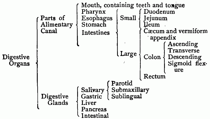
[pg 139]Coats of the Alimentary Canal.—The walls of the alimentary
canal, except at the mouth, are distinct from the surrounding tissues
and consist in most places of at least three layers, or coats, as
follows:

digestive system. 1. Mouth. 2. Soft palate. 3. Pharynx. 4. Parotid
gland. 5. Sublingual gland. 6. Submaxillary gland. 7. Esophagus. 8.
Stomach. 9. Pancreas. 10. Vermiform appendix. 11. Cæcum. 12. Ascending
colon. 13. Transverse colon. 14. Descending colon. 15. Sigmoid flexure.
16. Rectum. 17. Ileo-cæcal valve. 18. Duct from liver and pancreas. 19.
Liver.
Diagram does not show comparative length of the small intestine.
1. An inner coat, or lining, known
as the mucous membrane. This membrane is not confined to the alimentary
canal, but lines, as we have seen, the different air passages. It
covers, in fact, all those internal surfaces of the body that connect
with the external surface. It derives its name from the substance which
it secretes, called mucus. In
structure it resembles the skin, being continuous with the skin where
cavities open to the surface. It is made up of two layers—a thick
underlayer which contains blood vessels, nerves, and glands, and a thin
surface layer, called the epithelium.
The epithelium, like the cuticle, is without blood vessels, nerves, or
glands.
2. A middle coat, which is
muscular and which forms a continuous layer throughout the canal, except
at the mouth. (Here its place is taken by the strong muscles of
mastication which are separate and distinct from each other.) As a[pg 140] rule the muscles of this coat are
involuntary. They surround the canal as thin sheets and at most places
form two distinct layers. In the inner layer the fibers encircle the
canal, but in the outer layer they run longitudinally, or lengthwise,
along the canal.57
3. An outer or serous coat, which is limited to those
portions of the canal that occupy the abdominal cavity. This coat is not
found above the diaphragm. It is a part of the lining membrane of the
cavity of the abdomen, called

peritoneum. 1. Transverse colon. 2. Duodenum. 3. Small intestine.
4. Pancreas.
The Peritoneum.—The peritoneum is
to the abdominal cavity what the pleura is to the thoracic cavity. It
forms the outer covering for the alimentary canal and other abdominal
organs and supplies the inner lining of the cavity itself. It is also
the means of holding these organs in place, some of them being suspended
by it from the abdominal walls (Fig. 64). By the secretion of a small
amount of liquid, it prevents friction of the parts upon one
another.
Digestive Glands.—The glands which
provide the different fluids for acting on the foods derive their
constituents from the blood. They are situated either in the mucous
membrane or at convenient places outside of the[pg 141] canal and pass their liquids into
it by means of small tubes, called ducts. In the canal the food and the
digestive fluids come in direct contact—a condition which the dissolving
processes require. Each kind of fluid is secreted by a special kind of
gland and is emptied into the canal at the place where it is needed.
The Digestive Processes.—Digestion
is accomplished by acting upon the food in different ways, as it is
passed along the canal, with the final result of reducing it to the form
of a solution. Several distinct processes are necessary and they occur
in such an order that those preceding are preparatory to those that
follow. These processes are known as mastication, insalivation, deglutition, stomach digestion,
and intestinal digestion. As the
different materials become liquefied they are transferred to the blood,
and substances not reduced to the liquid state are passed on through the
canal as waste. The first two of the digestive processes occur in
The Mouth.—This is an oval-shaped
cavity situated at the very beginning of the canal. It is surrounded by
the lips in front, by the cheeks on the sides, by the hard palate above
and the soft palate behind, and by the tissues of the lower jaw below.
The mucous membrane lining the mouth is, soft and smooth, being covered
with flat epithelial cells. The external opening of the mouth is guarded
by the lips, and the soft palate forms a movable partition between the mouth and the pharynx. In a
condition of repose the mouth space is practically filled by the teeth
and the tongue, but the cavity may be enlarged and room provided for
food by depressing the lower jaw.
The mouth by its construction is well adapted to carrying on the
processes of mastication and insalivation. By the first process the
solid food is reduced, by the cutting[pg 142] and grinding action of the teeth,
to a finely divided condition. By the second, the saliva becomes mixed
with the food and is made to act upon it.

A. Section of a single molar. 1.
Pulp. 2. Dentine. 3. Enamel. 4. Crown. 5. Neck. 6. Root. B. Teeth in position in lower jaw. 1.
Incisors. 2. Canine. 3. Biscuspids. 4. Molars. C. Upper and lower teeth on one side. 1. Incisors. 2.
Canines. 3. Biscuspids. 4. Molars. 5. Wisdom. D. Upper and lower incisor, to show gliding contact.
Accessory Organs of the Mouth.—The
work of mastication and insalivation is accomplished through organs
situated in and around the mouth cavity. These comprise:
1. The Teeth.—The teeth are set in
the upper and lower jaws, one row directly over the other, with their
hardened surfaces facing. In reducing the food, the teeth of the lower
jaw move against those of the upper, while the food is held by the
tongue and cheeks between the grinding surfaces. The front teeth are
thin and chisel-shaped. They do not meet so squarely as do the back
ones, but their edges glide over each other, like the blades of
scissors—a condition that adapts them to cutting off and separating the
food (D, Fig. 65). The back teeth are
broad and irregular, having surfaces that are adapted to crushing and
grinding.
Each tooth is composed mainly of a bone-like substance, called dentine, which surrounds a central space,
containing blood vessels and[pg 143] nerves,
known as the pulp cavity. It is set
in a depression in the jaw where it is held firmly in place by a bony
substance, known as cement. The part
of the tooth exposed above the gum is the crown, the part surrounded by the gum is the neck, and the part which penetrates into
the jaw is the root (A, Fig. 65). A hard, protective material,
called enamel, covers the exposed
surface of the tooth.
The teeth which first appear are known as the temporary, or milk, teeth and are twenty in number, ten in
each jaw. They usually begin to appear about the sixth month, and they
disappear from the mouth at intervals from the sixth to the thirteenth
year. As they leave, teeth of the second, or permanent, set take their place. This set has thirty-two
teeth of four different kinds arranged in the two jaws as follows:
In front, above and below, are four chisel-shaped teeth, known as the
incisors. Next to these on either
side is a tooth longer and thicker than the incisors, called the canine. Back of these are two short,
rounded and double pointed teeth, the bicuspids, and back of the bicuspids are three heavy teeth
with irregular grinding surfaces, called the molars (B and C, Fig. 65). Since the molar farthest
back in each jaw is usually not cut until maturity, it is called a wisdom tooth. The molars are known as the
superadded permanent teeth because they do not take the place of milk
teeth, but form farther back as the jaw grows in length.

showing directions of muscular fibers in tongue.
2. The Tongue.—The tongue is a
muscular organ whose fibers extend through it in several directions
(Fig. 66). Its structure adapts it to a variety of movements. During
mastication the tongue transfers the food from one part of the mouth to
another, and, with the aid of the cheeks, holds the food between the
rows of teeth. (By an outward pressure from the tongue and an inward
pressure from the cheek the food is kept between the grinding surfaces.)
The tongue has functions in addition to these and is a most useful
organ.
[pg 144]3. The Muscles of Mastication.—These are attached to the lower
jaw and bring about its different movements. The masseter muscles, which are the heavy muscles in the
cheeks, and the temporal muscles,
located in the region of the temples, raise the lower jaw and supply the
force for grinding the food. Small muscles situated below the chin
depress the jaw and open the mouth.

glands and the ducts connecting them with the mouth.
4. The Salivary Glands.—These
glands are situated in the tissues surrounding the mouth, and
communicate with it by means of ducts (Fig. 67). They secrete the
saliva. The salivary glands are six in number and are arranged in three
pairs. The largest, called the parotid glands, lie, one on either side, in front of and
below the ears. A duct from each gland passes forward along the cheek
until it opens in the interior of the mouth, opposite the second molar
tooth in the upper jaw. Next in size to the parotids are the submaxillary glands. These are located,
one on either side, just below and in front of the triangular bend in
the lower jaw. The smallest of the salivary glands are the sublingual. They are situated in the
floor of the mouth, on either side, at the front and base of the tongue.
Ducts from the submaxillary and sublingual glands open into the mouth
below the tip of the tongue.
The Saliva and its Uses.—The saliva
is a transparent and somewhat slimy liquid which is slightly alkaline.
It[pg 145] consists chiefly of water (about
99 per cent), but in this are dissolved certain salts and an active
chemical agent, or enzyme, called ptyalin, which acts on the starch. The ptyalin changes
starch into a form of sugar (maltose), while the water in the saliva
dissolves the soluble portions of the food. In addition to this the
saliva moistens and lubricates the food which it does not dissolve, and
prepares it in this way for its passage to the stomach. The last is
considered the most important use of the saliva, and dry substances,
such as crackers, which require a considerable amount of this liquid,
cannot be eaten rapidly without choking. Slow mastication favors the
secretion and action of the saliva.
Deglutition.—Deglutition, or
swallowing, is the process by which food is transferred from the mouth
to the stomach. Though this is not, strictly speaking, a digestive
process, it is, nevertheless, necessary for the further digestion of the
food. Mastication and insalivation, which are largely mechanical,
prepare the food for certain chemical processes by which it is
dissolved. The first of these occurs in the stomach and to this organ
the food is transferred from the mouth. The chief organs concerned in
deglutition are the tongue, the pharynx, and the esophagus.
The Pharynx is a round and somewhat
cone-shaped cavity, about four and one half inches in length, which lies
just back of the nostrils, mouth, and larynx. It is remarkable for its
openings, seven in number, by means of which it communicates with other
cavities and tubes of the body. One of these openings is into the mouth,
one into the esophagus, one into the larynx, and one into each of the
nostrils, while two small tubes (the eustachian) pass from the upper
part of the pharynx to the middle ears.
The pharynx is the part of the food canal that is crossed[pg 146] by the passageway for the air. To
keep the food from passing out of its natural channel, the openings into
the air passages have to be carefully guarded. This is accomplished
through the soft palate and epiglottis, which are operated somewhat as
valves. The muscular coat of the pharynx is made up of a series of
overlapping muscles which, by their contractions, draw the sides
together and diminish the cavity. The mucous membrane lining the pharynx
is smooth, like that of the mouth, being covered with a layer of flat
epithelial cells.
The Esophagus, or gullet, is a tube
eight or nine inches long, connecting the pharynx with the stomach. It
lies for the most part in the thoracic cavity and consists chiefly of a
thick mucous lining surrounded by a heavy coat of muscle. The muscular
coat is composed of two layers—an inner layer whose fibers encircle the
tube and an outer layer whose fibers run lengthwise.
Steps in Deglutition.—The process
of deglutition varies with the kind of food. With bulky food it consists
of three steps, or stages, as follows: 1. By the contraction of the
muscles of the cheeks, the food ball, or bolus, is pressed into the
center of the mouth and upon the upper surface of the tongue. Then the
tongue, by an upward and backward movement, pushes the food under the
soft palate and into the pharynx.
2. As the food passes from the mouth, the pharynx is drawn up to
receive it. At the same time the soft palate is pushed upward and
backward, closing the opening into the upper pharynx, while the
epiglottis is made to close the opening into the larynx. By this means
all communication between the food canal and the air passages is
temporarily closed. The upper muscles of the pharynx now contract upon
the food, forcing it downward and into the esophagus.
3. In the esophagus the food is forced along by the successive
contractions of muscles, starting at the upper end of the tube, until
the stomach is reached.
Swallowing is doubtless aided to some extent by the force of
gravity. [pg 147]That it is independent of this
force, however, is shown by the fact that one may swallow with the
esophagus in a horizontal position, as in lying down.

Glands. A. Single gland showing
the two kinds of secreting cells and the duct where the gland opens on
to the surface. B. Inner surface of
stomach magnified. The small pits are the openings from the glands.
The Stomach.—The stomach is the
largest dilatation of the alimentary canal. It is situated in the
abdominal cavity, immediately below the diaphragm, with the larger
portion toward the left side. Its connection with the esophagus is known
as the cardiac orifice and its
opening into the small intestine is called the pyloric orifice. It varies greatly in size in different
individuals, being on the average from ten to twelve inches at its
greatest length, from four to five inches at its greatest width, and
holding from three to five pints. It has the coats common to the canal,
but these are modified somewhat to adapt them to its work.
The mucous membrane of the stomach
is thick and highly developed. It contains great numbers of minute
tube-shaped bodies, known as the gastric
glands (Fig. 68). These are of two general kinds and secrete large
quantities of a liquid called the gastric juice. When the stomach is
empty, the mucous membrane is thrown into folds which run lengthwise
over the inner surface. These disappear, however, when the walls of the
stomach are distended with food.
[pg 148]The muscular coat consists of three separate layers which are named,
from the direction of the fibers, the circular layer, the longitudinal
layer, and the oblique layer (Fig. 69).
The circular layer becomes quite thick at the pyloric orifice,
forming a distinct band which serves as a valve.
. The layer of Longitudinal fibers removed.")
stomach (from Morris’ Human
Anatomy). The layer of Longitudinal fibers removed.
The outer coat of the stomach, called the serous coat, is a continuation of the peritoneum, the
membrane lining the abdominal cavity.
Stomach Digestion.—In the stomach
begins the definite work of dissolving those foods which are insoluble
in water. This, as already stated, is a double process. There is first a
chemical action in which the insoluble are changed into soluble
substances, and this is followed immediately by the dissolving action of
water. The chief substances digested in the stomach are the proteids.
These, in dissolving, are changed into two soluble substances, known
as[pg 149] peptones and proteoses.
The digestion of the proteids is, of course, due to the
Gastric Juice.—The gastric juice is
a thin, colorless liquid composed of about 99 per cent of water and
about 1 per cent of other substances. The latter are dissolved in the
water and include, besides several salts, three active chemical
agents—hydrochloric acid, pepsin, and rennin. Pepsin is the enzyme which acts upon proteids, but it is
able to act only in an acid medium—a condition which is supplied by the
hydrochloric acid. Mixed with the
hydrochloric acid it converts the proteids into peptones and
proteoses.
Other Effects of the Gastric
Juice.—In addition to digesting proteids, the gastric juice brings
about several minor effects, as follows:
1. It checks, after a time, the digestion of the starch which was
begun in the mouth by the saliva.58 This is due to the presence of the
hydrochloric acid, the ptyalin being unable to act in an acid
medium.
2. While there is no appreciable action on the fat itself, the
proteid layers that inclose the fat particles are dissolved away (Fig.
79), and the fat is set free. By this means the fat is broken up and
prepared for a special digestive action in the small intestine.
3. Dissolved albumin, like that in milk, is curded, or coagulated, in
the stomach. This action is due to the rennin. The curded mass is then acted upon by the pepsin
and hydrochloric acid in the same manner as the other proteids.
[pg 150]4. The hydrochloric acid acts on certain of the insoluble mineral
salts found in the foods and reduces them to a soluble condition.
5. It is also the opinion of certain physiologists that cane sugar
and maltose (double sugars) are converted by the hydrochloric acid into
dextrose and levulose (single sugars).
After a variable length of time, the contents of the stomach is
reduced to a rather uniform and pulpy mass which is called chyme. Portions of this are now passed at
intervals into the small intestine.
Muscular Action of the Stomach.—The
muscles in the walls of the stomach have for one of their functions the
mixing of the food with the gastric juice. By alternately contracting and relaxing, the different layers
of muscle keep the form of the stomach changing—a result which agitates
and mixes its contents. This action varies in different parts of the
organ, being slight or entirely absent at the cardiac end, but quite
marked at the pyloric end.
Another purpose of the muscular coat is to empty the stomach into the
small intestine. During the greater part of the digestive period the
muscular band at the pyloric orifice is contracted. At intervals,
however, this band relaxes, permitting a part of the contents of the
stomach to be forced into the small intestine. After the discharge the
pyloric muscle again contracts, and so remains until the time arrives
for another discharge.
In addition to emptying the stomach into the small intestine, these
muscles also aid in emptying the organ upward and through the esophagus
and mouth, should occasion require. Vomiting in case of poisoning, or if
the food for some reason fails to digest, is a necessary though
unpleasant operation. It is accomplished by the[pg 151] contraction of all the muscles of
the stomach, together with the contraction of the walls of the abdomen.
During these contractions the pyloric valve is closed, and the muscles
of the esophagus and pharynx are in a relaxed condition.59

stomach into small intestine. Illustration also shows arrangement
of mucous membrane in the two organs. D. Bile duct.
The Small Intestine.—This division
of the alimentary canal consists of a coiled tube, about twenty-two feet
in length, which occupies the central, lower portion of the abdominal
cavity (Fig. 71). At its upper extremity it connects with the pyloric
end of the stomach (Fig. 70), and at its lower end it joins the large
intestine. It averages a little over an inch in diameter, and gradually
diminishes in size from the stomach to the large intestine. The first
eight or ten inches form a short curve, known as the duodenum. The upper two fifths of the
remainder is called the jejunum, and
the lower three fifths is known as the ileum. The ileum joins that part of the large intestine
known as the cæcum, and at their place of union is a marked constriction
which prevents material from passing from the large into the small
intestine (Fig. 73). This is known as the ileo-cæcal valve.
The mucous membrane of the small
intestine is richly supplied with blood vessels and contains glands that
secrete[pg 152] a digestive fluid known as the
intestinal juice. The membrane is
thrown into many transverse, or circular, folds which increase its
surface and also prevent materials from passing too rapidly through the
intestine. One important respect in which the small intestine differs
from all other portions of the food canal is that its surface is covered
with great numbers of minute elevations known as the villi. The purpose
of these is to aid in the absorption of the nutrients as they become
dissolved (Chapter XI).
The muscular coat of the small
intestine is made up of two distinct layers—the inner layer consisting
of circular fibers and the outer of longitudinal fibers. These muscles
keep the food materials mixed with the juices of the small intestine,
but their main purpose is to force the materials undergoing digestion
through this long and much-coiled tube.
The outer, or serous, coat of the
small intestine, like that of the stomach, is an extension from the
general lining of the abdominal cavity, or peritoneum. In fact, the
intestine lies in a fold of the peritoneum, somewhat as an arm in a
sling, while the peritoneum, by connecting with the back wall of the
abdominal cavity, holds this great coil of digestive tubing in place
(Fig. 64). The portion of the peritoneum which attaches the intestine to
the wall of the abdomen is called the mesentery.
Most of the liquid acting on the food in the small intestine is
supplied by two large glands, the liver and the pancreas, that connect
with it by ducts.

in position.
The Liver is situated immediately
below the diaphragm, on the right side (Figs. 71 and 72), and is the
largest gland in the body. It weighs about four pounds and is separated
into two main divisions, or lobes. It is complex in structure and
differs from the other glands in several particulars. It receives blood
from two distinct sources—the portal vein[pg 154] and the hepatic artery. The portal vein collects the blood from
the stomach, intestines, and spleen, and passes it to the liver. This
blood is loaded with food materials, but contains little or no oxygen.
The hepatic artery, which branches
from the aorta, carries to the liver blood rich in oxygen. In the liver
the portal vein and the hepatic artery divide and subdivide, and finally
empty their blood into a single system of capillaries surrounding the
liver cells. These capillaries in turn empty into a single system of
veins which, uniting to form the hepatic
veins (two or three in number), pass the blood into the inferior
vena cava (Fig. 72).

liver. Diagram showing the connection of the liver with the large
blood vessels and the food canal.
The liver secretes daily from one to two pounds of a liquid called
bile. A reservoir for the bile is
provided by a small, membranous sack, called the gall bladder, located on the underside of the liver. The
bile passes from the gall bladder, and from the right and left lobes of
the liver, by three separate ducts. These unite to form a common tube
which, uniting with the duct from the pancreas, empties into the
duodenum. Though usually described as a digestive gland, the liver has
other functions of equal or greater importance (Chapter XIII).
[pg 155]The
Bile is a golden yellow liquid, having a slightly alkaline reaction
and a very bitter taste. It consists, on the average, of about 97 per
cent of water and 3 per cent of solids.60 The solids include bile
pigments, bile salts, a substance called cholesterine, and mineral
salts. The pigments (coloring matter) of the bile are derived from the
hemoglobin of broken-down red corpuscles (page 27).
Much about the composition of the bile is not understood. It is
known, however, to be necessary to digestion, its chief use being to aid
in the digestion and absorption of fats. It is claimed also that the
bile aids the digestive processes in some general ways—counteracting the
acid of the gastric juice, preventing the decomposition of food in the
intestines, and stimulating muscular action in the intestinal walls. No
enzymes have been discovered in the bile.
The Pancreas is a tapering and
somewhat wedge-shaped gland, and is so situated that its larger
extremity, or head, is encircled by the duodenum. From here the more
slender portion extends across the abdominal cavity nearly parallel to
and behind the lower part of the stomach. It has a length of six or
eight inches and weighs from two to three and one half ounces. Its
secretion, the pancreatic juice, is emptied into the duodenum by a duct
which, as a rule, unites with the duct from the liver.
The Pancreatic Juice is a colorless
and rather viscid liquid, having an alkaline reaction. It consists of
about 97.6 per cent of water and 2.4 per cent of solids. The solids
include mineral salts (the chief of which is sodium carbonate) and four
different chemical agents, or enzymes,—trypsin, amylopsin, steapsin, and
a milk-curding enzyme. These active constituents make of the pancreatic
juice the[pg 156] most important of the digestive
fluids. It acts with vigor on all of the nutrients insoluble in water,
producing the following changes:
1. It converts the starch into maltose, completing the work begun by
the saliva. This action is due to the amylopsin,61 which is similar to ptyalin but is more
vigorous.
2. It changes proteids into peptones and proteoses, completing the
work begun by the gastric juice. This is accomplished by the trypsin, which is similar to, but more
active than, the pepsin.
3. It digests fat. In this work the active agent is the steapsin.
The necessity of a milk-curding enzyme, somewhat similar to the
rennin of the gastric juice, is not understood.
Digestion of Fat.—Several theories
have been proposed at different times regarding the digestion and
absorption of fat. Among these, what is known as the “solution theory”
seems to have the greatest amount of evidence in its favor. According to
this theory, the fat, under the influence of the steapsin, absorbs water
and splits into two substances, recognized as glycerine and fatty acid.
This finishes the process so far as the glycerine is concerned, as this
is soluble in water; but the fatty acid, which (from certain fats) is
insoluble in water,62 requires further treatment. The fatty acid is now
supposed to be acted on in one, or both, of the following ways: 1. To be
dissolved as fatty acid by the action of the bile (since bile is
capable[pg 157] of dissolving it under certain
conditions). 2. To be converted by the sodium carbonate into a form of
soap which is soluble in water.
The emulsification of fat is known to occur in the small intestine.
By this process the fat is separated into minute particles which are
suspended in water, but not changed chemically, the mixture being known
as an emulsion. While this is
believed by some to be an actual process of digestion, the advocates of
the solution theory claim that it is a process accompanying and aiding
the conversion of fat into fatty acid and glycerine.63
The Intestinal Juice is a clear
liquid with an alkaline reaction, containing water, mineral salts, and
certain proteid substances that may act as enzymes. It assists in
bringing about an alkaline condition in the small intestine and aids in
the reduction of cane sugar and maltose to the simple sugars, dextrose
and levulose. Since it is difficult to obtain this liquid in sufficient
quantities for experimenting, its uses have not been fully determined.
Recent investigators, however, assign to it an important place in the
work of digestion.
Work of the Small Intestine.—The
small intestine is the most important division of the alimentary canal.
It serves as a receptacle for holding the food while it is being acted
upon; it secretes the intestinal juice and mixes the food with the
digestive fluids; it propels the food toward the large intestine; and,
in addition to all this, serves as an organ of absorption.
Digestion is practically finished in the small intestine, and a large
portion of the reduced food is here absorbed. There is always present,
however, a variable amount of material that is not digested. This,
together with a considerable volume of liquid, is passed into
[pg 158]The
Large Intestine.—The large intestine is a tube from five to six
feet in length and averaging about one and one half inches in diameter.
It begins at the lower right side of the abdominal cavity, forms a coil
which almost completely surrounds the coil of small intestine, and
finally terminates at the surface of the body (Figs. 2, 71 and 73). It
has three divisions, known as the cæcum, the colon, and the rectum.

small into large intestine. At the ileo-cæcal valve is the narrowest
constriction of the food canal.
The cæcum is the pouch-like
dilatation of the large intestine which receives the lower end of the
small intestine. It measures about two and one half inches in diameter
and has extending from one side a short, slender, and blind tube, called
the vermiform appendix. This
structure serves no purpose in digestion, but appears to be the rudiment
of an organ which may have served a purpose at some remote period in the
history of the human race. The cæcum gradually blends into the second
division of the large intestine, called the colon.
The colon consists of four parts,
described as the ascending colon, the transverse colon, the descending
colon, and the sigmoid flexure, or sigmoid colon. The first three
divisions are named from the direction of the movement of materials
through them and the last from its shape, which is similar to that of
the Greek letter sigma (Σ).
The rectum is the last division of
the large intestine[pg 159] It is a nearly straight tube,
from six to eight inches in length, and connects with the external
surface of the body.
The general structure of the large intestine is similar to that of
the small intestine, and, like the small intestine, it is held in place
by the peritoneum. It differs from the small intestine, however, in its
lining of mucous membrane and in the arrangement of the muscular coat.
The mucous membrane presents a smooth appearance and has no villi, while
the longitudinal layer of the muscular coat is limited to three narrow
bands that extend along the greater length of the tube (Fig. 74). These
bands are shorter than the coats, and draw the large intestine into a
number of shallow pouches, by which it is readily distinguished from the
small intestine (Fig. 71).

intestine, showing the coats. 1. Serous coat. 2. Circular layer of
muscle. 3. Submucous coat. 4. Mucous membrane. 5. Muscular bands
extending lengthwise over the intestine.
Work of the Large Intestine.—The
large intestine serves as a receptacle for the materials from the small
intestine. The digestive fluids from the small intestine continue their
action here, and the dissolved materials also continue to be absorbed.
In these respects the work of the large intestine is similar to that of
the small intestine. It does, however, a work peculiar to itself in that
it collects and retains undigested food particles, together with other
wastes, and ejects them periodically from the canal.
Work of the Alimentary Muscles.—The
mechanical part of digestion is performed by the muscles that encircle
the food canal. Their uses, which have already been mentioned in
connection with the different organs of[pg 160] digestion, may be here
summarized: They supply the necessary force for masticating the food.
They propel the food through the canal. They mix the food with the
different juices. At certain places they partly or completely close the
passage until a digestive process is completed. They may even cause a
reverse movement of the food, as in vomiting. All of the alimentary
muscles, except those around the mouth, are involuntary. Their work is
of the greatest importance.
Other Purposes of the Digestive
Organs.—The digestive organs serve other important purposes besides
that of dissolving the foods. They provide favorable conditions for
passing the dissolved material into the blood. They dispose of such
portions of the foods as fail, in the digestive processes, to be reduced
to a liquid state. A considerable amount of waste material is also
separated from the blood by the glands of digestion (especially the
liver), and this is passed from the body with the undigested portions of
food. Then the food canal (stomach in particular) is a means of holding,
or storing, food which is awaiting the processes of digestion.
Considering the number of these purposes, the digestive organs are
remarkably simple, both in structure and in method of operation.
HYGIENE OF DIGESTION
Many of the ills to which flesh is heir are due to improper methods
of taking food and are cured by observing the simple rules of eating.
Habit plays a large part in the process and children should, for this
reason, be taught early to eat properly. Since the majority of the
digestive processes are involuntary and the food, after being swallowed,
is practically beyond control, careful attention must be given to the
proper mastication of the food and to such other phases of digestion as
are under control.
Necessity for Thorough
Mastication.—Mastication prepares the food for the digestive
processes which follow. Unless the food has been properly masticated,
the digestive[pg 161] fluids in the stomach
and intestines cannot act upon it to the best advantage. When the food
is carefully chewed, a larger per cent of it is actually digested—a
point of importance where economy in the use of food needs to be
practiced.
A fact not to be overlooked is that one cannot eat hurriedly and
practice thorough mastication. The food must not be swallowed in lumps,
but reduced to a finely divided and pulpy mass. This requires time. The
one who hurries through the meal is necessarily compelled to bolt his
food. Thirty minutes is not too long to give to a meal, and a longer
period is even better.
Perhaps the most important result of giving plenty of time to the
taking of food is that of stimulating the
digestive glands to a proper degree of activity. That both the
salivary and gastric glands are excited by the sight, smell, and thought
of food and, through taste, by the presence of food in the mouth, has
been fully demonstrated. Food that is thoroughly masticated and relished
will receive more saliva and gastric juice, and probably more of other
juices, than if hastily chewed and swallowed. This has a most important
bearing upon the efficiency of the digestive processes.
Order of Taking Food.—There has
been evolved through experience a rather definite order of taking food,
which our knowledge of the process of digestion seems to justify. The
heavy foods (proteids for the most part) are eaten first; after which
are taken starchy foods and fats; and the meal is finished off with
sweetmeats and pastry.64 The scientific arguments for this order are
the following:
1. By receiving the first of the gastric flow the proteids can
begin[pg 162] digesting without delay. Since
these are the main substances acted on in the stomach, the time required
for their digestion is shortened by eating them first.
2. Sugar, being of the nature of predigested starch, quickly gets
into the blood and satisfies the
relish for food. The result of taking sugar first may be to cause
one to eat less than he needs and to diminish the activity of the
glands.
3. Fat or grease, if taken first, tends to form a coating over the
walls of the stomach and around the material to be digested. This
prevents the juices from getting to and mixing with the foods upon which
they are to act.
4. Starch following the proteids, for the most part, does not so
quickly come in contact with the gastric juice. This enables the ptyalin
of the saliva to continue its action for a longer time than if the
starch were eaten first.
Liquids during the Meal.—Liquids as
ordinarily taken during the meal are objectionable. They tend to
diminish the secretion of the saliva and to cause rapid eating. Instead
of eating slowly and swallowing the food only so fast as the glands can
supply the necessary saliva, the liquid is used to wash the food down.
Water or other drinks should be taken after the completion of the meal
or when the mouth is completely free from food. Even then it should be
taken in small sips. While the taking of a small amount of water in this
way does no harm, a large volume has the effect of weakening the gastric
juice. Most of the water needed by the body should be taken between
meals.
The State of Mind has much to do
with the proper digestion of the food. Worry, anger, fear, and other
disturbed mental states are known to check the secretion of fluids and
to interfere with the digestive processes. While the cultivation of
cheerfulness is important for its general hygienic effects, it is of
especial value in relation to digestion. Intense emotions, either during
or following the[pg 163] meal, should if possible be
avoided. The table is no place for settling difficulties or
administering rebuke. The conversation, on the other hand, should be
elevating and joy giving, thereby inducing a desirable reactionary
influence upon the digestive processes.
Care of the Teeth.—The natural
teeth are indispensable for the proper mastication of the food. Of
especial value are the molars—the teeth that grind the food. The
development of the profession of dentistry has made possible the
preservation of the teeth, even when naturally poor, as long as one has
need of them. To preserve the teeth they must be kept clean. They should
be washed at least once a day with a soft-bristled brush, and small
particles of food, lodged between them, should be removed with a wooden
pick. The biting of hard substances, such as nuts, should be avoided, on
account of the danger of breaking the enamel, although the chewing of
tough substances is considered beneficial.
Decayed places in the teeth should be promptly filled by the dentist.
It is well, even when decayed places are not known to exist, to have the
teeth examined occasionally in order to detect such places before they
become large. On account of the expense, pain, and inconvenience there
is a tendency to put off dental work which one knows ought to be done.
Perhaps in no other instance is procrastination so surely punished. The
decayed places become larger and new points of decay are started; and
the pain, inconvenience, and expense are increased proportionately.
The Natural Appetite should be
followed with reference to both the kind and the amount of food eaten.
No system of knowledge will ever be devised which can replace the
appetite as an aid in the taking of food. It is[pg 164] nature’s means of indicating the needs of the body. The
natural appetite may be spoiled, however, by overeating and by the use
of highly seasoned foods, or by indulging in stimulants during the meal.
It is spoiled in children by too free indulgence in sweetmeats. By
cultivating the natural appetite and heeding its suggestions, one has at
his command an almost infallible guide in the taking of food.
Preparation of Meals.—The cooking
of food serves three important purposes. It renders the food more
digestible, relieving the organs of unnecessary work; it destroys
bacteria that may be present in the food, diminishing the likelihood of
introducing disease germs into the body; and it makes the food more
palatable, thereby supplying a necessary stimulus to the digestive
glands. While the methods employed in the preparation of the different
foods have much to do with the ease with which they are digested and
with their nourishing qualities, the scope of our subject does not
permit of a consideration of these methods.
Quantity of Food.—Overeating and
undereating are both objectionable from a hygienic standpoint.
Overeating, by introducing an unnecessary amount of food into the body,
overworks the organs of digestion and also the organs of excretion. It
may also lead to the accumulation of burdensome fat and of harmful
wastes. On the other hand, the taking of too little food impoverishes
the blood and weakens the entire body. As a rule, however, more people
eat too much than too little, and to quit eating before the appetite is
fully satisfied is with many persons a necessary precaution. The power
of self-control, valuable in all phases of life, is indispensable in the
avoidance of overeating.
Frequency of Taking Food.—Eating between
meals is manifestly an unhealthful practice. The question has also been
raised as to whether the common habit of eating three times a day is
best suited to all classes of people. Many people of weak digestive
organs have been benefited by the plan of two meals a day, while others
adopt the plan of eating one heavy meal and two light ones. Either plan
gives the organs of digestion more time to rest and diminishes the
liability of overeating. On the other hand, those doing heavy muscular
work can hardly derive the energy which they need from less than three
good meals a day. Though no definite rule can be laid down, there is
involved a hygienic principle which all should follow: Meals should not overlap. The stomach
should be free from food taken at a previous meal before more is
introduced into it. When this principle is not observed, material
ferments in the stomach, causing indigestion and other disorders. It
should be noted, however, that the overlapping may be due to overeating
as well as to eating too frequently.
Dangers from Impure Food.—Food is
frequently the carrier of disease germs and for this reason requires
close inspection (page 128). Typhoid fever, a most dangerous disease, is
usually contracted through either impure food or impure water (Chapter
XXIII). One safeguard against disease germs, as stated above, is
thorough cooking. Too much care cannot be exercised with reference to
the water for drinking purposes. Water which is not perfectly clear,
which smells of decaying material, or which forms a sediment on standing
is usually not fit to drink. It can, however, be rendered comparatively
harmless by boiling. The objections which many people have to drinking
boiled water are removed when it is boiled the day before it is[pg 166] used, so as to give it time to
cool, settle, and replace the air driven off by the boiling.
Care of the Bowels.—In considering
the hygiene of the alimentary canal, the fact that it is used as a means
of separating the impurities from the body must not be overlooked.
Frequently, through lack of exercise, negligence in evacuating the
bowels, or other causes, a weakened condition of the canal is induced
which results in the retention of impurities beyond the time when they
should be discharged. This is a great annoyance and at the same time a
menace to the health.
In most cases this condition can be relieved, and prevented from
recurring, by observing the following habits: 1. Have a regular time
each day for evacuating the bowels. This is a most important factor in
securing the necessary movements. 2. Drink a cup of cold water on rising
in the morning and on retiring at night. 3. Eat generously of fruits and
other coarse foods, such as corn bread, oatmeal, hominy, cabbage, etc.
4. Practice persistently such exercises as bring the abdominal muscles
into play. These exercises strengthen indirectly the muscles of the
canal. 5. Avoid overwork, especially of the nervous system.
Alcohol and Digestion.—Though
exciting temporarily a greater flow of the digestive fluids, alcoholic
drinks taken in any but very small quantities are considered detrimental
to the work of digestion. Large doses retard the action of enzymes,
inflame the mucous lining of the stomach,65 and[pg 167] bring about a diseased condition
of the liver. It may be noted, however, that the bad effects of
alcoholic beverages upon the stomach, the liver, and the body in general
are less pronounced when these are taken as a part of the regular
meals.
Effects of Tea and Coffee.—In
addition to the stimulating agent caffeine, tea and coffee contain a
bitter, astringent substance, known as tannin. On account of the tannin
these beverages tend to retard digestion and to irritate the lining of
the stomach—effects that may be largely obviated by methods of
preparing tea and coffee which dissolve little of the tannin. (They
should be made without continued boiling or steeping.) The
caffeine may do harm through its stimulating effect upon the
nervous system (page 56) and through the introduction of a special
waste into the body. In chemical composition caffeine closely resembles
a waste, called uric acid, and in the body is converted into this
substance. If one is in a weakened condition, the uric acid may fail to
be oxidized to urea, as occurs normally, or to be thrown off as uric
acid. In this case it accumulates in the body, causing rheumatism and
related diseases. It thus happens that while some people may use tea and
coffee without detriment, others are injured by them.
Summary.—The main structure in the
digestive system is the alimentary canal. This provides cavities where
important dissolving processes take place, and tubes for joining these
cavities, while glands connecting with the canal supply the necessary
liquids for changing and dissolving the foods. The general plan of
digestion is that of passing the food through the canal, beginning with
the mouth, and of acting on it at various places, with the final result
of reducing most of it to the liquid state. The digestive fluids[pg 168] supply water which acts as a solvent and
carries the active chemical agents, or enzymes, that convert the
insoluble foods into substances that are soluble. The muscles in the
walls of the canal perform the mechanical work of digestion, while the
nervous system controls and regulates the activity of the various organs
concerned in this work.
Exercises.—1. State the general purpose of digestion. How does
digested food differ from that not digested?
2. Name all the divisions of the alimentary canal in the order in
which the food passes through them.
3. What other work besides digestion is carried on by the alimentary
canal?
4. What is gained by the mastication of the food? Why should
mastication precede the other processes of digestion?
5. What is the work of the tongue in digestion?
6. State the purposes served by the gastric juice.
7. Give reasons for regarding the small intestine as the most
important division of the food canal.
8. At what places, and by the action of what liquids, are fats,
proteids, and starch digested?
9. What enzymes are found in the pancreatic juice? What is the
digestive action of each?
10. Describe the work performed by the muscles of the stomach, the
mouth, the esophagus, and the small intestine.
11. What advantages are derived from the use of cooked food?
12. State the advantages of drinking pure water.
13. If all the food that one needs to take at a single meal can be
thoroughly masticated in fifteen minutes, why is it better to spend a
longer time at the table?
14. What is meant by the overlapping of meals? What bad results
follow? How avoided?
PRACTICAL WORK
Examine a dissectible model of the human abdomen (Fig. 75), noting
the form, location, and connection of the different organs. Find the
connection of the esophagus with the stomach, of the stomach with the
small intestine, and of the small intestine with the large
intestine.[pg 169] Sketch a general outline of the
cavity, and locate in this outline its chief organs.
Where it is desirable to learn something of the actual structure of
the digestive organs, the dissection of the abdomen of some small animal
is necessary. On account of unpleasant features likely to be associated
with such a dissection, however, this work is not recommended for
immature pupils.

contents.
Dissection of the Abdomen.
(Optional)—For individual study, or for a small class, a half-grown cat
is perhaps the best available material. It should be killed with
chloroform, and then stretched, back downward, on a board, the feet
being secured to hold it in place.
The teacher should make a preliminary examination of the abdomen to
see that it is in a fit condition for class study. If the bladder is
unnaturally distended, its contents may be forced out by slight
pressure. The following materials will be needed during the dissection,
and should be kept near at hand: a sharp knife with a good point, a pair
of heavy scissors, a vessel of water, some cotton or a damp sponge, and
some fine cord. During the dissection the specimen should be kept as
clean as possible, and any escaping blood should be mopped up with the
cotton or the sponge. The dissection is best carried out by observing
the following order:
1. Cut through the abdominal wall in the center of the triangular
space where the ribs converge. From here cut a slit downward to the
lower portion of the abdomen, and sideward as far as convenient. Tack
the loosened abdominal walls to the board, and proceed to study the
exposed parts. Observe the muscles in the abdominal walls, and the fold
of the peritoneum which forms an
apron-like covering over the intestines.
2. Observe the position of the stomach, liver, spleen, and
intestines, and then, by pushing the intestines to one side, find the
kidneys and the bladder.
3. Study the liver with reference to its location, size, shape, and
color. On the under side, find the gall bladder, from which a small tube
leads to the small intestine. Observe the portal vein as it passes into
[pg 170] the liver. As the liver is filled
with blood, neither it nor its connecting blood vessels should be cut at
this time.
4. Trace out the continuity of the canal. Find the esophagus where it
penetrates the diaphragm and joins the stomach. Find next the union of
the stomach with the small intestine. Then, by carefully following the
coils of the small intestine, discover its union with the large
intestine.
5. Within the first coil of the small intestine, as it leaves the
stomach, find the pancreas. Note its
color, size, and branches. Find its connection with the small
intestine.
6. Beginning at the cut portion of the abdominal wall, lift the thin
lining of the peritoneum and carefully follow it toward the back and
central portion of the abdomen. Observe whether it extends back of or in
front of the kidneys, the aorta, and the inferior vena cava. Find where
it leaves the wall as a double
membrane, the mesentery, which
surrounds and holds in place the large and small intestines. Sketch a
coil of the intestine, showing the mesentery.
7. Find in the center of the coils of small intestine a long, slender
body having the appearance of a gland. This is the beginning of the thoracic duct and is called the receptacle of the chyle. From this the
thoracic duct rapidly narrows until it forms a tiny tube difficult to
trace in a small animal.
8. Cut away about two inches of the small intestine from the
remainder, having first tied the tube on the two sides of the section
removed. Split it open for a part of its length, and wash out its
contents. Observe its coats. Place it in a shallow vessel containing
water, and examine the mucous membrane with a lens to find the villi. Make a drawing of this section,
showing the coats.
9. Study the connection of the small intestine with the large. Split
them open at the place of union, wash out the contents, and examine the
ileo-cæcal valve.
10. Observe the size, shape, and position of the kidneys. Do they lie
in front of or back of the peritoneum? Do they lie exactly opposite each
other? Note the connection of each kidney with the aorta and the
inferior vena cava by the renal artery and the renal vein. Find a
slender tube, the ureter, running
from each kidney to the bladder. Do the ureters connect with the top or
with the base of the bladder? Show by a sketch the connection of the
kidneys with the large blood vessels and the bladder.
[pg 171]To
demonstrate the Teeth.—Procure from the dentist a collection of
different kinds of teeth, both sound and decayed.
(a) Examine external surfaces of
different kinds of teeth, noting general shape, cutting or grinding
surfaces, etc. Make a drawing of an incisor and also of a molar.
(b) After soaking some of the
teeth for a couple of days in warm water saw one of them in two
lengthwise, and another in two crosswise, and smooth the cut surfaces
with fine emery or sand paper. Examine both kinds of sections, noting
arrangement and extent of dentine, enamel, and pulp. Make drawings.
(c) Examine a decayed tooth. Which
substance of the tooth appears to decay most readily? Why is it
necessary to cut away a part of the tooth before filling?
(d) Test the effect of acids upon
the teeth by leaving a tooth over night in a mixture of one part
hydrochloric acid to four parts water, and by leaving a second tooth for
a couple of days in strong vinegar. Examine the teeth exposed to the
action of acids, noting results.
To show the Importance of
Mastication.—Fill two tumblers each half full of water. Into one
put a lump of rock salt. Into the other place an equal amount of salt
that has been finely pulverized. Which dissolves first and why?
To illustrate Acid and Alkaline
Reactions.—To a tumbler half full of water add a teaspoonful of
hydrochloric or other acid, as vinegar. To a second tumbler half full of
water add an equal amount of cooking soda. Taste each liquid, noting the
sour taste of the acid, and the alkaline taste of the soda. Hold a piece
of red litmus paper in the soda solution, noting that it is turned blue.
Then hold a piece of blue litmus paper in the acid solution, noting that
it is turned red. Add acid to the soda solution, and soda to the acid
solution, until the conditions are reversed, testing with the red and
blue litmus papers.
Hold, for a minute or longer, a narrow strip of red litmus paper in
the mouth, noting any change in the color of the paper. Repeat, using
blue litmus paper. What effect, if any, has the saliva upon the color of
the papers? Has the mouth an acid or an alkaline reaction?
To show the Action of Saliva on
Starch.—1 (Optional). Prepare starch paste by mixing half a
teaspoonful of starch in half a pint of water and heating the mixture to
boiling. Place some of this in a test tube and thin it by adding more
water. Then add a small drop of[pg 172] iodine solution (page 136) to the
solution of starch. It should turn a deep blue color. This is the test
for starch.
Now collect from the mouth, in a clean test tube, two or three
teaspoonfuls of saliva. Add portions of this to small amounts of fresh
starch solution in two test tubes. Let the tubes stand for five or ten
minutes surrounded by water having about the temperature of the body.
Test for changes that have occurred as follows:
(a) To one tube add a little of
the iodine solution. If it does not turn blue, it shows that the starch
has been converted into some other substance by the saliva, (b) To the other tube add a few drops of a
very dilute solution of copper sulphate. Then add sodium (or potassium)
hydroxide, a few drops at a time, until the precipitate which first
forms dissolves and turns a deep blue. Then gradually heat the upper
portion of the liquid to boiling. If it turns an orange or yellowish red
color, the presence of a form of sugar (maltose or dextrose) is proved.
See page 136.
2. Hold some powdered starch in the mouth until it completely
dissolves and observe that it gradually acquires a sweetish taste. This
shows the change of starch into sugar.
To illustrate the Action of the Gastric
Juice.—Add to a tumbler two thirds full of water as much scale
pepsin (obtained from a drug store) as will stay on the end of the large
blade of a penknife. Then add enough hydrochloric acid to give a
slightly sour taste. Place in the artificial gastric juice thus prepared
some boiled white of egg which has been finely divided by pressing it
through a piece of wire gauze. Also drop in a single large lump. Keep in
a warm place (about the temperature of the body) for several hours or a
day, examining from time to time. What is the general effect of the
artificial gastric juice upon the egg?
To illustrate Effect of Alcohol upon
Gastric Digestion.—Prepare a tumbler half full of artificial
gastric juice as in the above experiment, and add 10 cubic centimeters
of this to each of six clean test tubes bearing labels. To five of the
tubes add alcohol from a burette as follows: (1) .5 c.c., (2) 1 c.c.,
(3) 1.5 c.c., (4) 2 c.c., and (5) 3 c.c., leaving one tube without
alcohol. Now add to each tube about 1/4 gram of finely divided white of
egg from the experiment above, and place all of the tubes in a beaker
half full of water. Keep the water a little above the temperature of the
body for several hours, examining the tubes at intervals to note the
progress of digestion. Inferences.
[pg 173]
CHAPTER XI – ABSORPTION, STORAGE, AND ASSIMILATION
The dissolved nutrients, to reach the cells, must be transferred from
the alimentary canal to the blood stream. This process is known as absorption. In general, absorption means
the penetration of a liquid into the pores of a solid, and takes place
according to the simple laws of molecular movements. The absorption of
food is, however, not a simple process, and the passage takes place
through an active (living) membrane.
Another difference is that certain foods undergo chemical change while
being absorbed.
Small Intestine as an Organ of
Absorption.—While absorption may occur to a greater or less extent
along the entire length of the alimentary canal, most of it takes place
at the small intestine. Its great length, its small diameter, and its
numerous blood vessels all adapt the small intestine to the work of
absorption. The transverse folds in the mucous membrane, by retarding
the food in its passage and by increasing the absorbing surface, also
aid in the process. But of greatest importance are the minute elevations
that cover the surface of the mucous membrane, known as
The Villi.—Each single elevation,
or villus, has a length of about one fiftieth of an inch and a diameter
about half as great (A, Fig. 76), and
contains the following essential parts:
1. An outer layer of epithelial cells, resting upon a connective
tissue support.
[pg 174]2. A small lymph tube, called a
lacteal, which occupies the center of
the villus and connects at the base with other lymph tubes, also called
lacteals (B, Fig. 76).
3. A network of capillaries.
The villi are structures especially adapted to the work of
absorption, and they are found only in the small intestine.
The mucous membrane in all parts of the canal, however, is capable of
taking up some of the digested materials.

A. Diagram of a small section of
mucous membrane of small intestine. 1. Villi. 2. Small glands, called
crypts.
B. Diagram showing structure of
villi. 1. Small artery. 2. Lacteal. 3. Villus showing termination of the
lacteal. 4. Villus showing capillaries. 5. Villus showing both the
lacteal and the capillaries. 6. Small vein. 7. Layer of epithelial
cells.
Work of Capillaries and Lacteals.—The
capillaries and lacteals act as receivers of material as it passes
through the layer of epithelial cells covering the mucous membrane. The
lacteals take up the digested fats,66 and the capillaries receive all
the other kinds of nutrients. These vessels do not, of course, retain
the absorbed materials, but pass them on. Their final destination is the
general circulation, which they reach by two well-defined channels, or
routes.
Routes to the Circulation.—The two
routes from the[pg 175] place of absorption to the
general circulation are as follows:
1. Route taken by the Fat.—The fat
is conveyed by the lacteals from the villi to the receptacle of the
chyle. At this place it mingles with the lymph from the lower parts of
the body, and with it passes through the thoracic duct to the left
subclavian vein. Here it enters the general circulation. Thus, to reach
the general circulation, the fat has to pass through the villi, the
lacteals, the receptacle of the chyle, and the thoracic duct (Fig. 77).
Its passage through these places, like the movements in all lymph
vessels, is slow, and it is only gradually admitted to the blood
stream.

routes from food canal to general circulation. See text.
2. Route of All the Nutrients except
Fat.—Water and salts and the digested proteids and carbohydrates,
in passing into the capillaries, mix there with the blood. But this
blood, instead of flowing directly to the heart, is passed through the
portal vein to the liver, where it enters a second set of capillaries and is brought very near the
liver cells. From the liver it is passed through the hepatic veins into
the inferior vena cava, and[pg 176] by these it is emptied into the
right auricle. This route then includes the capillaries in the mucous
membrane of the stomach and intestines, the branches of the portal vein,
the portal vein proper, the liver, and the hepatic veins (Fig. 77). In
passing through the liver, a large portion of the food material is
temporarily retained for a purpose and in a manner to be described later
(page 177).
Absorption Changes.—During
digestion the insoluble foods are converted into certain soluble
materials, such as peptones, maltose, and glycerine,—the conversion
being necessary to their solution. A natural supposition is that these
materials enter and become a part of the blood, but examination shows
them to be absent from this liquid. (See Composition of the Blood, page
30.) There are present in the blood, however, substances closely related
to the peptones, maltose, glycerine, etc.; substances which have in fact
been formed from them. During their transfer from the food canal, the
dissolved nutrients undergo changes, giving rise to the materials in the
blood. Thus are the serum albumin and serum globulin of the blood
derived from the peptones and proteoses; the dextrose, from the maltose
and other forms of sugar; and the fat droplets, from the glycerine,
fatty acid, and soluble soap.
While considerable doubt exists as to the cause of these changes and
as to the places also where some of them occur, their purpose is quite
apparent. The materials forming the dissolved foods, although adapted to
absorption, are not suited to the needs of the body, and if introduced
in this form are likely to interfere with its work.67 They are changed,
therefore, into the forms which the body can use.
[pg 177]A
Second Purpose of Digestion.—Comparing the digestive changes with
those of absorption, it is found that they are of a directly opposite
nature; that while digestion is a process of tearing down, or
separating,—one which reduces the food to a more finely divided
condition—there is in absorption a process of building up. From the
comparatively simple compounds formed by digestion, there are formed
during absorption the more complex compounds of the blood. The one
exception is dextrose, which is a simple sugar; but even this is
combined in the liver and the muscles to form the more complex compound
known as glycogen. (See Methods of Storage, below.) These facts have
suggested a second purpose of digestion—that of reducing foods to forms
sufficiently simple to enable the body to construct out of them the more
complex materials that it needs. Evidence that digestion serves such a
purpose is found in the fact that both proteids and carbohydrates are
reduced to a simpler form than is necessary for dissolving them.68
The Storage of Nutriment.—For some
time after the taking of a meal, food materials are being absorbed more
rapidly than they can be used by the cells. Following this is an
interval when the body is taking no food, but during which the cells
must be supplied with nourishment. It also happens that the total amount
of food absorbed during a long interval may be in excess of the needs of
the cells during that time; and it is always possible, as in disease,
that the quantity absorbed is not equal to that consumed. To provide
against emergencies, and to keep up a uniform supply of food to the
cells, it is necessary that the body store up nutrients in excess of its
needs.
Methods of Storage.—The general
plan of storage varies with the different nutrients as follows:
1. The carbohydrates are stored in
the form of glycogen. This, as
already stated (page 120), is a substance closely resembling starch. It
is stored in the cells of both the[pg 178] liver and the muscles, but mainly
in the liver (Fig. 78). It is a chief function of the liver to collect
the excess of dextrose from the blood passing through it, and to convert
it into glycogen, which it then stores within its cells. It does not,
however, separate all of the dextrose from the blood, a small amount
being left for supplying the immediate needs of the tissues. As this is
used, the glycogen in the liver is changed back to dextrose and,
dissolving, again finds its way into the blood. In this way, the amount
of dextrose in the blood is kept practically constant. The carbohydrates
are stored also by converting them into fat.

where is stored the glycogen. C.
Capillaries.

fat. The figure shows four connective tissue cells containing small
particles of fat. 1. Nucleus. 2. Protoplasm. 3. Fat. 4. Connective
tissue fibers.
2. The fat is stored for the most
part in the connective tissue. Certain of the connective tissue cells
have the property of taking fat from the blood and of depositing it
within their inclosing membranes (Fig. 79). When this is done to excess,
and the cells become filled with fat, they form the so-called adipose tissue. Most of this tissue is
found under the skin, between the muscles, and among the organs
occupying the abdominal cavity. If one readily takes on fat, it may also
collect in the[pg 179] connective tissue
around the heart. The stored-up fat is redissolved as needed, and enters
the blood, where it again becomes available to the active cells.
3. The proteids form a part of all
the tissues, and for this reason are stored in larger quantities than
any of the other food substances. The large amount of proteid found in
the blood may also be looked upon as storage material. The proteids in
the various tissues are spoken of as tissue proteids, and those in the blood as circulating proteids. The proteids of the
tissues serve the double purpose of forming a working part of the cell
protoplasm, and of supplying reserve food material. That they are
available for supplying energy, and are properly regarded as storage material, is shown by the rapid
loss of proteid in starving animals. When the proteids are eaten in
excess of the body’s need for rebuilding the tissues, they are supposed
to be broken up in such a manner as to form glycogen and fat, which may
then be stored in ways already described.
General Facts Relating to
Storage.—The form into which the food is converted for storage in
the body is that of solids—the form
that takes up the least amount of space. These solids are of such a
nature that they can be changed back into their former condition and, by
dissolving, reënter the blood.
Only energy-yielding foods are stored. Water and salts, though they
may be absorbed in excess of the needs of the body, are not converted
into other substances and stored away. Oxygen, as already stated (page
108), is not stored. The interval of storage may be long or short,
depending upon the needs of the body. In the consumption of stored
material the glycogen is used first, then as a rule the fat, and last of
all the proteids.
Storage in the Food Canal.—Not
until three or four hours have elapsed are all the nutrients, eaten at a
single meal, digested and passed into the body proper. The undigested
food is held in reserve, awaiting digestion, and [pg 180]is only gradually absorbed as this process takes place. It
may properly, on this account, be regarded as stored material. That such storage is of advantage is shown
by the observed fact that substances which digest quickly (sugar,
dextrin, “predigested foods,” etc.) do not supply the needs of the body
so well as do substances which, like starch and proteids, digest slowly.
Even substances digesting quite slowly (greasy foods and pastry), since
they can be stored longer in the food canal, may be of real advantage
where, from hard work or exposure, the body requires a large supply of
energy for some time. These “stay by” the laborer, giving him strength
after the more easily digested foods have been used up. Storage by the
food canal is limited chiefly to the stomach.
Regulation of the Food Supply to the
Cells.—The storage of food materials is made to serve a second
purpose in the plan of the body which is even more important than that
of supplying nourishment to the cells during the intervals when no food
is being taken. It is largely the means whereby the rate of supply of
materials to the cells is regulated. The cells obtain their materials
from the lymph, and the lymph is supplied from the blood. Should food
substances, such as sugar, increase in the blood beyond a low per cent,
they are converted into a form, like glycogen, in which they are held in
reserve, or, for the time being, placed beyond the reach of the cells.
When, however, the supply is reduced, the stored-up materials reënter
the blood and again become available to the cells. By this means their
rate of supply to the cells is practically constant.
We are now in a position to understand why carbohydrates, fats, and
proteids are so well adapted to the needs of the body, while other
substances, like alcohol, which[pg 181] may also liberate energy, prove
injurious. It is because foods are of such a chemical nature that they
are adapted in all respects to the body plan of taking up and using
materials, while the other substances are lacking in some
particular.
. B. Diagram showing disposition of alcohol if this substance is taken in quantity corresponding to that of the nutrients (F.M.W.). The alcohol thrown off as waste is unoxidized and yields no energy.")
illustrating the relation of nutrients and the non-relation of
these to alcohol. A. Inter-relation
and convertibility of proteids, fats, and carbohydrates (after
Hall).
B. Diagram showing disposition of
alcohol if this substance is taken in quantity corresponding to that of
the nutrients (F.M.W.). The alcohol thrown off as waste is unoxidized
and yields no energy.
Why Alcohol is not a Food.—If the
passage of alcohol through the body is followed, it is seen, in the
first place, that it is a simple liquid and undergoes no digestive
change; and in the second place, that it is rapidly absorbed from the
stomach in both weak and concentrated solutions. This introduces it
quickly into the blood, and once there, it diffuses rapidly into the
lymph and then into the cells. Since the body cannot store alcohol or
convert it into some nutrient that can be stored (Fig. 80), there is no way of[pg 182] regulating the amount that shall be present in the blood, or of
supplying it to the cells as their needs require. They must take it
in excess of their needs, regardless of the effect, at least until the
organs of excretion can throw off the surplus as waste. Compared with
proteid, carbohydrates, or fats, alcohol is an unmanageable substance in the body. Attempting to use it as
a food is as foolish as trying to burn gasolene or kerosene in an
ordinary wood stove. It may be done to a limited extent, but is an
exceedingly hazardous experiment. Not being adapted to the body method
of using materials, alcohol cannot be classed as a food.
Assimilation.—Digestion,
absorption, circulation, and storage of foods are the processes that
finally make them available to the cells in the different parts of the
body. There still remains another process for these materials to undergo
before they serve their final purposes. This last process, known as assimilation, is the appropriation of the
food material by the cell protoplasm. In a sense the storage of fat by
connective tissue cells and of glycogen by the liver cells is
assimilation. The term is limited, however, to the disposition of
material with reference to its final use. Whether all the materials used
by the cells actually become a part of the protoplasm is not known. It
is known, however, that the cells are the places where most of the
oxidations of the body occur and that materials taking part in these
oxidations must, at least, come in close contact with the protoplasm.
Assimilation, then, is the last event in a series of processes by which
oxygen, food materials, and cell protoplasm are brought into close and
active relations. The steps leading
up to assimilation are shown in Table II.
| TABLE II. THE PASSAGE OF MATERIALS TO THE CELLS | |||||
|---|---|---|---|---|---|
| MATERIALS | DIGESTION | ABSORPTION | ROUTE TO THE GENERAL CIRCULATION | STORAGE | CONDITION IN THE BLOOD |
| Proteids | Changed into proteoses and peptones by the action of the gastric and pancreatic juices. | In passing into the capillaries, the proteoses and peptones change into the proteids of the blood. | Through the portal vein to the liver and from there through the hepatic veins into the inferior vena cava. | Become a part of the protoplasm of all the cells. | As proteids in colloidal solution. |
| Fat | Changed into fatty acid, glycerine, and soluable soap by the bile and pancreatic juice. | In passing into the lacteals, the glycerine unites with the soluable soap and fatty acid to form the oil droplets of the blood. | Through the lacteals to the thoracic duct, by which it is emptied into the left subclavian vein. | As fat in the cells of collective tissue. | Chiefly as minute oil droplets. |
| Starch | Reduced to some of the different forms of sugar, as maltose, dextrose, etc. | Enters the capillaries as dextrose. | Through the portal vein, liver, hepatic veins, into inferior vena cava. | As glycogen chiefly by the liver, but to some extent by muscle cells. | As dextrose in solution. |
| Water | Undergoes no change. | Taken up by both the lacteals and capillaries, but to the greater extent by the capilaries. | Both routes, but mostly by way of the liver. | Is not stored in the sense that energy foods are. | As the water which serves as a carrier of all the other constituents of the blood. |
| Common salt | Undergoes no change. | Taken up by the capillaries without undergoing apparent change. | By way of portal vein, liver, and hepatic veins into inferior vena cava. | Not stored. | In solution. |
| Oxygen | Taken up by the capillaries at the lungs. | Already in the general circulation. | Is not stored. | United with the hemoglobin and to a small extent in solution in the plasma. | |
[pg 184]Tissue Enzymes.—The important part
played by enzymes in the digestion of the food has suggested other uses
for them in the body. It has been recently shown that many of the
chemical changes in the tissues are in all probability due to the
presence of enzymes. An illustration of what a tissue enzyme may do is
seen in the changes which fat undergoes. In order for the body to use up
its reserve fat, it must be transferred from the connective tissue
cells, where it is stored, to the cells of the active tissues where it
is to be used. This requires that it be reduced to the form of a
solution and that it reënter the blood. In other words, it must be redigested. For bringing about these
changes a substance identical in function with the steapsin of the
pancreatic juice has been shown to exist in several of the tissues.
Although this subject is still under investigation, it may be stated
with certainty that there are present in the tissues, enzymes that
change dextrose to glycogen and vice
versa, that break down and build up the proteids, and that aid in
the oxidations at the cells. The necessity for such enzymes is quite
apparent.
Summary.—The digested nutrients are
taken up by the capillaries and the lymph vessels and transferred by two
routes to the circulation. In passing from the alimentary canal into the
circulation the more important of the foods undergo changes which adapt
them to the needs of the body. Since materials are absorbed more rapidly
than they are used, means are provided for storing them and for
supplying them to the cells as their needs require. Capability of storage is an essential quality
of energy-yielding foods; and substances, such as alcohol, which
lack this quality are not adapted to the needs of the body. For causing
the chemical changes that occur in the storage of foods, as well as the
oxidations at the cells, the presence of active agents, or enzymes, is
necessary.
Exercises.—1. In what respects does
the absorption of food materials from the alimentary canal differ from
the absorption of a simple liquid by a solid?
[pg 185]2. In what different ways is the small intestine especially adapted
to the work of absorption?
3. What are the parts of a villus? What are the lacteals? Account for
the name.
4. What part is played by the capillaries and the lacteals in the
work of absorption? How does their work differ?
5. What changes, if any, take place in water, common salt, fat,
proteids, and carbohydrates during absorption?
6. What double purpose is served by the processes of digestion?
7. Trace the passage of proteids, fats, and carbohydrates from the
small intestine into the general circulation.
8. What is the necessity for storing nutrients in the body? Why is it
not also necessary to store up oxygen?
9. In what form and at what places is each of the principal nutrients
stored?
10. How is the rate of supply of food to the cells regulated? Why is
the body unable to regulate the supply of alcohol to the cells when this
substance is taken?
11. Explain Fig. 80, page 181. What becomes of the alcohol if this is
taken in any but very small quantities?
12. State the general purpose of enzymes in the body. Name the
enzymes found in each of the digestive fluids. What ones are found in
the tissues?
PRACTICAL WORK
Illustrate the ordinary meaning of the term “absorption” by bringing
the end of a piece of crayon in contact with water, or a piece of
blotting paper in contact with ink, noting the passage of the liquid
into the crayon or the paper. Show how absorption from the food canal
differs from this kind of absorption.
Show by a diagram similar to Fig. 77 the two routes by which the
foods pass from the alimentary canal into the blood stream.
[pg 186]
CHAPTER XII – ENERGY SUPPLY OF THE BODY
If one stops taking food, it becomes difficult after a time for him
to move about and to keep warm. These results show that food has some
relation to the energy of the body, for motion and heat are forms of
energy. The relation of oxygen to the supply of energy has already been
discussed (Chapter VIII). We are now to inquire more fully into the
energy supply of the body, and to consider those conditions which make
necessary the introduction of both food and oxygen for this purpose.
Kinds of Bodily Energy.—The healthy
body has at any time a considerable amount of potential, or reserve, energy,—energy which it is not using
at the time, but which it is able to use as its needs require. When put
to use, this energy is converted into such forms of kinetic energy69 as are indicated by the
different kinds of bodily power. These are as follows:
1. Power of Motion.—The body can
move itself from place to place and it can give motion to things about
it.
[pg 187]2. Heat Power.—The body keeps
itself warm and is able to communicate warmth to its surroundings.
3. Nervous Power.—Through the
nervous system the body exercises the power of control over its
different parts.
As motion, heat, and nervous power the body uses most of its
energy.
The Source of Bodily Energy.—As
already indicated, the energy of the body is supplied through the food
and the oxygen. These contain energy in the potential form, which
becomes kinetic (active) through their uniting with each other in the
body. Somewhat as the power of the steam engine is derived from the
combustion of fuel in the furnaces, the energy of the body is supplied
through the oxidations at the cells. How the food and oxygen come to
possess energy is seen by a study of the general methods by which energy
is stored up and used.

device for storing energy through gravity.
Simple Methods of Storing
Energy.—Energy is stored by converting the kinetic into the
potential form. Two of the simplest ways of doing this are the
following:
1. Storing of Energy through
Gravity.—On account of the attraction between the earth and all
bodies upon the earth, the mere lifting of a weight puts it in a
position where gravity can cause it to move (Fig. 81). As a consequence
the raising of bodies above the earth’s
surface is a means of storing energy—the energy remaining stored
until the[pg 188] bodies fall. As they fall, the
stored-up (potential) energy becomes kinetic and can be made to do
work.
2. Storing of Energy through
Elasticity.—Energy is stored also by doing work in opposition to
elasticity, as in bending a bow or in winding a clock spring. The
bending, twisting, stretching, or compressing of elastic substances puts
them in a condition of strain which
causes them to exert a pressure (called elastic force) that tends to
restore them to their former condition. Energy stored by this means
becomes active as the distorted or compressed substance returns to its
former shape or volume.
These simple methods of storing energy will serve to illustrate the
general principles upon which such storage depends:
1. To store energy, energy must be expended, or work done.
2. The work must be against some force, such as gravity or
elasticity, which can undo the work, i.e., bring about an effect
opposite to that of the work.
3. The stored energy becomes active (kinetic) as the force through
which the energy was stored undoes the work, or puts the substance upon
which the work was done into its former condition (gravity causing
bodies to fall, etc.).
These principles are further illustrated by the
Storing of Energy through Chemical
Means.—A good example of storing energy by chemical means is that
of decomposing water with electricity. If a current of electricity is
passed through acidulated water in a suitable apparatus (Fig. 82), the
water separates into its component gases, oxygen and hydrogen. These
gases now have power (energy) which they did not possess before they
were separated. The hydrogen will burn in the oxygen,[pg 189] giving heat; and if the two gases
are mixed in the right proportions and then ignited, they explode with
violence. This energy was derived from the electricity. It was stored by
decomposing the water.

by chemical means. Apparatus for decomposing water with
electricity.
Energy is stored by chemical means by causing it to do work in
opposition to the force of chemism, or chemical affinity. Instead of
changing the form of bodies or moving them against gravity, it overcomes
the force that causes atoms to unite and to hold together after they
have united. Since in most cases the atoms on separating from any given
combination unite at once to form other combinations, we may say that
energy is stored when strong chemical
combinations are broken up and weak ones formed. Energy stored by
this means becomes active when the atoms of weak combinations unite to
form combinations that are strong.70
How Plants store the Sun’s
Energy.—The earth’s supply of energy comes from the sun. While much
of this, after warming and lighting the earth’s surface, is lost by
radiation, a portion of it is stored up and retained. The sun’s energy
is stored both through the force of gravity71[pg 190] and by chemical means, the latter
being the more important of the two methods. Plants supply the means for
storing it chemically (Fig. 83). Attention has already been called to
the fact (page 112) that growing plants are continually taking carbon
dioxide into their leaves from the air. This they decompose, adding the
carbon to compounds in their tissues and returning the oxygen to the
air. It is found, however, that this process does not occur unless the
plants are exposed to sunlight. The sunlight supplies the energy for
overcoming the attraction between the atoms of oxygen and the atoms of
carbon, while the plant itself serves as the instrument through which
the sunlight acts. The energy for decomposing the carbon dioxide then
comes from the sun, and through the decomposition of the carbon dioxide
the sun’s energy is stored—becomes potential. It remains stored until
the carbon of the plant again unites with the oxygen of the air, as in
combustion.

device for storing energy from the sun. See text.
The Sun’s Energy in Food and
Oxygen.—Food is derived directly or indirectly from plants and
sustains the same relation to the oxygen of the air as do the plants
themselves. (The elements in the food have an attraction for[pg 191] the oxygen, but are separated
chemically from it.) On account of this relation they have potential
energy—the energy derived through the plant from the sun. When a person
eats the food and breathes the oxygen, this energy becomes the
possession of the body. It is then converted into kinetic energy as the
needs of the body require.

apparatus for illustrating transformation of energy. Potential
energy is converted into heat and heat into motion.
From the Sun to the Cells.—It thus
appears that the body comes into possession of energy, and is able to
use it, through a series of transferences and transformations that can
be traced back to the sun.72 Coming to the earth as kinetic energy, it
is transformed into potential energy and stored in the compounds of
plants and in the oxygen of the air. Through the food and the oxygen the
potential energy is transferred to the cells of the body. Then by the
uniting of the food and the oxygen at the cells (oxidation), the
potential becomes kinetic energy and is[pg 192] used by the body in doing its
work. The phrase “Child of the Sun” has sometimes been applied to man to
express his dependence upon the sun for his supply of energy.
Why Oxygen and Food are Both
Necessary.—The necessity for introducing both oxygen and food into
the body for the purpose of supplying energy is now apparent. The energy
which is used in the body is not the energy of food alone. Nor is it the
energy of oxygen alone. It belongs to both. It is due to their
attraction for each other and their condition of separation. It cannot,
therefore, become kinetic except through their union. To introduce one
of these substances into the body without the other, would neither
introduce the energy nor set it free. They must both be introduced into
the body and there caused to unite.
Bodily Control of Energy.—A fact of
importance in the supply of energy to the body is that the rate of
transformation (changing of potential to kinetic) is just sufficient for
its needs. It is easily seen that too rapid or too slow a rate would
prove injurious. The oxidations at the cells are, therefore, under such
control that the quantity of kinetic energy supplied to the body as a
whole, and to the different organs, is proportional to the work that is
done. This is attained, in part at least, through the ability of the
body to store up the food materials and hold them in reserve until they
are to be oxidized (page 180).
Animal Heat and Motion.—Most of the
body’s energy is expended as heat in keeping warm. It is estimated that
as much as five sixths of the whole amount is used in this way. The
proportion, however, varies with different persons and is not constant
in the same individual during different seasons of the year. This heat
is used in keeping the body at that temperature which is best suited to
[pg 193]carrying on the vital processes. All
parts of the body, through oxidation, furnish heat. Active organs,
however, such as the muscles, the brain, and the glands (especially the
liver), furnish the larger share. The blood in its circulation serves as
a heat distributer for the body and
keeps the temperature about the same in all its parts (page 33).
Next to the production of heat, in the consumption of the body’s
energy, is the production of motion. This topic will be considered in
the study of the muscular system (Chapter XV).
Some Questions of Hygiene.—The
heat-producing capacity of the body sustains a very important relation
to the general health. A sudden chill may result in a number of
derangements and is supposed to be a predisposing cause of colds. One’s capacity for producing heat
may be so low that he is unable to respond to a sudden demand for heat,
as in going from a warm room into a cold one. As a consequence, the body
is unable to protect itself against unavoidable exposures.
Impairment of the heat-producing
capacity is brought about in many ways. Several diseases do this
directly, or indirectly, to quite an extent. In health too great care in
protecting the body from cold is the most potent cause of its
impairment. Staying in rooms heated above a temperature of 70° F.,
wearing clothing unnecessarily heavy, and sleeping under an excess of
bed clothes, all diminish the power of the body to produce heat. They
accustom it to producing only a small amount, so that it does not
receive sufficient of what might be called heat-producing exercise. Lack of physical exercise in the
open air, as well as too much time spent in poorly lighted and
ventilated rooms, tends also to reduce one’s ability to produce heat.
Moreover, since most of the heat of the body comes[pg 194] from the union of oxygen and food
materials at the cells, a lack of either of these will interfere with
the production of heat.
Results of Exhaustion.—Through
overwork, or excesses in pleasurable pursuits, one may make greater
demands upon the energy of his body than it can properly supply. The
resulting condition, known as exhaustion, is not only a matter of temporary
inconvenience, but may through repetition lead to a serious impairment
of the health. It should be noted, in this connection, that the energy
of the body is spent in two general ways: first, in carrying on the
vital processes; and second, in the performance of voluntary activities.
Since, in all cases, there is a limit to one’s energy, it is easily
possible to expend so much in the voluntary activities that the amount
left is not sufficient for the vital processes. This leads to various
disturbances and, among other things, renders the body less able to
supply itself with energy.
The Problem of Increasing One’s
Energy.—Since the energy supply is kept up through the food and the
oxygen, it might be inferred that the introduction of these substances
into the body in larger amounts would increase the energy at one’s
disposal. This does not necessarily follow. Oxidation at the cells is
preceded by digestion, absorption, circulation, and assimilation. It is
followed and influenced by the removal of wastes from the body. A
careful study of the problem leads to the conclusion that while the
energy supply to the body does depend upon the introduction of the
proper amounts of food and oxygen, it also depends upon the efficiency
of the vital processes. The maximum amount of energy may, therefore, be
expected when the body is in a condition of perfect health. Hence, one
desiring to increase the amount of his energy must[pg 195] give attention to all those
conditions that improve the health.
Effect of Stimulants on the Energy
Supply.—In the effort to get out of the body as much as possible of
work or of pleasure, various stimulants, such as alcohol, tobacco, and
strong tea and coffee, have been used. Though these have the effect of
giving a temporary feeling of strength and of enabling the individual in
some instances to accomplish results which he could not otherwise have
brought about, the general effect of their use is to lessen, rather than
to increase, the sum total of bodily power. The student, for example,
who drinks strong coffee in order to study late at night is able to
command less energy on the day following. While enabling him to draw
upon his reserve of nervous power for the time being, the coffee
deprives him of sleep and needed rest.
The danger of stimulants, so far as energy is concerned, is this:
they tend to exhaust the bodily reserve so that there is not sufficient
left for properly running the vital processes. Evidences of their
weakening effect are found in the feeling of discomfort and lassitude
which result when stimulants to which the body has become accustomed are
withdrawn. Not until one gets back his bodily reserve is he able to work
normally and effectively. Increase in bodily energy comes through health
and not through the use of stimulants.
Summary.—The body requires a
continuous supply of energy. To obtain this supply, materials possessing
potential, or stored-up, energy are introduced into it. The free oxygen
of the air and the substances known as foods, on account of the chemical
relations which they sustain to each other, contain potential energy and
are utilized for supplying the body. So long as the foods are not
oxidized, the energy remains in the potential form, but in the process
of oxidation the potential energy is changed to kinetic energy and made
to do the work of the body.
Exercises.—1. In what different
ways does the body use energy?
2. Show that a stone lying against the earth has no energy, while the
same stone above the earth has energy.
[pg 196]3. How does potential energy
differ from kinetic energy?
4. What kind of energy is possessed by a bent bow? By a revolving
wheel? By a coiled spring? By the wind? By gunpowder?
5. How does decomposing water with electricity store energy?
6. Account for the energy possessed by the oxygen of the air and food
substances.
7. Trace the energy supply of the body back to the sun.
8. Why must both oxygen and food be introduced into the body in order
to supply it with energy?
9. How may overwork and overexercise diminish the energy supply of
the body?
10. How may one increase the amount of his energy?
PRACTICAL WORK
Suggested Experiments.—1. The
change of kinetic into potential energy may be shown by stretching a
piece of rubber, by lifting a weight, and by separating the armature
from a magnet.
2. The change of potential into kinetic energy may be shown by
letting weights fall to the ground, by releasing the end of a piece of
stretched rubber, and by burning substances.
3. The change of one form of kinetic energy to another may be
illustrated by rubbing together two pieces of wood until they are
heated, by ringing a bell, and by causing motion in air or in water by
heating them. If suitable apparatus is at hand, the transformation of
electrical energy into heat, light, sound, and mechanical motion can
easily be shown.
4. A weight connected by a cord with some small machine and made to
run it, will help the pupil to grasp the general principles in the
storage of energy through gravity. A vessel of water on a high support
from which the water is siphoned on to a small water wheel will serve
the same purpose.
5. The storing of energy by chemical means may be illustrated by
decomposing potassium chlorate with heat or by decomposing water by
means of a current of electricity.
6. Study the transfer of energy from the body to surrounding objects,
as in moving substances and lifting weights.
Fill a half gallon jar two thirds full of water and carefully take
the temperature with a chemical thermometer. Hold the hand in the water
for four or five minutes and take the temperature again. Inference.
[pg 197]
CHAPTER XIII – GLANDS AND THE WORK OF EXCRETION
In our study so far we have been concerned mainly with the
introduction of materials into the body. We are now to consider the
removal of materials from the body. The structures most directly
concerned in this work are known as
Glands.—As generally understood,
glands are organs that prepare special liquids in the body and pour them
out upon free surfaces. These liquids, known as secretions, are used for protecting exposed parts,
lubricating surfaces that rub against each other, digesting food, and
for other purposes. They differ widely in properties as well as in
function, but are all alike in being composed chiefly of water. The
water, in addition to being necessary to the work of particular fluids,
serves in all cases as a carrier of solid substances which are dissolved
in it.
General Structure of Glands.—While
the various glands differ greatly in size, form, and purpose, they
present striking similarities in structure. All glands contain the
following parts:
1. Gland, or secreting, cells. These are specialized cells for the work of secretion and are the
active agents in the work of the gland. They are usually cubical in
shape.
2. A basement membrane. This is a thin, connective tissue support
upon which the secreting cells rest.
3. A network of capillary and lymph vessels. These[pg 198] penetrate the tissues immediately
beneath the secreting cells.
4. A system of nerve fibers which terminate in the secreting cells
and in the walls of the blood vessels passing to the glands.
These structures—secreting cells, basement membrane, capillary and
lymph vessels, and nerve fibers—form the essential parts of all glands.
The capillaries and the lymph vessels supply the secreting cells with
fluid, and the nerves control their activities.
Kinds of Glands.—Glands differ from
one another chiefly in the arrangement of their essential parts.73 The
most common plan is that of arranging the parts around a central cavity
formed by the folding or pitting of an exposed surface. Many such glands
are found in the mucous membrane, especially that lining the alimentary
canal, and are most numerous in the stomach, where they supply the
gastric juice. If these glands have the general form of tubes, they are
called tubular glands; if sac-like in
shape, they are called saccular
glands. Both the tubular and the saccular glands may, by branching, form
a great number of similar divisions which are connected with one
another, and which communicate by a common opening with the place where
the secretion is used. This forms a compound gland which, depending on the structure of the
minute parts, may be either a compound
tubular or a compound saccular
gland. The larger of the compound saccular glands are also called racemose glands, on account of their
having the general form of a cluster, or raceme, similar[pg 199] to that of a bunch of
grapes. The general structure of the different kinds of glands is shown
in Fig. 85.

illustrating evolution of glands. A. Simple secreting surface. 1. Gland cells. 2. Basement
membrane. 3. Blood vessel. 4. Nerve. B. Simple tubular gland. C. Simple saccular gland. D. Compound tubular gland. E. Compound saccular gland. F. A compound racemose gland with duct passing to a free
surface. G. Relation of food canal to
different forms of glands. The serous coat has a secreting surface.
Nature of the Secretory Process.—At
one time the gland was regarded merely as a kind of filter which
separated from the blood the ingredients found in its secretions. Recent
study, however, of several facts relating to secretion has led to
important modifications of this view. The secretions of many glands are
known to contain substances that are not found in the blood, or, if
present, are there in exceedingly small amounts. Then again the cells of
certain glands have been found to undergo marked changes during the
process of secretion. If, for example, the[pg 200] cells of the pancreas be examined
after a period of rest, they are found to contain small granular bodies.
On the other hand, if they are examined after a period of activity, the
granules have disappeared and the cells themselves have become smaller
(Fig. 86). The granules have no doubt been used up in forming the
secretion. These and other facts have led to the conclusion that
secretion is, in part, the separation of materials without change from
the blood, and, in part, a process by which special substances are
prepared and added to the secretion. According to this view the gland
plays the double rôle of a filtering
apparatus and of a manufacturing
organ.
. A. After a period of rest. B. After a short period of activity. C. After a period of prolonged activity. In A and B the nuclei are concealed by the granules that accumulate during the resting period.")
from the pancreas (after Langley). A. After a period of rest. B. After a short period of activity. C. After a period of
prolonged activity. In A and B the nuclei are concealed by the
granules that accumulate during the resting period.
Kinds of Secretion.—In a general
way all the liquids produced by glands may be considered as belonging to
one or the other of two classes, known as the useful and the useless
secretions. To the first class belong all the secretions that serve some
purpose in the body, while the second includes all those liquids that
are separated as waste from the blood. The first are usually called true secretions, or secretions proper,
while the second are called excretions. The most important glands producing liquids of
the first class are those of digestion (Chapter X).
[pg 201]Excretory Work of Glands.—The process of removing wastes from
the body is called excretion. While
in theory excretion may be regarded as a distinct physiological act, it
is, in fact, leaving out the work of the lungs, but a phase of the work
of glands. From the cells where they are formed, the waste materials
pass into the lymph and from the lymph they find their way into the
blood. They are removed from the blood by glands and then passed to the
exterior of the body.
The Necessity for Excretion is
found in the results attending oxidation and other chemical changes at
the cells (page 107). Through these changes large quantities of
materials are produced that can no longer take any part in the vital
processes. They correspond to the ashes and gases of ordinary combustion
and form wastes that must be removed. The most important of these
substances, as already noted (page 110), are carbon dioxide, water, and
urea.74 A number of mineral salts are also to be included with the
waste materials. Some of these are formed in the body, while others,
like common salt, enter as a part of the food. They are solids, but,
like the urea, leave the body dissolved in water.
Waste products, if left in the body, interfere with its work (some
of, them being poisons), and if allowed to accumulate, cause death.
Their removal, therefore, is as important as the introduction of food
and oxygen into the body. The most important of the excretory glands
are
The Kidneys.—The kidneys are two
bean-shaped glands, situated in the back and upper portion of the
abdominal[pg 202] cavity, one on each side of the
spinal column. They weigh from four to six ounces each, and lie between
the abdominal wall and the peritoneum. Two large arteries from the
aorta, called the renal arteries,
supply them with blood, and they are connected with the inferior vena
cava by the renal veins. They remove
from the blood an exceedingly complex liquid, called the urine, the principal constituents of
which are water, salts of different kinds, coloring matter, and urea.
The kidneys pass their secretion by two slender tubes, the ureters, to a reservoir called the bladder (Fig. 87).
 1. The kidneys. 2. Ureters. 3. Bladder. 4. Aorta. 5. Inferior vena cava. 6. Renal arteries. 7. Renal veins.")
kidneys. (Back view.) 1. The kidneys. 2. Ureters. 3. Bladder. 4.
Aorta. 5. Inferior vena cava. 6. Renal arteries. 7. Renal veins.
Structure of the Kidneys.—Each
kidney is a compound tubular gland and is composed chiefly of the parts
concerned in secretion. The ureter serves as a duct for removing the
secretion, while the blood supplies the materials from which the
secretion is formed. On making a longitudinal section of the kidney, the
upper end of the ureter is found to expand into a basin-like enlargement
which is embedded in the concave side of the kidney. The cavity within
this enlargement is called the pelvis of
the kidney, and into it project a number of cone-shaped elevations
from the kidney substance, called the pyramids (Fig. 88).
From the summits of the pyramids extend great numbers of very small
tubes which, by branching, penetrate to[pg 203] all parts of the kidneys. These
are the uriniferous tubules, and they
have their beginnings at the outer margin of the kidney in many small,
rounded bodies called the Malpighian
capsules (A, Fig. 88). Each
capsule incloses a cluster of looped capillaries and connects with a
single tubule (Fig. 89). From the capsule the tubule extends toward the
concave side of the kidney and, after uniting with similar tubules from
other parts, finally terminates at the pyramid. Between its origin and
termination, however, are several convolutions and one or more loops or
turns. After passing a distance many times greater than from the surface
to the center of the kidney, the tubule empties its contents into the
expanded portion of the ureter.

of kidney. 1. Outer portion or cortex. 2. Medullary portion. 3.
Pyramids. 4. Pelvis. 5. Ureter. A.
Small section enlarged to show the tubules and their connection with the
capsules.
. a. Small artery entering capsule and forming cluster of capillaries within. e. Small vein leaving capsule and branching into c, a second set of capillaries, h. Beginning of uriniferous tubule.")
capsule highly magnified (Landois). a. Small artery entering capsule and forming cluster of
capillaries within. e. Small vein
leaving capsule and branching into c,
a second set of capillaries, h.
Beginning of uriniferous tubule.
[pg 204]The uriniferous tubules are lined
with secreting cells. These differ greatly at different places, but they
all rest upon a basement membrane and are well supplied with
capillaries. These cells provide one means of separating wastes from the
blood (Fig. 90).
. 6. Small vein leaving the capsule and branching into the capillaries (7) which surround the uriniferous tubules. 4. Small veins which receive blood from the second set of capillaries. 8. Tubule showing lining of secreting cells.")
illustrating renal circulation. 1. Branch from renal artery. 2.
Branch from renal vein. 3. Small artery branches, one of which enters a
Malpighian capsule (5). 6. Small vein leaving the capsule and branching
into the capillaries (7) which surround the uriniferous tubules. 4.
Small veins which receive blood from the second set of capillaries. 8.
Tubule showing lining of secreting cells.
Blood Supply to the Kidneys.—The
method by which the kidneys do their work is suggested by the way in
which the blood circulates through them. The renal artery entering each
kidney divides into four branches and these send smaller divisions to
all parts of the kidney. At the outer margin of the kidney, called the
cortex, the blood is passed through
two sets of capillaries. The first
forms the clusters in the Malpighian capsules and receives the blood
directly from the smallest arteries. The second forms a network around
the uriniferous tubules and receives the blood which has passed from the
capillary clusters into a system of small veins (Fig. 90). From the last
set of capillaries the blood is passed into veins which leave the
kidneys where the artery branches enter, uniting there to form the main
renal veins.
[pg 205]Work
of the Kidneys.—Why should the blood pass through two systems of
capillaries in the kidneys? This is because the separation of waste is
done in part by the Malpighian capsules and in part by the uriniferous
tubules. Water and salts are removed chiefly at the capsules, while the
remaining solid constituents of the urine pass through the secreting
cells that line the tubules. It was formerly believed that the kidneys
obtained their secretion by a process of filtration from the blood, but
this belief has been gradually modified. The prevailing view now is that
the processes of filtration and secretion are both carried on by the
kidneys,—that the capillary clusters in the Malpighian bodies serve as
delicate filters for the separation of water and salts, while the
secreting cells of the tubules separate substances by the process of
secretion.
On account of the large volume of blood passing through the kidneys
this liquid is still a bright red color as it flows into the renal veins
(Fig. 90). The kidney cells require oxygen, but the amount which they
remove from the blood is not sufficient to affect its color noticeably.
The blood in the renal veins, having given up most of its impurities and
still retaining its oxygen, is considered the purest blood in the
body.
Urea is the most abundant solid
constituent of the urine and is the chief waste product arising from the
oxidation of nitrogenous substances in the body. Although secreted by
the cells lining the uriniferous tubules, it is not formed in the
kidneys. The secreting cells simply separate it from the blood where it
already exists. The muscles also have been suggested as a likely source
of urea, for here the proteids are broken down in largest quantities;
but the muscles produce little if any urea. Its production has been
found to be the work of the liver. In
the muscular tissue, and in the other tissues as well, the proteids are
reduced to a lower order of compounds, such as the compounds of[pg 206] ammonia, which pass into the
blood and are then taken up by the liver. By the action of the liver
cells these are converted into urea and this is turned back into the
blood. From the blood the urea is separated by the secreting cells of
the kidneys.
Work of the Liver.—The liver,
already described as an organ of digestion (page 152), assists in the
work of excretion both by changing waste nitrogenous compounds into urea
and by removing from the blood the wastes found in the bile. While the
chief work of the liver is perhaps not that of excretion, its functions
may here be summarized. The liver is, first of all, a manufacturing organ, producing, as we
have seen, three distinct products—bile, glycogen, and urea. On account
of the nature of the urea and the bile, the liver is properly classed as
an excretory organ; but in the
formation of the glycogen it plays the part of a storage organ. Then, on account of the use made of the bile
after it is passed into the food canal, the liver is also classed as a
digestive organ. These different
functions make of the liver an organ of the first importance.
Excretory Work of the Food
Canal.—The glands connected with the food canal, other than the
liver, while secreting liquids that aid in digestion, also separate
waste materials from the blood. These are passed into the canal, whence
they leave the body with the undigested portions of the food and the
waste from the liver. Though the nature and quantity of the materials
removed by these glands have not been fully determined, recent
investigations have tended to enhance the importance attached to this
mode of excretion.
The Perspiratory Glands.—The
perspiratory, or sweat, glands are located in the skin. They belong to
the type of simple tubular glands and are very numerous over the[pg 207] entire surface of the body. A typical
sweat gland consists of a tube which, starting at the surface of the
cuticle, penetrates to the under portion of the true skin and there
forms a ball-shaped coil. The coiled extremity, which forms the
secreting portion, is lined with secreting cells and surrounded by a
network of capillaries. The portion of the tube passing from the coil to
the surface serves as a duct (Figs. 91 and 121).

section through a sweat gland. a. Outer layer of skin or cuticle. b. Dermis or true skin. d,
e. Sections of the tube forming the coiled portion of the gland.
c. Duct passing to the surface. The
other structures of the skin not shown.
The sweat glands secrete a thin, colorless fluid, called perspiration, or sweat. This consists
chiefly of water, but contains a small per cent of salts and of urea.
The excretory work of these glands seems not to be so great as was
formerly supposed, but they supplement in a practical way the work of
the kidneys and, during diseases of these organs, show an increase in
excretory function to a marked degree. The perspiration also aids in the
regulation of the temperature of the body (Chapter XVI).
Excretory Work of the Lungs.—While
the lungs cannot be regarded as glands, they do a work in the removal of
waste from the body which must be considered in the general process of
excretion. They are especially adapted to the removal of gaseous
substances from the blood, and it is through them that most of the
carbon dioxide leaves the body. The lungs[pg 208] remove also a considerable
quantity of water. This is of course in the gaseous form, being known as
water vapor.
Ductless Glands and Internal
Secretion.—Midway in function between the glands that secrete
useful liquids and those that remove waste materials from the blood is a
class of bodies, found at various places, known as the ductless glands. They are so named from
their having the general form of glands and from the fact that they have
no external openings or ducts. They prepare special materials which are
passed into the blood and which are supposed to exert some beneficial
effect either upon the blood or upon the tissues through which the blood
circulates. The most important of the ductless glands are the thyroid
gland, located in the neck; the suprarenal bodies, situated one just
over each kidney; and the thymus gland, a temporary gland in the upper
part of the chest. The spleen and the lymphatic glands (page 68) are
also classed with the ductless glands. The liver, the pancreas, and
(according to some authorities) the kidneys, in addition to their
external secretions, produce materials that pass into the blood. They
perform in this way a function like that of the ductless glands. The
work of glands in preparing substances that enter the blood is known as
internal secretion.
Quantity of Excretory Products.—If
the weight of the normal body be taken at intervals, after growth has
been attained, there will be found to be practically no gain or loss
from time to time. This shows that materials are leaving the body as
fast as they enter and that the tissues are being torn down as fast as
they are built up. It also shows that substances do not remain in the
body permanently, but only so long
perhaps as is necessary for them to give up their energy, or serve some
additional purpose in the ever changing protoplasm. The excretory organs
then remove from the body a quantity of material that is equal in weight
to the materials absorbed by the organs of digestion and respiration.
This is estimated for the average individual to be about five pounds
daily. The passage of waste from the body is summarized in Table
III.
| TABLE III. THE PASSAGE OF WASTE MATERIALS FROM THE BODY | ||||
|---|---|---|---|---|
| Materials | State | How Formed in the Body | Condition in the Blood | How Removed from the Blood |
| Carbon dioxide | Gas | By the oxidation of the carbon of proteids, carbohydrates, and fats. | Dissolved in the plasma and in loose combination with salts in the blood. | Separated from the blood at the alveoli of the lungs and then forced through the air passages into the atmosphere. |
| Urea | Solid | By the oxidation in the liver of nitrogenous compounds. | Dissolved in the plasma. | Removed by the uriniferous tubules of the kidneys and to a small extent by the perspiratory glands. |
| Water | Liquid | By the oxidation of the hydrogen of proteids, carbohydrates, and fats. Amount formed in the body is small. | As water. | Removed by all the organs of excretion, but in the largest quantities by the kidneys and the skin. |
| Salts | Solid | Dissolved in the plasma. | By the kidneys, liver, and skin. | |
HYGIENE
The separation of wastes from the body has such a close relation to
the health that all conditions affecting it should receive the most
careful attention. Their retention beyond the time when they should be
discharged undoubtedly does harm and is the cause of many bodily
disorders.
Value of Water.—As a rule the
work of excretion is aided by drinking freely of pure water. As water is the natural dissolver and
transporter of materials in the body, it is generally conceded by
hygienists and physicians that the taking of plenty of water is a
healthful practice. People do not as a rule drink a sufficient amount of
water, about three pints per day being required by the average adult, in
addition to that contained in the food. Most of the water should, of
course, be taken between meals, although the sipping of a small amount
during meals does not interfere with digestion. As stated elsewhere, the
taking of a cup of water on retiring at night and again on rising in the
morning is very generally recommended.
Protection of Kidneys and Liver.—The
kidneys and liver are closely related in their work and in many
instances are injured or benefited by the same causes. Both, as already
stated (page 124), are liable to injury from an excess of proteid food, especially meats, and also by a
condition of inactivity of the bowels (page 166). The free use of
alcohol also has an injurious effect on both of these organs.75 On the
other hand, increasing the activity of the skin has a beneficial effect
upon them, especially[pg 211] the kidneys. Exercise and
bathing, which tend to make the skin more active, are valuable aids both
in ridding the body of impurities and in lessening the work of the other
excretory organs. One having a disease of the kidneys, however, needs to
exercise great care in bathing on account of the bad results which
follow getting chilled.
Special Care after Certain
Diseases.—Certain diseases, as measles, diphtheria, scarlet fever,
and typhoid fever, sometimes have the effect of weakening the kidneys
(and other vital organs) and of starting disease in them. When this
occurs it is usually the result of exposure or of over-exertion while the
body is in a weakened condition. Severe chilling at such a time, by
driving blood from the surface to the parts within, often causes
inflammation of the kidneys. On recovering from any wasting disease one
should exercise great caution both in resuming his regular work and in
exposing his body to wet or cold.
Misunderstood Symptoms.—Pains in
the small of the back, an increase in the secretions of the kidneys, and
a sediment in the urine very naturally suggest some disorder of the
kidneys. It is a fact, however, that these symptoms have little or no
relation to the state of the kidneys and may occur when the kidneys are
in a perfectly healthy condition. The kidneys are not located in the
small of the back, but above this place, so that pains in this region
are evidently not from the kidneys, while the increase in the flow of
the urine may arise from a number of causes, one of which is an increase
of certain waste products passed into the blood. The symptoms referred
to are frequently the results of nervous exhaustion, resulting from
overstudy, worry, eye strain, or some other condition that overtaxes the
nervous system. When this is the case, relief is obtained through
resting the nerves. Actual[pg 212] disease of the kidneys can only
be determined through a chemical and microscopic examination of the
urine. To resort to some patent medicine for kidney trouble without
knowing that such trouble exists, as is sometimes done, is both foolish
and unhygienic.
Alcoholic Beverages and the Elimination
of Waste.—Causing as it does such serious diseases as cirrhosis of
the liver and Bright’s disease of the kidneys (footnote, page 210),
alcohol will greatly interfere in this way with the elimination of
waste. There is also evidence to the effect that it interferes with
waste elimination before the stage is reached of causing disease of
these organs. Researches have shown that alcohol increases the amount of
uric acid in the body and decreases the amount of urea found in the
urine. The conclusion to be drawn is that alcohol interferes in some way
with the change of the harmful uric acid into the comparatively harmless
urea—an interference which in some instances results in great harm. It
has also been shown that malted liquors, such as beer and ale, contain
substances which, like the caffein of tea and coffee (page 167), are
readily converted into uric acid.76 Wines contain acids which may also
act injuriously. The harm which such substances do is, of course,
additional to that caused by the alcohol.
Summary.—As a result of the
oxidations and other changes at the cells, substances are produced that
can no longer serve a purpose in the body. They are of the nature of
waste, and their continuous removal from the body is as necessary to the
maintenance of life as the introduction of food and oxygen. The organs
whose work it is to remove the waste, excepting the lungs, are glands;
and the material which they remove are of the nature of secretions. From
the cells, the waste passes through the lymph in the blood. From the
blood it is separated by the excretory organs and passed to the exterior
of the body.
Exercises.—1. What general purposes
are served by the glands in the body?
[pg 213]2. What are the parts common to
all glands? What purpose is served by each of these parts?
3. How do tubular glands differ in structure from saccular glands?
What is a racemose gland? Why so called?
4. Describe the nature of the secretory process.
5. What conditions render necessary the formation of waste materials
in the body? Why must these be removed?
6. How do the waste materials get from the cells to the organs of
excretion?
7. Show by a drawing the connections of the kidneys with the large
blood vessels and the bladder. Name parts of drawing.
8. In what do the uriniferous tubes have their beginning? In what do
they terminate? With what are they lined?
9. Why should the blood pass through two sets of capillaries in the
kidneys?
10. Bright’s disease of the kidneys affects the uriniferous tubes and
interferes with their work. What impurity is then left in the blood?
11. Trace water and salts from the Malpighian capsules to the
bladder, naming parts through which they pass.
12. Trace carbon dioxide from the cells to the outside
atmosphere.
13. How does the quantity of material introduced into the body
compare with that which is removed by the organs of excretion?
14. Name two ways of lessening the work of the kidneys.
15. Why is the drinking of plenty of pure water a healthful
practice?
PRACTICAL WORK
To suggest the Double Work of
Glands.—Prepare a simple filter by fitting a piece of porous paper
into a glass funnel. Through this pass pure water and also water having
salt dissolved in it and containing some sediment, as sand. The water
and the dissolved salt pass through, while the sediment remains on the
filter. Now substitute a fresh piece of paper in the funnel and drop on
its surface a little solid coloring matter, such as cochineal. Again
pass the liquid through the funnel. This time it comes through colored,
the color being added by the filter. Compare the filter and materials
filtered to the gland and the materials concerned in secretion (blood,
the liquid secreted, substances added by the gland, etc.).

suggesting the essential relation of the bodily activities. See Summary
of Part I, page 215, and Summary of Part II, page 413.
SUMMARY OF PART I
The body is an organization of different kinds of cells; it grows
through the growth and reproduction of these cells; and its life as a
whole is maintained by providing such conditions as will enable the
cells to keep alive. Of chief importance in the work of the body is a
nutrient fluid which supplies the cells with food and oxygen and
relieves them of waste. A moving portion of this fluid, called the
blood, serves as a transporting agent, while another portion, called the
lymph, passes the materials between the blood and the cells. Through
their effects upon the blood and the lymph, the organs of circulation,
respiration, digestion, and excretion minister in different ways to the
cells, and aid in the maintenance of life. By their combined action two
distinct movements are kept up in the body, as follows:
1. An inward movement which
carries materials from the outside of the body toward the cells.
2. An outward movement which
carries materials from the cells to the outside of the body.
Passing inward are the oxygen and
food materials in a condition to unite
with each other and thereby change their potential into kinetic
energy. Passing outward are the
oxygen and the elements that formed the food materials after having united at the cells and
liberated their energy.
As a final and all-important result, there is kept up a continuous series of chemical changes in
the cells. These liberate the energy, provide special substances needed
by the cells, and preserve the life of the body (Fig. 92).
In the chapters which follow, we are to consider the problem of
adjusting the body to and of bringing it into proper relations with its
surroundings.
[pg 216]
PART II: MOTION, COORDINATION, AND SENSATION
CHAPTER XIV – THE SKELETON
One necessary means of establishing proper relations between the body
and its surroundings is motion.77
Not only can the body move itself from place to place, but it is able to
move surrounding objects as well. In the production of motion three
important systems are employed—the muscular system, the nervous system,
and a system of mechanical devices which are found mainly in the
skeleton. The muscular system supplies the energy for operating the
mechanical devices, while the nervous system controls the movements.78
Although the skeleton serves other purposes, such as giving shape to the
body and protecting certain organs, its main use is that of an aid in
the production of motion.
[pg 217]Skeleton Tissues.—The tissues
employed in the construction of the skeleton are the osseous, the
cartilaginous, and the connective tissues. These are known as the
supporting tissues of the body. They form the bones, supply the elastic
pads at the ends of the bones, and furnish strong bands, called
ligaments, for fastening the bones together. The skeleton forms about 16
per cent of the weight of the body. Its tissues, being of a more durable
nature than the rest of the body, do not so readily decay. Especially is
this true of the osseous tissue, which may be preserved indefinitely,
after removal from the body, by simply keeping it dry.
The Bones.—The separate units, or
parts, of which the skeleton is constructed are called bones. They are
the hard structures that can be felt in all parts of the body, and they
comprise nearly the entire amount of material found in the prepared
skeleton. As usually estimated, the bones are 208 in number. They vary
greatly in size and shape in different parts of the body.
Composition and Properties of
Bones.—The most noticeable and important properties of the bones
are those of hardness, stiffness, and toughness. Upon these properties
the uses of the bones depend. These properties may, in turn, be shown to
depend upon the presence in osseous tissue of two essentially different
kinds of substance, known as the animal
matter and the mineral matter.
If a bone is soaked in an acid, the mineral matter is dissolved out, and
as a result it loses its properties of hardness and stiffness. (See
Practical Work.) This is because the mineral matter supplies these
properties, being composed of substances which are hard and closely
resemble certain kinds of rock. The chief materials forming the mineral
matter are calcium phosphate and calcium carbonate.
[pg 218]On the other hand, burning a bone destroys the animal matter. When
this is done the bone loses its toughness, and becomes quite brittle.
The property of toughness is, therefore, supplied by the animal matter.
This consists mainly of a substance called ossein, which may be dissolved out of the bones by boiling
them. Separated from the bones it is known as gelatine. The blood vessels and nerves in the bones, and
the protoplasm of the bone cells, are also counted in with the animal
matter.
, showing the gross structure.")
long bone (tibia), showing the
gross structure.
If a dry bone from a full-grown, but not old, animal be weighed
before and after being burned, it is found to lose about one third of
its weight. From this we may conclude that about one third of the bone
by weight is animal matter and two thirds is mineral matter. This
proportion, however, varies with age, the mineral matter increasing with
advance of years.
Gross Structure of Bones.—The gross
structure of the bones is best learned by studying both dry and fresh
specimens. (See Practical Work.) The ends of the bones are capped by a
layer of smooth, elastic cartilage, while all the remaining surface is
covered by a rather dense sheath of connective tissue, called the periosteum. Usually the central part[pg 219] of the long bones is hollow,
being filled with a fatty substance known as the yellow marrow. Around the marrow cavity the bone is very
dense and compact, but most of the material forming the ends is porous
and spongy. These materials are usually referred to as the compact substance and the cancellous, or spongy, substance of the bones (Fig. 93).
The arrangement of the compact and spongy substance varies with the
different bones. In the short bones (wrist and ankle bones, vertebræ,
etc.) and also in the flat bones (skull bones, ribs, shoulder blades,
etc.) there is no cavity for the yellow marrow, all of the interior
space being filled with the spongy substance. The red marrow, relations of which to the red corpuscles of the
blood have already been noted (page 27), occupies the minute spaces in
the spongy substance.

bone showing minute structure. Magnified. 1. Surface layer of bone.
2. Deeper portion. 3. Haversian canals from which pass the canaliculi.
4. A lacuna. Observe arrangement of lacunæ at surface and in deeper
portion.
Minute Structure of Bone.—A
microscopic examination of a thin slice of bone taken from the compact
substance shows this to be porous as well as the spongy substance. Two
kinds of small channels are found running through it in different
directions, known as the Haversian canals and the canaliculi (Fig. 94).
These serve the general purpose of distributing nourishment through the
bone. The Haversian canals are larger
[pg 220]than the canaliculi and contain
small nerves and blood vessels, chiefly capillaries (Fig. 95). They
extend lengthwise through the bone. The canaliculi are channels for conveying lymph. They pass out
from the Haversian canals at right angles, going to all portions of the
compact substance except a thin layer at the surface. In the surface
layer of the bone the canaliculi are in communication with the
periosteum.
. 1. Arterial capillary. 2. Venous capillary. 3. Nerve fibers. 4. Lymph vessel.")
Haversian canal and contents, highly magnified (after Schäfer). 1.
Arterial capillary. 2. Venous capillary. 3. Nerve fibers. 4. Lymph
vessel.
The Bone Cells.—Surrounding the
Haversian canals are thin layers of bone substance called the laminæ, and within these are great
numbers of irregular bodies, known as the lacunæ. The walls of the lacunæ are hard and dense, but
within each is an open space. In this lies a flattened body, having a
nucleus, which is recognized as the bone
cell, or the bone corpuscle (Fig. 96). It appears to be the work of
the bone cells to deposit mineral matter in the walls surrounding them
and in this way to supply the properties of hardness and stiffness to
the bones. The canaliculi connect with the lacunæ in all parts of the
bone, causing them to appear under the microscope like so many burs
fastened together by their projecting spines (Fig. 94).
")
removed from the lacuna and very highly magnified. (From Quain’s Anatomy.)
How the Bone Cells are
Nourished.—The bone cells, like all the other cells of the body,
are nourished by the lymph that escapes from the blood. This passes
through the canaliculi to the cells in the different parts of the bone,
as follows:
[pg 221]1. The cells in the
surface layer of the bone receive lymph from the capillaries in the
periosteum.79 It gets to them through the short canaliculi that run out
to the surface.
2. The cells within the interior of the bone receive their
nourishment from the small blood vessels in the Haversian canals. Lymph
from these vessels is conveyed to the cells through the canaliculi that
connect with the Haversian canals.
Plan and Purpose of the
Skeleton.—The framework of the body is such as to adapt it to a movable structure. Obviously the
different parts of the body cannot be secured to a foundation, as are
those of a stationary building, but must be arranged after a plan that
is conducive to motion. A moving structure, as a wagon or a bicycle, has
within it some strong central part to which the remainder is joined. The
same is true of the skeleton. That part to which the others are attached
is a long, bony axis, known as the spinal
column. Certain parts, as the ribs and the skull, are attached
directly to the spinal column, while others are attached indirectly to
it. The arrangement of all the parts is such that the spinal column is
made the central, cohering portion of the skeleton and also of the whole
body.
Besides the general arrangement of the parts of the skeleton, there
is such a grouping of the bones in each of its main divisions as will
enable them to serve definite purposes. In most places they form
mechanical devices for supplying special movements, and in certain
places they provide for the support or protection of important organs.
In most cases there is a definite combination of different bones,
forming what is called the bone group.

[pg 223]Bone
Groups.—On account of the close relation between the bones of the
same group, they cannot profitably be studied as individual bones, but
each must be considered as a part of the group to which it belongs. By
first making out the relation of a given bone to its group, its value to
the whole body can be determined. The most important of the groups of
bones are as follows:
1. The Spinal Column.—This group
consists of twenty-four similarly shaped bones, placed one above the
other, called the vertebræ, and two
bones found below the vertebræ, known as the sacrum and the coccyx (Fig.
98). These twenty-six bones supply the central axis of the body, support
the head and upper extremities, and inclose and protect the spinal
cord.

The upper seven vertebræ form the neck and are called the cervical vertebræ. They are smaller and
have greater freedom of motion than the others. The first and second
cervical vertebræ, known as the atlas
and the axis, are specially modified
to form a support for the head and provide for its movements. The head
rests upon the atlas, forming with it a hinge joint (used in nodding to
indicate “yes”); and the atlas turns upon an upward projection of the
axis forming a pivot joint (used in shaking the head to indicate
“no”).
[pg 224]The next twelve vertebræ, in order below the cervical, are known as
the thoracic vertebræ. They form the
back part of the framework of the thorax and have little freedom of
motion. The five vertebræ below the thoracic are known as the lumbar vertebræ. These bones are large
and strong and admit of considerable motion. Below the last lumbar
vertebra is a wedge-shaped bone which has the appearance of five
vertebræ fused together. This bone, known as the sacrum, connects with the large bones which form the pelvic
girdle. Attached to the lower end of the sacrum is a group of from two
to four small vertebræ, more or less fused, called the coccyx.

lumbar vertebra. A. From above.
B. From the side. 1. Body. 2, 3, 4,
5. Projections from the neural arch.
The Joining of the Vertebræ.—A
typical vertebra consists of a heavy, disk-shaped portion in front,
called the body, which is connected
with a ring-like portion behind, called the neural arch. The body and the neural arch together encircle
a round opening which is a part of the canal that contains the spinal
cord (Fig. 99). From the neural arch are seven bony projections, or
processes, three of which serve for the attachment of muscles and
ligaments, while the other four, two above and two below, are for the
interlocking of the vertebræ with each other. The separate vertebræ are
joined together in the spinal column, as follows:
a. Between the bodies of adjacent
vertebræ are disks of elastic cartilage. Each disk is about one fourth
of an inch thick and is grown [pg 225]tight
onto the face of the vertebra above and also onto the face of the
vertebra below. By means of these disks a very close connection is
secured between the vertebræ on the front side of the column.
b. On the back of the column, the
downward projections from the neural arch of each vertebra above fit
into depressions found in the neural arch of the vertebra below. This
interlocking of the vertebræ, which
is most marked in the lumbar region, strengthens greatly the back
portion of the column.
c. To further secure one bone upon
the other, numerous ligaments pass from vertebra to vertebra on all
sides of the column.
2. The Skull.—The skull is formed
by the close union of twenty-two irregular bones. These fall naturally
into two subgroups—the cranium and the face (Fig. 100). The cranium consists of eight thin, curved
bones which inclose the space, called the cranial cavity, that holds the brain. The face group, consisting of fourteen bones,
provides cavities and supports for the different organs of the face, and
supplies a movable part (the inferior maxillary) which, with the bones
above (superior maxillary), forms the machine for masticating the
food.
. The illustration shows most of the bones of the skull.")
(Huxley). The illustration shows most of the bones of the
skull.
3. The Thorax.—This group contains
twenty-four bones of similar form, called ribs, and a straight flat bone, called the sternum, or breastbone (Fig. 101). The
ribs connect with the spinal column behind, and all but the two lowest
ones connect with the sternum in front, and, by so doing, inclose the
thoracic cavity. As already stated (page 85),[pg 226] the bones of the thorax form a
mechanical device, or machine, for breathing. The ribs are so arranged
that the volume of the thorax is increased by elevating them and
diminished by depressing them, enabling the air to be forced into and
out of the lungs.

trunk.
4. The Shoulder and Pelvic
Girdles.—These groups form two bony supports—one at the upper and
the other at the lower portion of the trunk—which serve for the
attachment of the arms and legs (Fig. 101). The shoulder girdle is formed by four bones—two clavicles, or
collar bones, and two scapulæ, or shoulder blades. The clavicle on
either side connects with the upper end of the sternum and serves as a
brace for the shoulder, while the
scapula forms a socket for the humerus (the large bone of the arm) and
supplies many places for the attachment of muscles.
The pelvic girdle consists of two
large bones of irregular shape, called the innominate bones. They connect behind with the sacrum and
in front they connect, through a small pad of cartilage, with each
other. On the inside of the girdle is a smooth, basin-shaped support for
the contents of the abdomen, but on the outside the bones are rough[pg 227] and irregular and provide many
places for the attachment of muscles and ligaments. Each innominate bone
has a deep, round socket into which the end of the femur (the long bone
of the leg) accurately fits.
5. The Arm and Hand Groups.—A long
bone, the humerus, connects the arm
with the shoulder and gives form to the upper arm. In the forearm are
two bones, the radius and the ulna, which connect at one end with the
humerus and at the other with the bones of the wrist (Fig. 102).

arm and leg.
A group of eight small, round bones is found in the wrist, known as
the carpal bones. These are arranged
in two rows and are movable upon one another. Five straight bones, the
metacarpals, connect with the wrist
bones and form the framework for the palm of the hand. Attached to the
metacarpals are the bones of the fingers and thumb. These form an
interesting group of fourteen bones, called the phalanges of the fingers (Fig. 102).
The bones of the hand provide a mechanical device, or machine, for
grasping, and the arm serves as a device for moving this grasping
machine from place to place. The work of the arm, in this respect, is
not unlike that of a revolving crane upon the end of which is a
grab-hook. The hand without the arm to move it about would be of little
use.
[pg 228]6. The Leg and Foot Groups.—These correspond in form and
arrangement to the bones of the arm and hand. Since, however, the leg
and foot are used for purposes different from those of the arm and hand,
certain differences in structure are to be found. The patella, or kneepan, has no corresponding
bone in the arm; and the carpus, or
ankle, which corresponds to the wrist, contains seven instead of eight
bones. The bones of the foot and toes are the same in number as those of
the hand and fingers, but they differ greatly in size and form and have
less freedom of motion. The femur,
which gives form to the thigh, is the longest bone of the body. The tibia, or shin bone, and the fibula, the slender bone by its side,
give form to the lower part of the leg (Fig. 102).
The legs are mechanical devices (walking machines) for moving the
body from place to place. The feet serve both as supports for the body
and as levers for pushing the body forward. By their attachment to the
legs they may be placed in all necessary positions for supporting and
moving the body.
The different bone groups are shown in Fig. 97 and named in Table
IV.
Adaptation to Special Needs.—When
any single bone is studied in its relation to the other members of the
group to which it belongs or with particular reference to its purpose in
the body, its adaptation to some special place or use is at once
apparent. Each bone serves some special purpose, and to this purpose it
is adapted by its form and structure. Long bones, like the humerus and
femur, are suited to giving strength, form, and stiffness to certain
parts, while irregular bones, like the vertebræ and the pelvic bones,
are fitted for supporting and protecting organs. Others, like the wrist
and ear bones, make possible a peculiar kind of motion, and still
others, like the ribs, are adapted to more than one purpose. The vast
differences in shape, size, structure, and surface among the various
bones are but the conditions that adapt them to particular forms of
service in the body.
[pg 229]TABLE IV – The Principal Bones and their Grouping in the Body
I. AXIAL SKELETON
| |||||||||||||||||||||||||||||||||||||||||||||||||||||||||||||||
II. APPENDICULAR SKELETON
|
ARTICULATIONS
Any place in the body where two or more bones meet is called an
articulation, or joint. At the place of meeting the bones are firmly
attached to each other, thereby securing the necessary coherence of the
skeleton. The large number of bones, and consequently of articulations,
are necessary for the different movements of the body and also on
account of the manner in which the skeleton develops, or grows.
Articulations are classed with reference to their freedom of motion, as
movable, slightly movable, and immovable articulations.
Most of the immovable
articulations are found in the skull. Here irregular, tooth-like
projections from the different bones enable them to interlock with one
another, while they are held firmly together by a thin layer of
connective tissue. The wavy lines formed by articulations of this kind
are called sutures (Fig. 100).
The best examples of joints that are slightly, but not freely, movable are found in the front of the spinal column. The
cartilaginous pads between the vertebræ permit, by their elasticity, of
a slight bending of the column in different directions. These movements
are caused, not by one bone gliding over another, but by compressions
and extensions of the cartilage. Between the vertebræ in the back of the
spinal column, however, there is a slight movement of the bone surfaces
upon one another.
Structure of the Movable Joints.—By
far the most numerous and important of the joints are those that are
freely movable. Such joints are strongly constructed and endure great
strain without dislocation, and yet their parts move over each other
easily and without friction. The ends of the bones are usually enlarged
and have specially formed[pg 231] projections or depressions which
fit into corresponding depressions or elevations on the bones with which
they articulate. In addition to this the articular surfaces are quite
smooth and dense, having no Haversian canals, and they are covered with
a layer of cartilage. Strong ligaments pass from one bone to the other
to hold each in its place (A, Fig.
103). Some of these consist simply of bands, connecting the joint on its
different sides, while others form continuous sheaths around the
joint.

inside view of knee joint. 1. Tendons. 2. Ligaments. 3. Cartilage.
4. Space containing synovial fluid. This space is lined, except upon the
articular surfaces, by the synovial membrane.
The interior of the joint, except where the bone surfaces rub upon
each other, is covered with a serous lining, called the synovial membrane (B, Fig. 103). This secretes a thick, viscid liquid, the synovial fluid, which prevents friction.
The synovial membrane does not cover the ends of the bones, but passes
around the joint and connects with the bones at their edges so as to
form a closed sac in which the fluid is retained.
Kinds of Movable Joints.—The
different kinds of movable joints are the ball and socket joint, the
hinge joint, the pivot joint, the condyloid joint, and the gliding
joint. These are constructed and admit of motion, as follows:
1. In the ball and socket joint
the ball-shaped end of one bone fits into a cup-shaped cavity in another
bone, called the socket. The best examples of such joints are[pg 232] found at the hips and shoulders.
The ball and socket joint admits of motion in all directions.
2. In the hinge joint the bones
are grooved and fit together after the manner of a hinge. Hinge joints
are found at the elbows and knees and also in the fingers. The hinge
joint gives motion in but two directions—forward and backward.
3. A pivot joint is formed by the
fitting of a pivot-like projection of one bone into a ring-like
receptacle of a second bone, so that one, or the other, is free to turn.
A good example of the pivot joint is found at the elbow, where the
radius turns upon the humerus. Another example is the articulation of
the atlas with the axis vertebra as already noted. The pivot joint
admits of motion around an axis.
4. The condyloid joint is formed
by the fitting of the ovoid (egg-shaped) end of one bone into an
elliptical cavity of a second bone. Examples of condyloid joints are
found at the knuckles and where the wrist bones articulate with the
radius and ulna. They move easily in two directions, like hinge joints,
and slightly in other directions.
5. Gliding joints are formed by
the articulation of plain (almost flat) surfaces. Examples of gliding
joints are found in the articulations between the bones of the wrist and
those of the ankle. They are the simplest of the movable joints and are
formed by one bone gliding, or slipping, upon the surface of
another.
The Machinery of the Body.—A
machine is a contrivance for directing energy in doing work. A sewing
machine, for example, so directs the energy of the foot that it is made
to sew. Through its construction the machine is able to produce just
that form of motion needed for its work, and no other forms, so that
energy is not wasted in the production of useless motion. The places in
machines where parts rub or[pg 233] turn upon each other are called
bearings, and extra precautions are
taken in the construction and care of the bearings to prevent
friction.
The body cannot properly be compared to any single machine, but must
be looked upon as a complex organization which employs a number of
different kinds of machines in carrying on its work. The majority of
these machines are found in the skeleton. The bones are the parts that
are moved, and the joints serve as bearings. Connected with the bones
are the muscles that supply energy, and attached to the muscles are the
nerves that control the motion. Other parts also are required for
rendering the machines of the body effective in doing work. These are
supplied by the tissues connected with the bones and the muscles.
HYGIENE OF THE SKELETON
Of chief concern in the hygiene of the skeleton is the proper adjustment of its parts. The efficiency
of any of the body machines is impaired by lack of proper adjustment.
Not only this, but because of the fact that the skeleton forms the
groundwork of the whole body—muscles, blood vessels, nerves, everything
in fact, being arranged with reference to it—any lack of proper
adjustment of the bones interferes generally with the arrangement and
work of tissues and organs. The displaced bones may even compress blood
vessels and nerves and interfere, in this way, with the nourishment and
control of organs remote from the places where the displacements occur.
For these reasons the proper adjustment of the different parts of the
skeleton supplies one of the essential conditions for preserving the
health.
Hygienic Importance of the Spinal
Column.—What has been said about the adjustment of the skeleton in
general applies with particular force to the spinal column. The spinal
column serves both as the central axis of the body and as the container
of the spinal cord. Thirty-one pairs[pg 234] of nerves pass between the
vertebræ to connect the spinal cord with different parts of the body,
and two important arteries (the vertebral) pass through a series of
small openings in the bones of the neck to reach the brain. Unnatural
curves of the spine throw different parts of the body out of their
natural positions, diminish the thoracic and abdominal cavities, and,
according to the belief of certain physicians, compress the nerves that
pass from the cord to other parts of the body. Slightly misplaced
vertebræ in the neck, by compressing the vertebral arteries, may also
interfere with the supply of blood
")
Mosher)
")
in writing. (From Pyle’s Personal
Hygiene.)
How the Skeleton becomes
Deformed—We are accustomed to look upon the skeleton as a rigid
framework which can get out of its natural form only through severe
strain or by violence. This view is far from being correct. On account
of their necessary freedom of motion, the bones, especially those of the
spinal column, are easily slipped from their normal positions; and where
improper attitudes are frequently[pg 235] assumed, or continued through
long periods of time, the skeleton gradually becomes deformed (Fig.
104). For example, the habit of always sleeping on the same side with a
high pillow may develop a bad crook in the neck; and the ugly curves,
assumed so frequently in writing 80 (Fig. 105), and also in standing,
when the weight is shifted too much on one foot, may become permanent.
Then the habit of reclining in a chair with the hips resting on the
front of the seat often deforms the back and causes a drooping of the
shoulders. In fact, slight displacements of the vertebræ come about so
easily through incorrect positions,
that they may almost be said to “occur of themselves” where active
measures are not taken to preserve the natural form of the body. The
very few people who have perfectly formed bodies show to what an extent
has been overlooked an essential law of hygiene.
Prevention of Skeletal
Deformities.—Those deformities of the skeleton that are acquired
through improper positions are prevented by giving sufficient attention
to the positions assumed in sitting, standing, and sleeping, and also to
the posture in various kinds of work. In sitting the trunk should be
erect and the hips should touch the back of the chair. One should not
lounge in the ordinary chair. In standing the body should be erect, the
shoulders back and down, the chest pushed slightly up and forward, and
the chin slightly depressed, while the weight should, as a rule, rest
about equally on the two feet. The habit of leaning against some object
when standing (the pupil in[pg 236] reciting often leans on his desk)
should be avoided. In sleeping the pillow should be of the right
thickness to support the head on a level with the spinal column and
should not be too soft. If one sleeps on his back, no pillow is
required. It is best not to acquire the habit of sleeping always on the
same side.
Where one is compelled by his work to assume harmful positions, these
should be corrected by proper exercises, and by cultivating opposing
positions during the leisure hours. Much is to be accomplished through
those forms of physical exercise which develop the muscles whose work it
is to keep the body in an upright position.
School Furniture.—It has long been
observed that school children are more subject to curvature of the spine
and other deformities of the skeleton than the children who do not
attend school. While this is due largely to faulty positions assumed by
the pupils at their work, it has been suggested that the school
furniture may be in part to blame for these positions. Investigations of
this problem have shown that most of the school desks and seats in use
in our public schools are unhygienically constructed, in that they force pupils into unnatural positions.
School seats should support the pupil in a natural position, both in the
use of his books and in writing, and there are many arguments in favor
of the so-called “adjustable” school furniture. Fig. 106 shows the seat
and desk designed by the Boston, Mass., Schoolhouse Commission after
much study and experimenting and used in the Boston schools. This
furniture, which provides a seat adjustable for height, having a back
rest also adjustable for height, and a desk which is likewise provided
with a vertical adjustment, supplies all essential hygienic
requirements. It is to be hoped that school furniture of this character
may in the near future come into general use.

Boston, Mass.
[pg 237]Correction of Skeletal Deformities.—It is, of course, easier
to prevent deformities of the skeleton by giving attention to proper
positions, than to correct them after they have occurred. It should also
be noted that severe deformities cannot be corrected by the individual
for himself, but these must come under the treatment of specialists in
this line of medical work. In mild cases of spinal curvature, drooping
of the head, and round shoulders, the individual can benefit his condition. By working to “substitute a
correct attitude for the faulty one,”81 he can by persistence bring
about marked improvements. It is better, however, to have the advice and
aid of a physical director, where this is possible. It should also be
borne in mind that the correction of skeletal deformities requires
effort through a long period of time, especially where the deformities
are pronounced; and one lacking the will power to persist will not
secure all the results which he seeks.
“Setting Up” Exercises.—The
splendid carriage of students from military schools shows what may be
accomplished in securing erectness of form where proper attention is
given to this matter. The military student gets his fine form partly
through his exercises in handling arms, but mainly through his so-called
“setting up” drill. As a suggestion to one desiring to improve the form
of his body, a modification of the usual “setting up” drill is here
given:
1. Standing erect, with the heels together, the feet at an angle of
45°, and hands at the sides, bring the arms to a horizontal position in
front, little fingers touching and nails down. From this position raise
the hands straight over the head, bringing the palms gradually together.
Then with a backward sweeping movement, return the hands again to the
sides. Repeat several times.
2. With the feet as in the above exercise, bring the hands and the
arms to a level with the shoulders, palms down, elbows bent, middle
fingers of the two hands touching, and the extended thumbs touching the
chest. Keeping the palms down and the arms on a level with the[pg 238] shoulders, extend the hands as
far sideward and backward as possible, returning each time to the first
position. As the hands move out, inhale deeply (through the nose), and
as they are brought back, exhale quickly (through the mouth). Repeat
several times.
3. With the arms at the sides and the feet side by side and touching,
bring the hands in a circular movement to a vertical position over the
head, and lock the thumbs. Keeping the knees straight and the thumbs
locked, bend forward, letting the hands touch the ground if possible,
and then bring the body and hands again to the vertical position. Then
by a backward sweeping movement, return the hands again to the sides.
Repeat.
While these exercises may be practiced whenever convenient, it is
best to set apart some special time each day for them, as on retiring at
night or on rising in the morning.
Hygienic Footwear.—A necessary aid
to erectness of position in standing and walking is a properly fitting
shoe. Heels that are too high tilt the body unnaturally forward, and
shoes that cause any kind of discomfort in walking lead to unnatural
positions in order to protect the feet. Shoes should fit snugly, being
neither too large nor too small. Many shoes, however, are unhygienically
constructed, and no attempt should be made to wear them. Certainly is
this true of styles that approach the “French heel” or the “toothpick
toe” (Fig. 107). However, many styles of shoes are manufactured that are
both hygienic and neat fitting. Rubber heels, on account of their
elasticity, are to be preferred to those made of leather.

footwear.
The Skeleton in Childhood and Old
Age.—Certain peculiarities are found to exist in the bones of
children and of old people which call for special care of the skeleton
during the first and last periods of life. The bones of children are
soft, lacking mineral matter, and are liable to become bent[pg 239] For this reason, children who are
encouraged to walk at too early an age may bend the thigh bones, causing
the too familiar “bow-legs.” These bones may also be bent by having
children sit on benches and chairs which are too high for the feet to
reach the floor, and which do not provide supports for the feet.
Wholesome food, fresh air, sunlight, and exercise are also necessary to
the proper development of the bones of children. Where these natural
conditions are lacking, as in the crowded districts of cities, children
often suffer from a disease known as “rickets,” on account of which
their bones are unnaturally soft and easily bent.
On account of the accumulation of mineral matter, the bones of
elderly people become brittle and are easily broken, and from lack of
vigor of the bone cells they heal slowly after such injuries occur. This
makes the breaking of a bone by an aged person a serious matter. Old
people should, as far as possible, avoid liabilities to falls, such as
going rapidly up and down stairs, or walking on icy sidewalks, and
should use the utmost care in getting about. In old people also the
cartilage between the bones softens, increasing the liability of getting
misshaped. Special attention, therefore, should be given to erectness of
form, and to such exercises as tend to preserve the natural shape of the
body.
Treatment of Fractures.—A fractured
bone always requires the aid of a surgeon, and no time should be lost in
securing his services. In the meantime the patient should be put in a
comfortable position, and the broken limb supported above the rest of
the body. Though the breaking of a bone is not, as a rule, a serious
mishap, it is necessary that the very best skill be employed in setting
it. Any failure to bring the ends of the broken bone into their
normal[pg 240] relations permanently deforms the
limb and interferes with its use.
Dislocations and
Sprains.—Dislocations, if they be of the larger joints, also
require the aid of the surgeon in their reduction and sometimes in their
subsequent treatment. Simple dislocations of the finger joints, however,
may be reduced by pulling the parts until the bones can be slipped into
position.
A sprain, which is an overstrained
condition of the ligaments surrounding a joint, frequently requires very
careful treatment. When the sprain is at all serious, a physician should
be called. Because of the limited supply of blood to the ligaments, they
are slow to heal, and the temptation to use the joint before it is fully
recovered is always great. Massage82 judiciously applied to a sprained
joint, by bringing about a more rapid change in the blood and the lymph,
is beneficial both in relieving the pain, and in hastening recovery.
Summary.—The skeleton, or framework
of the body, is a structure which is movable as a whole and in most of
its parts. It preserves the form of the body, protects important organs,
and supplies the mechanical devices, or machines, upon which the muscles
act in the production of motion. The skeleton is adapted to its purposes
through the number and properties of the bones, and through the
cartilage and connective tissue associated with the bones. The places
where the different bones connect one with another are known as joints,
and most of these admit of motion. The preservation of the natural form
of the skeleton is necessary, both for its proper action and for the
health of the body.
[pg 241]Exercises.—1. State the main
purpose of the skeleton. What is the necessity for so many bones in its
construction?
2. How may the per cent of animal and of mineral matter in a bone be
determined?
3. What properties are given the bones by the animal matter? What by
the mineral matter?
4. Locate the bone cells. What is their special function?
5. State the plan by which nourishment is supplied to the bone cells
in different parts of the bone.
6. Give the uses of the periosteum.
7. State the purpose of the Haversian canals. Of the canaliculi.
8. Give functions of the spinal column.
9. Name the different materials used in the construction of a joint
and the purpose served by each.
10. Name four mechanical devices, or machines, found in the skeleton
and state the purpose served by each.
11. Name one or more of the body machines not located in the
skeleton.
12. Of what advantage is the peculiar shape of the lower jaw? Of the
ribs? Of the bones of the pelvic girdle?
13. State the importance of preserving the natural form of the
skeleton. How are unnatural curves produced in the spinal column?
14. How may slight deformities of the skeleton be corrected?
15. What different systems are employed in the body in the production
of motion? What is the special function of each?
PRACTICAL WORK
To obtain clear ideas of the form and functions of the bones, a
careful examination of a prepared and mounted skeleton is necessary.
Many of the bones, however, may be located and their general form made
out from the living body. Bones of the lower animals may also be studied
to advantage.
Experiments to show the Composition of
Bone.—1. Examine a slender bone, like that in a chicken’s leg. Note
that it resists bending and is difficult to break. Note also that it is
elastic—that, when slightly bent, it will spring back.
2. Soak such a bone over night in a mixture of one part hydrochloric
acid and four parts water. Then ascertain by bending, stretching,
and[pg 242] twisting what properties the bone
has lost. The acid has dissolved out the mineral matter.
3. Burn a small piece of bone in a clear gas flame, or on a bed of
coals, until it ceases to blaze and turns a white color. Can the bone
now be bent or twisted? What properties has it lost and what retained?
What substance has been removed from the bone by burning?
Observation on the Gross Structure of
Bone.—1. Procure a long, dry bone. (One that has lain out in the
field until it has bleached will answer the purpose excellently.) Test
its hardness, strength, and stiffness. Saw it in two a third of the
distance from one end, and saw the shorter piece in two lengthwise.
Compare the structure at different places. Find rough elevations on the
outside for the attachment of muscles, and small openings into the bone
for the entrance of blood vessels and nerves. Make drawings to represent
the sections.
2. Procure a fresh bone from the butcher shop. Note the difference
between it and the dry bone. Examine the materials surrounding the sides
and covering the ends of the bone. Saw through the enlarged portion at
the end and examine the red marrow. Saw through the middle of the bone
and observe the yellow marrow.
To show the Minute Structure of the
Bone.—Prepare a section of bone for microscopic study as follows:
With a jeweler’s saw cut as thin a slice as possible. Place this upon a
good-sized whetstone, not having too much grit, and keeping it wet rub
it under the finger, or a piece of leather, until it is thin enough to
let the light shine through. The section may then be washed and examined
with the microscope. If the specimen is to be preserved for future
study, it may be mounted in the usual way, but with hard balsam. Prepare and study both
transverse and longitudinal sections, making drawings. The sections
should be prepared from bones that are thoroughly dry but which have not
begun to decay.
To show the Structure of a
Joint.—Procure from a butcher the joint of some small animal (hog
or sheep). Cut it open and locate the cartilage, synovial membrane, and
ligaments. Observe the shape and surface of the rubbing parts and the
strength of the ligaments.
[pg 243]
CHAPTER XV – THE MUSCULAR SYSTEM
As already stated, the skeleton, the nervous system, and the muscular
system are concerned in the production of motion. The skeleton and the
nervous system, however, serve other purposes in the body, while the
muscular system is devoted exclusively to the production of motion. For
this reason it is looked upon as the special motor system. The muscular tissue is the most abundant of
all the tissues, forming about 41 per cent of the weight of the
body.
Properties of Muscles.—The ability
of muscular tissue to produce motion depends primarily upon two
properties—the property of irritability and the property of
contractility. Irritability is that
property of a substance which enables it to respond to a stimulus, or to
act when acted upon. Contractility is
the property which enables the muscle when stimulated to draw up,
thereby becoming shorter and thicker (a condition called contraction),
and when the stimulation ceases, to return to its former condition (of
relaxation). The property of contractility enables the muscles to
produce motion. Irritability is a condition necessary to their control
in the body.
Kinds of Muscular Tissue.—Three
kinds of muscular tissue are found in the body. These are known as the
striated, or striped, muscular
tissue; the non-striated, or plain,
muscular tissue; and the muscular tissue
of the heart. These are made up of different kinds of muscle cells
and act in different ways to cause motion. The [pg 244]striated muscular tissue far exceeds the others in amount
and forms all those muscles that can be felt from the surface of the
body. The non-striated muscle is found in the walls of the food canal,
blood vessels, air passages, and other tubes of the body; while the
muscular tissue of the heart is confined entirely to that organ.
Striated Muscle Cells.—The cells of
the striated muscles are slender, thread-like structures, having an
average length of 1-1/2 inches (35 millimeters) and a diameter of about
1/400 of an inch (60 μ). Because of their great length they are
called fibers, or fiber cells. They are marked by a number of dark,
transverse bands, or stripes, called striations,83 which seem to divide
them into a number of sections, or disks (Fig. 108). A thin sac-like
covering, called the sarcolemma,
surrounds the entire cell and just beneath this are a number of
nuclei.84

muscle cell highly magnified, showing striations and nuclei.
Attached to the cell is the termination of a nerve fiber.
Within the sarcolemma are minute fibrils and a semiliquid substance,
called the sarcoplasm. At each end
the cell tapers to a point from which the sarcolemma appears to continue
as a fine thread, and this, by attaching itself to the inclosing sheath,
holds the cell in place. Most of the muscle cells receive, at some
portion of their length, the termination of a nerve fiber. This
penetrates the sarcolemma and spreads out upon a kind of disk, having
several nuclei, known as the end
plate.
[pg 245]The
“Muscle-organ.”—We must distinguish between the term “muscle” as
applied to the muscular tissue and the term as applied to a working
group of muscular tissue, which is an organ. In the muscle, or
muscle-organ, is found a definite grouping of muscle fibers such as will
enable a large number of them to act together in the production of the
same movement. An examination of one of the striated muscles shows the
individual fibers to lie parallel in small bundles, each bundle being
surrounded by a thin layer of connective tissue. (See Practical Work.)
These small bundles are bound into larger ones by thicker sheaths and
these in turn may be bound into bundles of still larger size (Fig. 109).
The sheaths surrounding the fiber bundles are connected with one another
and also with the outer covering of the muscle, known as

a section of a muscle, showing the perimysium and the bundles of fiber
cells.
")
in position. The tendons connect at one end with the bones and at
the other end with the fiber cells and perimysium. (See text.)
The Perimysium.—The plan of the
muscle-organ is revealed through a study of the perimysium. This is not
limited to the surface of the muscle, as the name suggests, but properly
includes the sheaths that surround the bundles of fibers. Furthermore,[pg 246] the surface perimysium and
that within the muscle are both continuous with the strong, white cords,
called tendons, that connect the
muscles with the bones. By uniting with the bone at one end and blending
with the perimysium and fiber bundles at the other, the tendon forms a
very secure attachment for the muscle. The perimysium and the tendon are
thus the means through which the fiber cells in any muscle-organ are
made to pull together upon the same
part of the body (Fig. 110).
Purpose of Striated Muscles.—The
striated muscles, by their attachments to the bones, supply motion to
all the mechanical devices, or machines, located in the skeleton.
Through them the body is moved from place to place and all the external
organs are supplied with such motion as they require. Because of the
attachment of the striated muscles to the skeleton, and their action
upon it, they are called skeletal
muscles. As most of them are under the control of the will, they are
also called voluntary muscles. They
are of special value in adapting the body to its surroundings.
Structure of the Non-striated
Muscles.—The cells of the non-striated muscles differ from those of
the striated muscles in being decidedly spindle-shaped and in having but
a single well-defined nucleus (Fig. 111). Furthermore, they have no
striations, and their connection with the nerve fibers is less marked.
They are also much smaller than the striated cells, being less than one
one-hundredth of an inch in length and one three-thousandth of an inch
in diameter.
In the formation of the non-striated muscles, the cells are attached
to one another by a kind of muscle cement to form thin sheets or slender
bundles. These differ from the striated muscles in several particulars.
They are of a pale, whitish color, and they have no tendons. Instead
of[pg 247] being attached to the bones, they
usually form a distinct layer in the walls of small cavities or of tubes
(Fig. 111). Since they are controlled by the part of the nervous system
which acts independently of the will, they are said to be involuntary. They contract and relax
slowly.
 the layer of non-striated cells. B. Three non-striated cells highly magnified.")
muscle cells. A. Cross section
of small artery magnified, showing (1) the layer of non-striated cells.
B. Three non-striated cells highly
magnified.
Work of the Non-striated
Muscles.—The work of the non-striated muscles, both in purpose and
in method, is radically different from that of the striated. They do not
change the position of parts of the
body, as do the striated muscles, but they alter the size and shape of the parts which they surround. Their purpose, as a
rule, is to move, or control the movement of, materials within cavities
and tubes, and they do this by means of the pressure which they exert. Examples of their action have
already been studied in the propulsion of the food through the
alimentary canal and in the regulation of the flow of blood through the
arteries (pages 159 and 49). While they do not contract so quickly, nor
with such great force as the striated muscles, their work is more
closely related to the vital processes.
Structure of the Heart Muscle.—The
cells of the heart combine the structure and properties of the striated
and the non-striated muscle cells, and form an intermediate type between
the two. They are cross-striped like the striated cells, and are nearly
as wide, but are rather short (Fig. 112). Each cell has a well-defined
nucleus, but the sarcolemma is absent. They are placed end to end to
form fibers, and[pg 248] many of the cells have branches
by which they are united to the cells in neighboring fibers. In this way
they interlace more or less with each other, but are also cemented
together. They contract quickly and with great force, but are not under
control of the will. Muscular tissue of this variety seems excellently
adapted to the work of the heart.
.")
from the heart, highly magnified (after Schäfer).
The Muscular Stimulus.—The
inactive, or resting, condition of a muscle is that of relaxation. It
does work through contracting. It becomes active, or contracts, only
when it is being acted upon by some force outside of itself, and it
relaxes again when this force is withdrawn. Any kind of force which, by
acting on muscles, causes them to contract, is called a muscular stimulus. Electricity, chemicals
of different kinds, and mechanical force may be so applied to the
muscles as to cause them to contract. These are artificial stimuli. So far as known, muscles are stimulated
naturally in but one way. This is
through the nervous system. The nervous system supplies a stimulus
called the nervous impulse, which
reaches the muscles by the nerves, causing them to contract. By means of
nervous impulses, all of the muscles (both voluntary and involuntary)
are made to contract as the needs of the body for motion require.
Energy Transformation in the
Muscle.—The muscle serves as a kind of engine, doing work by the
transformation of potential into kinetic energy. Evidences of this are
found in the changes that accompany contraction. Careful study shows
that during any period of contraction oxygen and food materials are
consumed, waste products, such as carbon dioxide, are produced, and heat
is[pg 249] liberated. Furthermore, the blood supply to the muscle is such that
the materials for providing energy may be carried rapidly to it and the
products of oxidation as rapidly removed. Blood vessels penetrate the
muscles in all directions and the capillaries lie very near the
individual cells (Fig. 113). Provision is made also, through the nervous
system, for increasing the blood
supply when the muscle is at work. From these facts, as well as from the
great force with which the muscle contracts, one must conclude that the
muscle is a transformer of
energy—that within its protoplasm, chemical changes take place
whereby the potential energy of oxygen and food is converted into the
kinetic energy of motion.

Plan of Using Muscular Force.—Two
difficulties have to be overcome in the using of muscular force in the
body. The first of these is due to the fact that the muscles exert their
force only when they contract. They
can pull but not push. Hence, in order to bring about the opposing
movements85 of the body, each muscle must work against some force that
produces a result directly opposite to that which the muscle produces.
Some of the muscles (those of breathing) work against the elasticity of
certain parts of the body; others (those that hold the body in an
upright position), to some extent against gravity; and others[pg 250] (the non-striated muscle in
arteries), against pressure. But in most cases, muscles work against muscles.

pair that operates the forearm. For names of these muscles, see
Fig. 119.
The striated, or skeletal, muscles are nearly all arranged after the
last-named plan. As a rule a pair of muscles is so placed, with
reference to a joint, that one moves the part in one direction, and the
other moves it in the opposite direction. From the kinds of motion which
the various muscle pairs produce, they are classified as follows:
1. Flexors and Extensors.—The
flexor muscles bend and the extensors straighten joints (Fig. 114).
2. Adductors and Abductors.—The
adductors draw the limbs into positions parallel with the axis of the
body and the abductors draw them away.
3. Rotators (two kinds).—The
rotators are attached about pivot joints and bring about twisting
movements.
4. Radiating and Sphincter Muscles.
—The radiating muscles open and the sphincter muscles close the
natural openings of the body, such as the mouth.
The pupil should locate examples of the different kinds of muscle
pairs in his own body.
Exchange of Muscular Force for
Motion.—The second difficulty to be overcome in the use of muscular
force in the body is due to the fact that the muscles contract through
short distances, while it is
necessary for most of them to move portions of the body through long distances. It may be easily shown
that the longest muscles of the body do not shorten more than three or
four inches during[pg 251] contraction. To bring about the
required movements of the body, which in some instances amount to four
or five feet, requires that a large proportion of the muscular force be
exchanged for motion. The machines of the skeleton, while providing for
motion in definite directions, also provide the means whereby strong forces, acting through short distances, are made to produce
movements of less force, through long distances. The mechanical device
employed for this purpose is known as
The Lever.—The lever may be
described as a stiff bar which turns about a fixed point of support,
called the fulcrum. The force applied
to the bar to make it turn is called the power, and that which is lifted or moved is termed the weight. The weight, the power, and the
fulcrum may occupy different positions along the bar and this gives rise
to the three kinds of levers, known as levers of the first class, the
second class, and the third class (Fig. 115). In levers of the first class the fulcrum occupies a
position somewhere between the power and the weight. In the second class the weight is between the
fulcrum and the power. In the third
class the power is between the fulcrum and the weight.

levers. I. Two levers of first class showing fulcrums in different
positions. II. Lever of second class. III. Lever of third class. F. Fulcrum. P. Power. W. Weight. a. Power-arm. b. Weight-arm.
Application to the Body.—In the
body the bones serve as levers; the turning points, or fulcrums, are
found at the joints; the muscles supply the power; and parts of the[pg 252] body, or things to be lifted,
serve as weights. For these levers to increase the motion of the muscles, it is necessary that
the muscles be attached to the bones near
the joints, and that the parts to be moved be located at some
distance from the joints. In other words the (muscle) power-arm must be
shorter than the (body)
weight-arm.86
Examining Fig. 116, it is seen that the distances moved by the power
and weight vary as their respective distances from the fulcrum. That is
to say, if the weight is twice as far from the fulcrum as the power, it
will move through twice the distance, and if three times as far, through
three times the distance. Thus the muscles, by acting through short
distances (on the short arms of levers), are able to move portions of
the body (located on the long arms) through long distances. Can all
three classes of levers be used in this way in the body?

producing levers. Diagrams show relative distances moved by the
power and weight in levers having the power nearer the fulcrum than is
the weight. F. Fulcrum. P, P’. Power. W, W’. Weight.
Classes of Levers found in the
Body.—Practically all of the levers of the body belong either to
the first class or the third class. In both of these the muscle power
can be applied to the short arm of the lever, thereby moving the body
weight through a longer distance than the muscle contracts (Fig. 116).
In the levers of the second class, however, the weight occupies this
position, being situated between the
power and fulcrum (Fig. 117). The weight,[pg 253] therefore, cannot move farther than the power in this lever. It must
always move a shorter distance. While such a lever is of great advantage
in lifting heavy weights outside of the body, it cannot be used for
increasing the motion of the muscles. For this reason no well-defined
levers of the second class are present in the body.87

levers. Diagrams show relative distances moved by the power and
weight in levers having the weight nearer the fulcrum than is the power.
F. Fulcrum. P, P’. Power. W, W’.
Weight.
 is forced through the distance FE.")
foot lever. F. Fulcrum at ankle
joint. W. Body weight expressed as
pressure against the earth. While the muscle power acts through the
distance ab, the fulcrum support
(body) is forced through the distance FE.
Loss of Muscular Force.—Using a
small spring balance for measuring the power, a light stick for a lever,
and a small piece of metal for a weight, and arranging these to
represent some lever of the body (as the[pg 254] forearm), it is easily shown that
the gain in motion causes a corresponding loss in muscular power. (See
Practical Work.) If, for example, the balance is attached two inches
from the fulcrum and the weight twelve inches, the pull on the balance
is found to be six times greater than the weight that is being lifted.
If other positions are tried, it is found that the power exerted in each
case is as many times greater than the weight as the weight-arm is times
longer than the power-arm.
Applying this principle to the levers of the body, it is seen that
the gain in motion is at the expense of muscular force, or, as we say,
muscular force is exchanged for
motion. This exchange is greatly to the advantage of the body; for
while the ability to lift heavy weights is important, the ability to
move portions of the body rapidly and through long distances is much
more to be desired.
Important Muscles.—There are about
five hundred separate muscles in the body. These vary in size, shape,
and plan of attachment, to suit their special work. Some of those that
are prominent enough to be felt at the surface are as follows:
Of the head: The temporal, in the temple, and the masseter, in the cheek. These muscles are
attached to the lower jaw and are the chief muscles of mastication.
Of the neck: The sterno-mastoids, which pass between the
mastoid processes, back of the ears, and the upper end of the sternum.
They assist in turning the head and may be felt at the sides of the neck
(Fig. 119).
Of the upper arm: The biceps on the front side, the triceps behind, and the deltoid at the upper part of the arm
beyond the projection of the shoulder.

Of the forearm: The flexors of the fingers, on the front[pg 256] side, and the extensors of the fingers, on the back of
the forearm (Fig. 119).
Of the hand: The adductor pollicis between the thumb and
the palm.
Of the trunk: The pectoralis major, between the upper front
part of the thorax and the shoulder; the trapezius, between the back of the shoulders and the spine;
the rectus abdominis, passing over
the abdomen from above downward; and the erector spinæ, found in the small of the back.
Of the hips: The glutens maximus, fastened between the
lower back part of the hips and the upper part of the femur.
Of the upper part of the leg: The
rectus femoris, the large muscle on
the front of the leg which connects at the lower end with the
kneepan.
Of the lower leg: The tibialis anticus on the front side,
exterior to the tibia, and the gastrocnemius, the large muscle in the calf of the leg.
This is the largest muscle of the body, and is connected with the heel
bone by the tendon of Achilles (Fig.
119).
The use of these muscles is, in most instances, easily determined by
observing the results of their contraction.
HYGIENE OF THE MUSCLES
The hygiene of the muscles is almost expressed by the one word exercise. It is a matter of everyday
knowledge that the muscles are developed and strengthened by use, and
that they become weak, soft, and flabby by disuse. The effects of
exercise are, however, not limited to the large muscles attached to the
skeleton, but are apparent also upon the involuntary muscles, whose work
is so closely related to the vital processes. While it is true that
exercise cannot be applied directly to the involuntary muscles, it is
also true that exercise of the voluntary muscles causes[pg 257] a greater activity on the part of
those that are involuntary and is indirectly a means of exercising
them.
Exercise and Health.—In addition to
its effects upon the muscles themselves, exercise is recognized as one
of the most fundamental factors in the preservation of the health.
Practically every process of the body is stimulated and the body as a
whole invigorated by exercise properly taken. On the other hand, a lack
of exercise has an effect upon the entire body somewhat similar to that
observed upon a single muscle. It becomes weak, lacks energy, and in
many instances actually loses weight when exercise is omitted. This
shows exercise to supply an actual need and to be in harmony with the
nature and plan of the body.
How Exercise benefits the Body.—In
accounting for the healthful effects of exercise, it must be borne in
mind that the body is essentially a motion-producing structure.
Furthermore, its plan is such that the movements of its different parts
aid indirectly the vital processes. The student will recall instances of
such aid, as, for example, the assistance rendered by muscular
contractions in the circulation of the blood and lymph, due to the
valves in veins and lymph vessels, and the assistance rendered by
abdominal movements in the propulsion of materials through the food
canal. A fact not as yet brought out, however, is that exercise stimulates nutritive changes in the
cells, thereby imparting to them new vigor and vitality. While this
effect of exercise cannot be fully accounted for, two conditions that
undoubtedly influence it are the following:
1. Exercise causes the blood to circulate more rapidly.
2. Exercise increases the movement of the lymph through the lymph
vessels.
The increase in the flow of the blood and the lymph[pg 258] causes changes to take place more
rapidly in the liquids around the cells, thereby increasing the supply
of food and oxygen, and hastening the removal of waste.
One should plan for Exercise.—Since
exercise is demanded by the nature and plan of the body, to neglect it
is a serious matter. People do not purposely omit exercise, but from
lack of time or from its interference with the daily routine of duties,
the needed amount is frequently not taken. Especially is this true of
students and others who follow sedentary occupations. People of this
class should plan for exercise as they plan for the other great needs of
the body—food, sleep, clothing, etc. It is only by making a sufficient
amount of muscular work or play a regular part of the daily program that
the needs of the body for exercise are adequately supplied.
Amount and Kind of Exercise.—The
amount of exercise required varies greatly with different individuals,
and definite recommendations cannot be made. For each individual also
the amount should vary with the physical condition and the other demands
made upon the energy. One in health should exercise sufficiently to keep
the muscles firm to the touch and the body in a vigorous condition.
Of the many forms of exercise from which one may choose, the question
is again one of individual adaptability and convenience. While the
different forms of exercise vary in their effects and may be made to
serve different purposes, the consideration of these is beyond the scope
of an elementary text. As a rule one will not go far wrong by following
his inclinations, observing of course the conditions under which
exercise is taken to the best advantage.
General Rules for Healthful
Exercise.—That exercise may secure the best results from the
standpoint of health, a number of conditions should be observed: 1. It
should[pg 259] not be excessive or carried to
the point of exhaustion. Severe physical exercise is destructive to both
muscular and nervous tissues. 2. It should, if possible, be of an
interesting nature and taken in the open air. 3. It should be
counter-active, that is, calling into play those parts of the body that
have not been used during the regular work.88 4. It should be directed
toward the weak rather than toward the strong parts of the body. 5. When
one is already tired from study, or other work, it should be taken with
moderation or omitted for the time being. (For exercise of the heart
muscle and the muscular coat of the blood vessels see pages 55 and
57.)
Massage.—In lieu of exercise taken
in the usual way, similar effects are sometimes obtained by a systematic
rubbing, pressing, stroking, or kneading of the skin and the muscles by
one trained in the art. This process, known as massage, may be gentle or
vigorous and is subject to a variety of modifications. Massage is
applied when one is unable to take exercise, on account of disease or
accident, and also in the treatment of certain bodily disorders. A weak
ankle, wrist, or other part of the body, or even a bruise, may be
greatly benefited by massage. The flow of blood and lymph is stimulated,
causing new materials to be passed to the affected parts and waste
materials to be removed. Massage, however, should never be applied to a
boil, or other infected sore. The effect in this case would be to spread
the infection and increase the trouble.
Summary.—Motion is provided for in
the body mainly through the muscle cells. These are grouped into working
parts, called muscles, which in turn are attached to the movable parts
of the body. The striated muscles, as a[pg 260] rule, are attached to the
mechanical devices found in the skeleton, and bring about the voluntary,
movements. The non-striated muscles surround the parts on which they
act, and produce involuntary movements. Both, however, are under the
control of the nervous system. To bring about the opposing movements of
the body, the striated muscles are arranged in pairs; and to increase
their motion, the bones are used as levers. Physical exercise is
necessary both for the development of the muscles and for the health and
vigor of the entire body.
Exercises.—1. Compare the striated
and non-striated muscles with reference to structure, location, and
method of work.
2. In what respects is the muscular tissue of the heart like the
striated, and in what respects like the non-striated, muscular
tissue?
3. If muscles could push as well as pull, would so many be needed in
the body? Why?
4. Locate muscles that work to some extent against elasticity and
gravity.
5. Locate five muscles that act as flexors; five that act as
extensors; two that act as adductors; and two as abductors. Locate
sphincter and radiating muscles.
6. By what means does the nervous system control the muscles?
7. Give proofs of the change of potential into kinetic energy during
muscular contraction.
8. Define the essential properties of muscular tissue and state the
purpose served by each.
9. Describe a lever. For what general purpose are levers used in the
body? What other purpose do they serve outside of the body?
10. Why are levers of the second class not adapted to the work of the
body?
11. Name the class of lever used in bending the elbow; in
straightening the elbow; in raising the knee; in elevating the toes; and
in biting. Why is one able to bite harder with the back teeth than with
the front ones when the same muscles are used in both cases?
12. Measure the distance from the middle of the palm of the hand to
the center of the elbow joint. Find the attachment of the tendon of the
biceps muscle to the radius and measure its distance to the[pg 261] center of the elbow joint. From
these distances calculate the force with which the biceps contracts in
order to support a weight of ten pounds on the palm of the hand.
13. How does exercise benefit the health? How does a short walk
“clear the brain” and enable one to study to better advantage?
14. When exercisers taken for its effects upon the health, what
conditions should be observed?
PRACTICAL WORK
The reddish muscle found in a piece of beef is a good example of
striated muscle. The clear ring surrounding the intestine of a cat
(shown by cross section) and the outer portion of the preparation from
the cow’s stomach, sold at the butcher shop under the name of tripe, are good examples of non-striated
muscular tissue. The heart of any animal, of course, shows the heart
muscle.
To show the Structure of Striated
Muscle.—Boil a tough piece of beef, as a cut from the neck, until
the connective tissue has thoroughly softened. Then with some pointed
instrument, separate the main piece into its fiber bundles and these in
turn into their smallest divisions. The smallest divisions obtainable
are the muscle cells or fibers.
To show Striated Fibers.—Place a
small muscle from the leg of a frog in a fifty-per-cent solution of
alcohol and leave it there for half a day or longer. Then cover with
water on a glass slide, and with a couple of fine needles tease out the
small muscle threads. Protect with a cover glass and examine with a
microscope, first with a low and then with a high power. The striations,
sarcolemma, and sometimes the nuclei and nerve plates, may be
distinguished in such a preparation.
To show Non-striated Cells.—Place
a clean section of the small intestine of a cat in a mixture of one part
of nitric acid and four parts of water and leave for four or five hours.
Thoroughly wash out the acid with water and separate the muscular layer
from the mucous membrane. Cover a small portion of the muscle with water
on a glass slide and tease out, with needles, until it is as finely
divided as possible. Examine with a microscope, first with a low and
then with a high power. The cells appear as very fine, spindle-shaped
bodies.
To illustrate Muscular Stimulus and
Contraction.—Separate the muscles at the back of the thigh of a
frog which has just been killed and draw the large sciatic nerve to the
surface. Cut this as high up as possible and, with a sharp knife and a
small pair of scissors, dissect it[pg 262]
out to the knee. Now cut out entirely the large muscle of the calf of
the leg (the gastrocnemius), but leave attached to it the nerve, the
lower tendon, and the bones of the knee. Mount on an upright support, as
shown in Fig. 120, and fasten the tendon to a lever below by a thread or
small wire hook:

for demonstrating properties of muscles.
1. Lay the nerve over the ends of the wires from a small battery
which are attached to the support at A, and arrange a second break in the circuit at B. At this place the battery circuit is
made and broken either by a telegraph key or by simply touching and
separating the wires. Note that the muscle gives a single contraction,
or twitch, both when the current is made and when it is broken.
2. Remove the current and pinch the end of the nerve, noting the
result. With very fine wires, connect the battery directly to the ends
of the muscle. Stimulate by making and breaking the current as before.
In this experiment the muscle cells are stimulated by the direct action
of the current and not by the current acting on the nerve.
3. With the wires attached to either the muscle or the nerve, make
and break the current in rapid succession. This causes the muscle to
enter into a second contraction before it has relaxed from the first,
and if the shocks follow in rapid succession, to continue in the
contracted state. This condition, which represents the method of
contraction of the muscles in the body, is called tetanus.
NOTE.—In these experiments a twitching of the muscle is frequently
observed when no stimulus is being applied. This is due to the drying
out of the nerve and is prevented by keeping it wet with a physiological
salt solution. (See footnote, page 38.)
To show the Action of Levers.—With
a light but stiff wooden bar, a spring balance, and a wedge-shaped
fulcrum, show:
1. The position of the weight, the fulcrum, and the power in the
different classes of levers, and also the weight-arm and the power-arm
in each case.
2. The direction moved by the power and the weight respectively in
the use of the different classes of levers.
3. That when the power-arm and weight-arm are equal, the power equals
the weight and moves through the same distance.
[pg 263]4. That when the power-arm is longer than the weight-arm,
the weight is greater, but moves through a shorter distance than the
power.
5. That when the weight-arm is longer than the power-arm, the power
is greater and moves through a shorter distance than the weight.
To show the Loss of Power in the Use of
the Body Levers.—Construct a frame similar to, but larger than,
that shown in Fig. 120, (about 12 inches high), and hang a small spring
balance (250 grams capacity) at the place where the muscle is attached.
Fasten the end of a lever to the upright piece, at a point on a level
with the end of the balance hook. (The nail or screw used for this
purpose must pass loosely through the lever, and serve as a pivot upon
which it can turn.) The lever should consist of a light piece of wood,
and should have a length at least three times as great as the distance
from the hook to the turning point. Connect the balance hook with the
lever by a thread or string, and then hang upon it a small body of known
weight. Note the amount of force exerted at the balance in order to
support the weight at different places on the lever. At what point is
the force just equal to the weight? Where is it twice as great? Where
three times? Show that the force required to support the weight
increases proportionally as the weight-arm and as the distance through
which the weight may be moved by the lever. Apply to the action of the
biceps muscle in lifting weights on the forearm.
A Study of the Action of the Biceps
Muscle.—Place the fingers upon the tendon of the biceps where it
connects with the radius of the forearm. With the forearm resting upon
the table, note that the tendon is somewhat loose and flaccid, but that
with the slightest effort to raise the forearm it quickly tightens. Now
transfer the fingers to the body of the muscle, and sweep the forearm
through two or three complete movements, noting the changes in the
length and thickness of the muscle. Lay the forearm again on the table,
back of hand down, and place a heavy weight (a flatiron or a hammer)
upon the hand. Note the effort required to raise the weight, and then
shift it along the arm. Observe that the nearer it approaches the elbow
the lighter it seems. Account for the difference in the effort required
to raise the weight at different places. Does the effort vary as the
distance from the tendon?
[pg 264]
CHAPTER XVI – THE SKIN
Protective coverings are found at all the exposed surfaces of the
body. These vary considerably at different places, each being adapted to
the conditions under which it serves. The most important ones are the
skin, which covers the entire
external surface of the body; the mucous
membrane, which lines all the cavities that communicate by openings
with the external surface; and the serous
membrane, which, including the synovial membranes, lines all the
closed cavities of the body. In addition to the protection which it
affords, the skin is one of the means by which the body is brought into
proper relations with its surroundings. It is because of this function
that we take up the study of the skin at this time.
The Skin is one of the most complex
structures of the body, and serves several distinct purposes. It is
estimated to have an area of from 14 to 16 square feet, and to have a
thickness which varies from less than one eighth to more than one fourth
of an inch. It is thickest on the palms of the hands and the soles of
the feet, the places where it is most subject to wear. It is made up of
two distinct layers—an outer layer called the epidermis, or cuticle, and an inner layer called the dermis, or cutis vera (Fig. 121).
The Dermis.—This is the thicker and
heavier of the two layers, and is made up chiefly of connective tissue.
The network of tough fibers which this tissue supplies, [pg 265] forms the essential body of the dermis and gives
to it its power of resistance. It is on account of the connective tissue
that the skins of animals can be converted into leather by tanning. A
variety of structures, including blood and lymph vessels, oil and
perspiratory glands, hair follicles, and nerves, are found embedded in
the connective tissue (Fig. 122). These aid in different ways in the
work of the skin.

skin magnified, a, b. Epidermis,
b. Pigment layer. c. Papillæ, d. Dermis. e. Fatty
tissue. f, g, h. Sweat gland and
duct. i, k. Hair and follicle. l. Oil gland.
On the outer surface of the dermis are numerous [pg 266]elevations, called papillæ. These average about one
one-hundredth of an inch in height, and one two hundred and fiftieth of
an inch in diameter. They are most numerous on the palms of the hands,
the soles of the feet, and the under surfaces of the fingers and toes.
At these places they are larger than in other parts of the body, and are
closely grouped, forming the parallel curved ridges which cover the
surfaces. Each papilla contains a loop of capillaries and a small nerve,
and many of them are crowned with touch corpuscles (page 342).

section of skin showing its different structures.
The Epidermis is much thinner than
the dermis. It is made up of several layers of cells which are flat and
scale-like at the surface, but are rounded in form where the epidermis
joins the dermis. The epidermis has the appearance of being moulded onto the dermis, filling up the
depressions between the papillæ and having corresponding irregularities
(Fig. 121). No blood vessels are found in the epidermis, its nourishment
being derived from the lymph which reaches it from the dermis. Only the
part next to the dermis is made up of living cells. These are active, however, in the formation
of new cells, which take the place of those that are worn off at the
surface. Some of the cells belonging to the inner layer of epidermis
contain pigment granules, which give
the skin its color (Fig.[pg 267] 121). The
epidermis contains no nerves and is therefore non-sensitive. The hair
and the nails are important modifications of the epidermis.
A Hair is a slender cylinder,
formed by the union of epidermal cells, which grows from a kind of pit
in the dermis, called the hair
follicle. The oval and somewhat enlarged part of the hair within
the follicle is called the root, or
bulb, and the uniform cylinder beyond
the follicle is called the shaft.
Connected with the sides of the follicles are the oil, or sebaceous,
glands (Figs. 121 and 122). These secrete an oily liquid which
keeps the hair and cuticle soft and pliable. Attached to the inner ends
of the follicles are small, involuntary muscles whose contractions cause
the roughened condition of the skin that occurs on exposure to cold.
A Nail is a tough and rather horny
plate of epidermal tissue which grows from a depression in the dermis,
called the matrix. The back part of
the nail is known as the root, the
middle convex portion as the body,
and the front margin as the free edge
(Fig. 123). Material for the growth of the nail is derived from the
matrix, which is lined with active epidermal cells and is richly
supplied with blood vessels. Cells added to the root cause the nail to
grow in length (forward) and cells added to the under surface cause it
to grow in thickness. The cuticle adheres to the nail around its entire
circumference so that the covering over the dermis is complete.

of finger showing nail in position.
Functions of the Skin.—The chief
function of the skin is that of protection. It is able to protect the
body on account of the tough connective tissue in the dermis, the
non-sensitive cells of the epidermis, and also by the touch[pg 268] corpuscles and their connecting
nerve fibers. This protection is of at least three kinds, as
follows:
1. From mechanical injuries such
as might result from contact with hard, rough, or sharp objects. The
main quality needed for resisting mechanical injuries is toughness, and this is supplied both by
the epidermis and by the connective tissue of the dermis.
2. From chemical injuries caused
by contact with various chemical agents, as acids, alkalies, and the
oxygen of the air. The epidermis, being of such a nature as to resist to
a considerable extent the action of chemical agents, affords protection
from these substances. 89
3. From disease germs which are
everywhere present. The epidermis is the main protective agent against
attacks of germs, but should the epidermis be broken, they meet with
further resistance from the fluids of the dermis and the white
corpuscles of the blood.
4. From an excessive evaporation of
liquid from the surface of the body. In the performance of this
function, the skin is an important means of keeping the tissues soft and
the blood and lymph from becoming too concentrated.
Other Functions of the
Skin.—Through the perspiratory glands the skin is an organ of excretion. While the secretion
from a single gland is small, the waste that leaves the body through all
of the perspiratory glands is considerable 90 (page 206). By means of
the nerves terminating in the touch corpuscles, the skin serves as the
organ of touch, or feeling (Chapter
XX). To a slight extent also the skin[pg 269] may absorb liquid substances, these
being taken up by the blood and lymph vessels, and perform a respiratory
function, throwing off carbon dioxide. But the most important function
of the skin, in addition to protection, is that of serving as
An Organ of Adaptation.—Forming, as
it does, the boundary between the body and its physical environment, the
skin is perhaps the most important agent through which the body is
adapted to its immediate surroundings. Evidence of this is found in the
great variety of influences which are able to affect the body through
their action upon the nerves in the skin, and in the changes which the
epidermis undergoes on exposure. The latter function is especially
marked in the lower animals, the coverings of epidermal tissue (hair,
scales, feathers, etc.) adapting each species to the physical conditions
under which it lives. In man the most striking example of adaptation
through the skin is seen in the variations in the quantity of blood
circulating through it, corresponding to the changes in the temperature
outside of the body. These variations are of great importance, having to
do with the
Maintenance of the Normal
Temperature.—It is necessary to the continuance of life that the
temperature of the body be kept at a nearly uniform degree, called the
normal temperature, which is about
98.6° F. The maintenance of the normal temperature depends mainly upon
four conditions: the chemical changes at the cells, the circulation of
the blood, the nervous system, and the
skin. The chemical changes produce the heat, the blood in its
circulation distributes the heat over the body, and the nervous system
controls the heat-producing and distributing processes (page 320). The
skin is the chief means by which the body[pg 270] gets rid of an excess of heat
and, by so doing, avoids overheating. 91
How the Skin cools the Body.—The
skin is a means of ridding the body of an excess of heat in at least two
ways:
1. By the conduction and radiation of
heat from its surface as from a stove. This goes on all the time,
but varies with the amount of heat brought to the surface by the
blood.
2. By the evaporation of the
perspiration. It is a well-established and easily demonstrated
principle that liquids in evaporating use up heat.(See Practical Work.)
It is also a matter of everyday experience that the perspiration has a
cooling effect upon the body and that its flow increases with the amount
of heat to be gotten rid of. The quantity of perspiration secreted, and
of heat disposed of through its evaporation, also varies with the amount
of blood circulating through the skin.
Temperature Regulation by the
Skin.—Variations in the quantity of blood circulating through the
skin enable this organ to throw off just the right amount of heat for
keeping the body at the normal temperature. If it is necessary for the
body to rid itself of an excess of heat, the quantity of blood
circulating in the skin is increased. This brings the blood near the
surface, where more heat can be radiated and where it may cause an
increase in the perspiration. On the other hand, if the body is in
danger of losing too much heat, the circulation diminishes in the skin
and increases in the internal organs. This stops the rapid loss of heat
from the surface. The skin in this work[pg 271] is of
course made to cooperate with other parts of the body. That it is not
the only organ concerned in regulating the escape of heat is seen in the
results that follow sensations either of chilliness or of heat at the
surface.
Effects of Heat and Cold
Sensations.—Sensations, or feelings, of heat and cold are made
possible through the nerves which connect the brain with the temperature corpuscles, found in the skin
(page 343). As the warm blood recedes from the skin, a sensation of cold
is felt, but when the blood returns, there is again the feeling of
warmth. The sensation of cold prompts one to seek a warmer place, or to
put on more clothing; while the sensation of heat, if it be oppressive,
leads to activities of an opposite kind. Prompted in this way by the
sensations from the skin, one voluntarily supplies the external
conditions, such as clothing and heat, that affect the body
temperature.
Alcohol and the Regulation of
Temperature.—Alcohol, through its effect upon the nervous system,
interferes seriously with the regulation of the body temperature. By
dilating the capillaries, it increases the circulation in the skin and
leads to an undue loss of heat. At the same time the excess of blood in
the skin causes a feeling of warmth
which has led to the erroneous belief that alcohol is a heat producer.
If taken on a cold day, it deceives one about his true condition and
leads to a wasting of heat when it should be carefully economized. Not
only is alcohol of no value in maintaining the body temperature, but if
taken during severe exposure to cold, it becomes a menace to life
itself. Arctic, explorers and others exposed to severe cold have found
that they withstand cold far better when no alcohol at all is
used.92
HYGIENE OF THE SKIN
Much of the hygiene of the skin is included in the problems of
keeping it warm and clean. It is kept warm by clothing; bathing is the
method of keeping it clean.
Clothing should be warm and
loose-fitting. Woolen fabrics are to be preferred in winter to cotton
because, being poorer conductors of heat, they afford better protection
from the cold. But wool fails to absorb the perspiration rapidly from
the skin and to pass it to the outside where it is evaporated. This,
together with its tendency to irritate, makes woolen clothing somewhat
objectionable for wearing next to the skin. This objection, however, is
obviated by woolen underwear which is lined by a thin weaving of
cotton.
Bathing.—The solid material from
the perspiration, which is left on the skin, together with the oil from
the oil glands and the dirt from the outside, tends to close up the
pores and develop offensive odors. Keeping the skin clean is, for these
reasons, necessary from both a health and a social standpoint. While one
should always keep clean, the frequency of the bath will depend upon the
season, the occupation of the individual, and the nature and amount of
the perspiration. As to the kind of bath to be taken and the precautions
to be observed, no specific rules can be laid down. These must be
determined by[pg 273] the facilities at hand and by the
health and natural vigor of the bather. Severe chilling of the body
should be avoided, especially by those in delicate health. If a hot bath
is taken, one should dash cold water over the body on finishing. One
should then quickly dry and rub the body with a coarse towel. The dash
of cold water closes the pores of the skin and lessens the liability of
taking cold.
The Tonic Bath.—The cold bath has
been found to have a beneficial effect upon the general health beyond
its effect upon the skin. When taken with care as to the length of time
and the degree of cold, decided tonic effects are observed on the
circulation and on the nervous system. The rapid changes of temperature
vigorously exercise the non-striated muscles of the blood vessels (page
57) and the nerves controlling them. The irritability of the nervous
system in general is also lessened. For this reason the cold bath is one
of the best means of keeping both mind and body in good condition during
the warm months. Sponging off the body with cold or tepid water before
retiring is also an excellent aid in securing sound sleep during the hot
summer nights.
Danger from the cold bath arises through the shock to the nervous
system and the loss of heat from the body. It is avoided by using water
whose temperature is not too low and by limiting the time spent in the
bath. A brisk rubbing with a coarse towel should always follow the cold
bath. People past middle age are, as a rule, not benefited by the cold
bath; and those in delicate health, especially if inclined toward
rheumatism, are likely to be affected injuriously by it.
Care of the Complexion.—A good
complexion is a natural accompaniment of good health and depends
primarily[pg 274] upon two conditions—a
clear skin and an active circulation of the blood through it. Clearness
of the skin depends largely upon the elimination of waste material from
the body, and where the solid wastes are not effectively removed through
the natural channels (the liver, kidneys, and bowels), blotches,
sallowness of the skin, and skin eruptions are likely to result. In
seeking to clear the complexion, attention must be given to all those
agencies that favor the elimination of waste, and especially should
there be a free and thorough evacuation of the bowels each day. The
general health should also be looked after, attention being given to
exercise, fresh air, proper food,93 sufficient sleep, etc.
Bathing is the chief means employed for increasing the circulation in
the skin, although exercise which is sufficiently vigorous to cause one
to perspire freely is a valuable aid. A daily bath of warm or hot water,
finished off with a dash of cold, followed by a thorough rubbing of the
entire surface, and this by a kneading of the skin with the thumbs and
fingers, will in most cases bring about the desired results. A little
olive oil, thoroughly worked into the skin during the kneading process,
is beneficial where one lacks flesh or where the skin is dry and thin.
The olive oil is also beneficial where the baths are exhausting or
render one susceptible to cold. In rubbing and kneading, the skin should
not be bruised or irritated.
The much advertised “complexion beautifiers” which are applied
directly to the face frequently have the effect of clogging the pores
and of causing eruptions of the skin. [pg 275]On the other hand, certain
authorities state that the cold cream preparations may be of advantage
in giving the skin a desired softness, and that when judiciously used
(the face being cleansed after each application) they do no harm. Of the
different kinds of face powder those prepared from rice are considered
the least injurious.
Treatment of Skin Wounds.—Skin
wounds which may not be serious in themselves frequently become so
through getting infected with germs. Blood poisoning often results from
such infections, one of the worst forms being tetanus, or lockjaw. A wound should be kept clean, and if
it shows signs of infection, it should be washed with some antiseptic
solution. Or, it may be cleansed with pure warm water and then covered
with some antiseptic ointment,94 of which there are a number on the
market. A weak solution of carbolic acid (one part acid to twenty-five
parts of water) makes an excellent antiseptic wash. It may be used not
only for cleansing wounds, but also in counteracting the poisonous
effects that follow the bites of insects.
A wound resulting from the bite of an animal (cat or dog), even
though slight, should receive more serious attention, and as soon as
possible after the occurrence. Such wounds should be cauterized, and for
this purpose pure carbolic, acid (undiluted with water) may be used. A
wooden toothpick is dipped into the acid and this is worked about in the
wound. The acid is then washed out with warm water. A deep wound from a
rusty nail or[pg 276] a thorn should be treated in the
same manner and should be kept open, not being allowed to heal at the
surface first. If one has reason to believe he has been bitten by a mad
dog, the wound should be cauterized as above, and a physician should be
summoned at once. Deep wounds from explosives, or other causes, should
also receive the attention of the physician. Many cases of lockjaw
result every year from wounds inflicted by the toy pistols,
firecrackers, etc., used in our Fourth of July celebrations. These are
due to the embedding in the skin or flesh of small solid particles on
which are lockjaw germs. Wounds of this nature should, of course,
receive the attention of the physician.
Care of the Nails.—Relief from a
blood blister under the nail is secured by boring a small hole through
the nail with the sharp point of a sterilized penknife (page 38). This
simple bit of surgery not only relieves the pain, but is frequently the
only means of saving the nail. Ingrown toe nails are relieved by
scraping a broad strip in the middle of the nail until very thin. This
relieves the pressure, preventing the sides of the nail from being
forced into the toe. While the finger nails should be trimmed in a
curve, corresponding to the end of the finger, it is recommended that
the toe nails be cut straight across (Fig. 124), as this method
diminishes the pressure from the shoe and keeps the nails from
ingrowing. Shoes that pinch the toes should, of course, not be worn
(page 238).

toes.
Care of the Hair.—Occasional
washing of the hair is beneficial, but too much wetting causes decay of
the hair roots, which leads to its falling out. The worst enemy of the
hair is dandruff. A method of removing dandruff which is highly
recommended is that of rubbing olive oil[pg 277] into the scalp and later of
removing this with a cleansing shampoo. The olive oil is placed on the
scalp with a medicine dropper and thoroughly rubbed in with the fingers.
After three or four hours the hair is washed with soap and water (any
good toilet soap will do) and rinsed with pure water. The hair is then
dried, the surplus water being removed with a coarse towel. Where the
dandruff is very troublesome, this treatment may be given once or twice
a week; but in mild cases once a month is sufficient. Massage of the
scalp, by increasing the circulation at the hair roots, is beneficial,
but irritation by a fine-tooth comb, a stiff hair brush, or by other
means should be avoided. Frequent brushing and combing, however, are
necessary both for the good appearance of the hair and for spreading the
oil secreted by the glands at the hair roots.
Summary.—The skin forms the
external covering of the body and also serves additional purposes. It is
a most important agency in adapting the body to its physical
surroundings, as shown by the part which it plays in the regulation of
the body temperature. The skin should be kept clean and active, and skin
wounds, even though small, should be guarded against infection.
Exercises.—1. Name an example of
each of the protective coverings of the body.
2. Compare the dermis and the epidermis with reference to thickness,
composition, and function.
3. To what is the color of the skin due? How is the color of the skin
affected by the sunlight?
4. What modifications of the epidermis are found on our bodies? What
are found on the body of a chicken?
5. What different kinds of protection are provided by the skin?
6. How does the perspiration cool the body?
7. What change occurs in the circulation in the skin when the body is
becoming too cold? When becoming too warm? What is the purpose of these
changes?
[pg 278]8. How does alcohol cause one to
feel warm when he may be losing too
much of his heat?
9. What precaution should be observed by one in poor health, in
taking a bath?
10. How may the cold bath be a means of improving the general
health?
PRACTICAL WORK
Observations on the Skin and its
Appendages.—Examine the palm of the hand with a lens. Note the
small ridges which correspond to the rows of papillæ beneath the
cuticle. In these find small pits, which are the openings of the sweat
glands.
2. Examine the epidermis on the back of the hand and palm. At which
place is it thickest and most resisting? Is it of uniform thickness over
the palm? Try picking it with a pin at the thickest place, noting if
pain is felt. Inference?
3. Examine a finger nail. Is the free edge or the root the thickest?
Trim closely the thumb nail and the nail of the middle finger of one
hand and try to pick up a pin, or other minute object, from a smooth,
hard surface. The result indicates what use of the nails? Suggest other
uses.
4. Examine with a microscope under a low power hairs from a variety
of animals, as the horse, dog, cat, etc., noting peculiarities of form
and surface.
To illustrate Cooling Effects of
Evaporation.—1. Wet the back of the hand and move it through the
air to hasten evaporation. Observe that, as the hand dries, a sensation
of cold is felt. Repeat the experiment, using ether, alcohol, or
gasolene instead of the water, noting the differences in results. These
liquids evaporate faster than water.
2. Wet the bulb of a thermometer with alcohol or water. Move it
through the air to hasten evaporation. Note and account for the fall of
the mercury.
[pg 279]
CHAPTER XVII – STRUCTURE OF THE NERVOUS SYSTEM
Coördination and Adjustment.—If we
consider for a moment the movements of the body, we cannot fail to note
the coöperation of organs, one with another. In the simple act of
whittling a stick one hand holds the stick and the other the knife,
while the movements of each hand are such as to aid in the whittling
process. Examples of coöperation are also found in the taking of food,
in walking, and in the performance of different kinds of work. Not only
is coöperation found among the external organs, but our study of the
vital processes has shown that the principle of coöperation is carried
out by the internal organs as well. The fact that all the activities of
the body are directed toward a common purpose makes the coöperation of
its parts a necessity. The term “coördination” is employed to express
this coöperation, or working together, of the different parts of the
body.
A further study of the movements of the body shows that many of them
have particular reference to things outside of it. In going about one
naturally avoids obstructions, and if anything is in the way he walks
around or steps over it. Somewhat as a delicate instrument (the
microscope for example) is altered or adjusted, in order to adapt it to
its work, the parts of the body, and the body as a whole, have to be adjusted to their surroundings. This is
seen in the attitude assumed in sitting and in standing, in the position
of the hands for different kinds[pg 280] of work, in the variations of the
circulation of the blood in the skin, and in the movements for
protecting the body.95
Work of the Nervous System.—How are
the different activities of the body controlled and coördinated? How is
the body adjusted to its surroundings? The answer is found in the study
of the nervous system. Briefly speaking, the nervous system controls,
coördinates, and adjusts the different parts of the body by fulfilling
two conditions:
1. It provides a complete system of connections throughout the body,
thereby bringing all parts into communication.
2. It supplies a means of controlling action (the so-called impulse)
which it passes along the nervous connections from one part of the body
to another.
The present chapter deals with the first of these conditions; the
chapter following, with the second.
The Nerve Skeleton.—If all the
other tissues are removed, leaving only the nervous tissue, a complete
skeleton outline of the body still remains. This nerve skeleton, as it
has been called, has the general form of the framework of bones, but
differs from it greatly in the fineness of its structures and the extent
to which it represents every portion of the body. An examination of a
nerve skeleton, or a diagram of one (Fig. 125), shows the main
structures of the nervous system and their connection with the different
parts of the body.
Corresponding to the skull and the spinal column is a central nervous
axis, made up of two parts, the brain
and the spinal cord. From this
central axis white, cord-like bodies emerge and pass to different parts
of the body. [pg 281]These are called nerve trunks, and the smaller branches
into which they divide are called nerves. The nerves also undergo division until they
terminate as fine thread-like structures in all parts of the body. The
distribution of nerve terminations, however, is not uniform, as might be
supposed, but the skin and important organs like the heart, stomach, and
muscles are the more abundantly supplied. On many of the nerves are
small rounded masses, called ganglia,
and from many of these small nerves also emerge. At certain places the
nerves and ganglia are so numerous as to form a kind of network, known
as a plexus.

nerve skeleton. The illustration shows the extent and general
arrangement of the nervous tissue. A.
Brain. B. Spinal cord. N. Nerve trunks and nerves. G. Ganglia.
It is through these structures—brain and spinal cord, nerve trunks
and nerves, ganglia and nerve terminations—that connections are
established between all parts of the body, but more especially between
the surface of the body and the organs within.
The Neurons, or Nerve Cells.—While
a hasty examination of the nerve skeleton is sufficient to show the
connection[pg 282] of the nervous system with all
parts of the body, no amount of study of its gross structures reveals
the nature of its connections or suggests its method of operation.
Insight into the real nature of the nervous system is obtained only
through a study of its minute structural elements. These, instead of
being called cells, as in the case of the other tissues, are called neurons. The use of this term, instead of
the simpler one of nerve cell, is the result of recent advances in our
knowledge of the nervous system.96
. The central thread in the axon is the axis cylinder.")
mon-axonic neuron (greatly enlarged except as to length). The
central thread in the axon is the axis cylinder.
The neurons are in all respects cells. They differ widely, however,
from all the other cells of the body and are, in some respects, the most
remarkable of all cells. They are characterized by minute extensions, or
prolongations, which in some instances extend to great distances. Though
the neurons in certain parts of the body differ greatly in form and size
from those in other parts of the body, most of them may be included in
one or the other of two classes, known as mon-axonic neurons and di-axonic neurons.
Mon-axonic Neurons.—Neurons of
this[pg 283] class consist of three distinct
parts, known as the cell-body, the dendrites, and the axon (Fig.
126).
The cell-body has in itself the
form of a complete cell and was at one time so described. It consists of
a rounded mass of protoplasm, containing a well-defined nucleus. The
protoplasm is similar to that of other cells, but is characterized by
the presence of many small granules and has a slightly grayish
color.
The dendrites are short extensions
from the cell-body. They branch somewhat as the roots of a tree and form
in many instances a complex network of tiny rootlets. Their protoplasm,
like that of the cell-body, is more or less granular. The dendrites
increase greatly the surface of the cell-body, to which they are related
in function.
The axon, or nerve fiber, is a
long, slender extension from the cell-body, which connects with some
organ or tissue. It was at one time described as a distinct nervous
element, but later study has shown it to be an outgrowth from the
cell-body. The mon-axonic neurons are so called from their having but a
single axon.
Di-axonic Neurons.—Neurons
belonging to this class have each a well-defined cell-body and two
axons, but no parts just like the dendrites of mon-axonic neurons. The
cell-body is smooth and rounded, and its axons extend from it in
opposite directions (Fig. 127).

di-axonic neuron. The diagram shows only the conducting portion of
the axon, or axis cylinder.
Structure of the Axon.—The axon, or
nerve fiber, has practically the same structure in both classes of
neurons, being composed in most cases of three distinct parts. In[pg 284] the center, and running the
entire length of the axon, is a thread-like body, called the axis cylinder (Fig. 126). The axis
cylinder is present in all axons and is the part essential to their
work. It may be considered as an extension of the protoplasm from the
cell-body. Surrounding the axis cylinder is a thick, whitish-looking
layer, known as the medullary sheath,
and around this is a thin covering, called the primitive sheath, or neurilemma. The medullary sheath and
the primitive sheath are not, strictly speaking, parts of the nerve
cell, but appear to be growths that have formed around it. Certain of
the axons have no primitive sheath and others are without a medullary
sheath.97
Form and Length of Axons.—Where the
axons terminate they usually separate into a number of small divisions,
thereby increasing the number of their connections. Certain axons are
also observed to give off branches before the place of termination is
reached (Fig. 131). These collateral branches, by distributing
themselves in a manner similar to the main fiber, greatly extend the
influence of a single neuron.
In the matter of length, great variation is found among the axons in
different parts of the body. In certain parts of the brain, for example,
are fibers not more than one one-hundredth of an inch in length, while
the axons that pass all the way from the spinal cord to the toes have a
length of more than three feet. Between these extremes practically all
variations in length are found.
Arrangements of the
Neurons.—Nowhere in the body do the neurons exist singly, but they
are everywhere connected with each other to form the different
structures observed in the nerve skeleton. Two general plans of
connection are to be observed, known as the anatomical and the
physiological, or, more simply speaking, as the “side-by-side” and
“end-to-end” plans. The side-by-side[pg 285] plan is seen in that
disposition of the neurons which enables them to form the nerves and the
ganglia, as well as the brain and spinal cord. The end-to-end
connections are necessary to the work which the neurons do.
Side-by-side Connections.—On
separating the ganglia and nerves into their finest divisions, it is
found that the nerves consist of axons, while the ganglia are made up
mainly of cell-bodies and dendrites. The axons lie side by side in the
nerve, being surrounded by the same protective coverings, while the
cell-bodies form a rounded mass or cluster, which is the ganglion (Fig.
128). But the axons, in order to connect with the cell-bodies, must
terminate within the ganglion, so that they too form a part of it. To
some extent, also, axons pass through ganglia with which they make no
connection. The neurons in the brain and spinal cord also lie side by
side, but their arrangement is more complex than that in the nerves and
ganglia.

illustrating arrangement of neurons. A, B. Ganglia and short segments of nerves. 1. Ganglion. 2.
Nerve. In the ganglion of A are
end-to-end connections of different neurons; in the ganglion of B are the cell-bodies of di-axonic
neurons. C. Section of a nerve trunk.
1. Epineurium consisting chiefly of connective tissue. 2. Bundles of
nerve fibers. 3. Covering of fiber bundle, or perineurium. 4. Small
artery and vein.
[pg 286]The side-by-side arrangement of the neurons shows clearly the
structure of the ganglia and nerves. The nerve is seen to be a bundle of
axons, or nerve fibers, held together by connective tissue, while the
ganglion is little more than a cluster of cell-bodies. Their connection
is necessarily very close, for the same group of neurons will form, with
their axons, the nerve, and, with their cell-bodies, the ganglion (Fig.
128).
End-to-end Connections.—These
consist of loose end-to-end unions of the fiber branches of certain
neurons with the dendrites of other neurons. The purpose of such
connections is to provide the means of communication between different
parts of the body. There appears to be no actual uniting of the fiber
branches with the dendrites, but they come into relations sufficiently
close to establish conduction
pathways, and these extend throughout the body (Fig. 129). They
connect all parts of the body with the brain and spinal cord, while
connections within the brain and cord bring the parts into communication
with each other.

nerve path starting at the skin, extending through the spinal cord,
and passing out to muscles. A division of this path also reaches the
brain.
[pg 287]Nature of the Nervous System.—The
nervous system represents the sum total of the neurons in the body. In
some respects it may be compared to the modern telephone system. The
neurons, like the electric wires, connect different places with a
central station (the brain and spinal cord), and through the central
station connections are established between the different places in the
system. As the separate wires are massed together to form cables, the
neurons are massed to form the gross structures of the nervous system.
The nervous system, however, is so radically different from anything
found outside of the animal body that no comparison can give an adequate
idea of it. We now pass to a study of the gross structures observed in
the nerve skeleton.
Divisions of the Nervous
System.—While all of the nervous structures are very closely
blended, forming one complete system for the entire body, this system
presents different divisions which may, for convenience, be studied
separately. As physiologists have become better acquainted with the
human nervous system, different schemes of classification have been
proposed. The following outline, based upon the location of the
different parts, presents perhaps the simplest view of the entire group
of nervous structures:
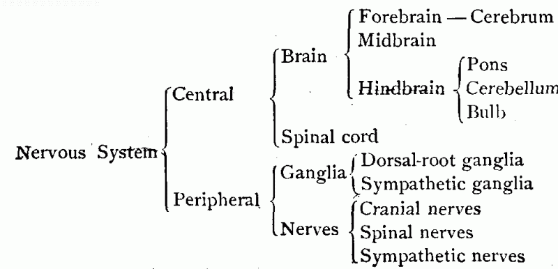
[pg 288]The
Central Division.—This division of the nervous system lies within
the cranial and spinal cavities, and consists of the brain and the
spinal cord. The brain occupying the cranial cavity and the spinal cord
in the spinal cavity connect with each other through the large opening
at the base of the skull to form one continuous structure. The brain and
cord are the most complicated portions of the nervous system, and the
ones most difficult to understand.

divisions of brain.
The Brain.—The brain, which is the
largest mass of nervous tissue in the body, weighs in the average sized
man about 50 ounces, and in the average sized woman about 44 ounces.98
It may be roughly divided into three parts, which are named from their
positions (in lower animals) the forebrain, the midbrain, and the
hindbrain (Fig. 130). The forebrain consists almost entirely of a single
part, known as
The Cerebrum.—The cerebrum
comprises about seven eighths of the entire brain, and occupies all the
front, middle, back, and upper portions of the cranial cavity, spreading
over and concealing, to a large extent, the parts beneath. The surface
layer of the cerebrum is called the cortex. This is made up largely of cell-bodies, and has a
grayish appearance.99 The cortex is greatly increased in[pg 289] area by the presence everywhere
of ridge-like convolutions, between
which are deep but narrow depressions, called fissures. The interior of the cerebrum consists mainly of
nerve fibers, or axons, which give it a whitish appearance. These fibers
connect with the cell-bodies in the cortex (Fig. 131).
The cerebrum is a double organ, consisting of two similar divisions,
called the cerebral hemispheres.
These are separated by a deep groove, extending from the front to the
back of the brain, known as the median
fissure. The hemispheres, however, are closely connected by a great
band of underlying nerve fibers, called the corpus callosum.

drawing of a neuron from cerebral cortex. a. Short segment of the axis cylinder with collateral
branches.
At the base of the cerebrum three large masses of cell-bodies are to
be found. One of these, a double mass, occupies a central position
between the hemispheres, and is called the optic thalami. The other two occupy front central positions
at the base of either hemisphere, and are known as the corpora striata, or the striate
bodies.
The Midbrain is a short, rounded,
and compact body that lies immediately beneath the cerebrum, and
connects[pg 290] it with the hindbrain. On account
of the great size of the cerebrum, the midbrain is entirely concealed
from view when the other parts occupy their normal positions. However,
if the cerebrum is pulled away from the hindbrain, it is brought into
view somewhat as in Fig. 130.
The midbrain carries upon its back and upper surface four small
rounded masses of cell-bodies, called the corpora quadrigemina. The upper two of these bodies are
connected with the eyes; the lower two appear to have some connection
with the organs of hearing. On the front and under surface, the midbrain
separates slightly as if to form two pillars, which are called the crura cerebri, or cerebral peduncles.
These contain the great bundles of nerve fibers that connect the
cerebrum with the parts of the nervous system below.
The Hindbrain lies beneath the back
portion of the cerebrum, and occupies the enlargement at the base of the
skull. It forms about one eighth of the entire brain, and is composed of
three parts—the cerebellum, the pons, and the bulb.
The Cerebellum is a flat and
somewhat triangular structure with its upper surface fitting into the
triangular under surface of the back of the cerebrum. It is divided into
three lobes—a central lobe and two lateral lobes—and weighs about two
and one half ounces. In its general form and appearance, as well as in
the arrangement of its cell-bodies and axons, the cerebellum resembles
the cerebrum. It differs from the cerebrum, however, in being more
compact, and in having its surface covered with narrow, transverse
ridges instead of the irregular and broader convolutions (Fig. 132).
The Pons, or pons Varolii, named
from its supposed resemblance to a bridge, is situated in front of the
cerebellum, and is readily recognized as a circular expansion which
extends forward from that body. It consists largely of[pg 291] bands of nerve fibers, between
which are several small masses of cell-bodies. The fibers connect with
different parts of the cerebellum and with parts above.

brain viewed from below. C.
Cerebrum. Cb. Cerebellum. M. Midbrain. P. Pons. B. Bulb. I-XII.
Cranial nerves.
The Bulb, or medulla oblongata, is,
properly speaking, an enlargement of the spinal cord within the cranial
cavity. It is somewhat triangular in shape, and lies immediately below
the cerebellum. It contains important clusters of cell-bodies, as well
as the nerve fibers that pass from the spinal cord to the brain.
[pg 292]The
Spinal Cord.—This division of the central nervous system is about
seventeen inches in length and two thirds of an inch in diameter. It
does not extend the entire length of the spinal cavity, as might be
supposed, but terminates at the lower margin of the first lumbar
vertebra.100 It connects at the upper end with the bulb, and terminates
at the lower extremity in a number of large nerve roots, which are
continuous with the nerves of the hips and legs (Fig. 133). Two deep
fissures, one in front and the other at the back, extend the entire
length of the cord, and separate it into two similar divisions. These
are connected, however, along their entire length by a central band
consisting of both gray and white matter.

cord, showing on one side the nerves and ganglia with which it is
closely related in function. A. Bulb.
B. Cervical enlargement. C. Lumbar enlargement. D. Termination of cord. E. Nerve roots that occupy the spinal
cavity below the cord. P. Pons. D.G. Dorsal root ganglia. S.G. Sympathetic ganglia. N. Nerve trunks to upper and lower
extremities.
The arrangement of the neurons of the spinal cord is just the reverse
of[pg 293] that in the cerebrum—the center
being occupied by a double column of cell-bodies, which give it a
grayish appearance, while the fibers occupy the outer portion of the
cord, giving it a whitish appearance.
The spinal cord is not uniform in thickness, but tapers slightly,
though not uniformly, from the upper toward the lower end. At the places
where the nerves from the arms and legs enter the cord two enlargements
are to be found, the upper being called the cervical and the lower the lumbar enlargement. These, on account of the difference in
length between the cord and the spinal cavity, are above—the lower one
considerably above—the places where the limbs which they supply join the
trunk (Fig. 133).
Arrangement of the Neurons of the Brain
and Cord.—The cell-bodies in the brain and spinal cord are
collected into groups, and their fibers extend from these groups to
places that may be near or remote. Guided by the white and gray colors
of the nervous tissue, and also by the structures revealed by the
microscope, physiologists have made out three general schemes in the
grouping of cell-bodies, as follows:
1. That of surface distribution,
the cell-bodies forming a thin but continuous layer over a given
surface. This is the plan in the cerebrum and cerebellum, and here are
found devices for increasing the surface: the cerebrum having
convolutions, the cerebellum transverse ridges.
2. That of collections of cell-bodies
into rounded masses. Such masses are found in the bulb, the pons,
the midbrain, and the base of the cerebrum.
3. That of arrangement in a continuous
column. This is the plan in the spinal cord. It matters not at what
place the spinal cord be cut, a central area of gray matter, resembling
in form the capital letter H, is always found.
The fibers connecting with the cell-bodies in the brain and spinal
cord are gathered into bundles or tracts, and these pass through
different parts somewhat as follows:
1. In the cerebrum they extend in
three general directions, forming three classes of fibers. The first
connect different localities in the same hemisphere, and are known as
association fibers (A, Fig. 134). The second make connection
between the two hemispheres, and form[pg 294]
the corpus callosum. These are known as commissural fibers (C,
Fig. 134). The third connect the cerebrum with the parts of the nervous
system below, and are called projection fibers (P,
Fig. 134).
2. In the cerebellum both
association and commissural fibers are found. Bands of fibers, passing
upward toward the cerebrum and downward toward the cord, connect this
part of the brain with other parts of the nervous system.

cerebral hemisphere, showing fiber tracts. A. Association fibers. C. Commissural fibers. P. Projection fibers. The cell-bodies with which the fiber
bundles connect are in the surface layer or cortex.
3. In the midbrain, bulb, and spinal
cord fibers are found: first, that connect these parts with the
cerebrum101 and cerebellum above; second,[pg 295]
that pass into and become a part of the nerves of the body; and third,
that connect the opposite sides of these parts together.
The Peripheral Division.—The
peripheral division of the nervous system includes all the nervous
structures found outside of the brain and spinal cord. These consist of
the cranial, spinal, and sympathetic nerves, and of various small
ganglia, all of which are closely connected with the central system.
Spinal Nerves and Dorsal-root
Ganglia.—The spinal nerves comprise a group of thirty-one pairs,
which connect the spinal cord with different parts of the trunk, with
the upper, and with the lower extremities. Each nerve joins the cord by
two roots, these being named from their positions the ventral, or anterior, root and the dorsal, or posterior, root. The two roots
blend together within the spinal cavity to form a single nerve trunk,
which passes out between the vertebræ. On the dorsal root of each
spinal nerve is a small ganglion which is named, from its position, the
dorsal-root ganglion. (Consult Figs.
133 and 135, and also Fig. 125.)
Double Nature of Spinal
Nerves.—Charles Bell, in 1811, made the remarkable discovery that
each spinal nerve is double in function. He found the portion connecting
with the cord by the dorsal root to be concerned in the production of feeling and the portion
connecting by the ventral root to be concerned in the production of motion. In keeping with
these functions, the two divisions of the nerve are made up of different
kinds of fibers, as follows:
1. The dorsal-root divisions, of the fibers of di-axonic neurons, the
cell-bodies of which form the dorsal-root ganglia (Fig. 135).
2. The ventral-root divisions, of the fibers of mon-axonic[pg 296] neurons, the cell-bodies of which
are in the gray matter of the cord.
The first convey impulses to the cord and are called afferent neurons;102 the second convey
impulses from the cord and are known as efferent neurons. Thus, by forming a part of the nerve
pathways between the skin and the brain, the dorsal divisions of these
nerves aid in the production of feeling; and by completing pathways to
the muscles, the ventral divisions aid in the production of motion
(Figs. 129, 135, and 141).

spinal nerves with the cord. On the right is shown a nerve pathway
from the skin to the muscle. A division of this pathway reaches the
brain.
The Cranial Nerves.—From the under
front surface of the brain, twelve pairs of nerves emerge and pass to
the head, neck, and upper portions of the trunk. These, the cranial
nerves, have names suggestive of their function or distribution and, in
addition, are given numbers which indicate the order in which they leave
the brain (Fig. 136). Unlike the spinal nerves, the cranial nerves
present great variety among themselves, scarcely any two of them being
alike in function or in their connection with different parts of the
body. Several of them have to do with the special senses, and are for
this reason very important. They[pg 297] connect the brain with the
different parts of the head, neck, and trunk, as follows:
1. The first pair (olfactory
nerves; nerves of smell; afferent) connect with the mucous membrane of
the nostrils (Fig. 136).
2. The second pair (optic nerves;
nerves of sight; afferent) connect with the retina of the eyes.
3. The third, fourth, and sixth pairs (motores oculi; control muscles of the eyes; efferent)
connect with the internal and external muscles of the eyeballs (Fig.
136).
. See also Fig. 132.")
suggesting the distribution and functions of the cranial nerves
(Colton). See also Fig. 132.
4. The fifth pair (trigeminal
nerves; nerves of feeling[pg 298] to the face, of taste to the
front of the tongue, and of control of muscles of mastication; afferent
and efferent) connect with the skin of the face, the mucous membrane of
the mouth, the teeth, and the muscles of mastication.
5. The seventh pair (facial
nerves; control muscles that give the facial expressions; efferent)
connect with the muscles just beneath the skin of the face.
6. The eighth pair (auditory
nerves; nerves of hearing; afferent) connect with the internal ear.
7. The ninth pair (glossopharyngeal nerves; nerves of taste to back of tongue
and of muscular control of pharynx; afferent and efferent) connect with
the back surface of the tongue and with the muscles of the pharynx.
8. The tenth pair (vagus, or
pneumogastric, nerves; nerves of feeling and of muscular control;
afferent and efferent) connect with the heart, larynx, lungs, and
stomach. They have the widest distribution of any of the cranial
nerves.
9. The eleventh pair (spinal
accessory nerves; control muscles of neck; efferent) connect with
the muscles of the neck.
10. The twelfth pair (hypoglossal
nerves; control muscles of the tongue; efferent) connect with the
muscles of the tongue.
Sympathetic Ganglia and Nerves.—The
sympathetic ganglia are found in different parts of the body, and vary
in size from those which are half an inch in diameter to those that are
smaller than the heads of pins. The largest and most important ones are
found in two chains which lie in front, and a little to either side, of
the spinal column, and extend from the neck to the region of the pelvis
(Figs. 125 and 133). The number of ganglia in each of these chains is
about twenty-four. They are connected[pg 299] on either side by the right and
left sympathetic nerves which extend vertically from ganglion to
ganglion. In addition to the ganglia forming these chains, important
ones are found in the head (outside of the cranial cavity) and in the
plexuses of the thorax and the abdomen.
The sympathetic ganglia receive nerves from the central division of
the nervous system, but connect with glands, blood vessels, and the
intestinal walls through fibers from their own cell-bodies. Some of
these latter fibers join the spinal nerves, and some blend with each
other to form small sympathetic nerves.
Protection of Brain and Spinal
Cord.—On account of their delicate structure, the brain and spinal
cord require the most complete protection. In the first place, they are
surrounded by the bones of the head and spinal column; these not only
shield them from the direct effects of physical force, but by their
peculiar construction prevent, to a large degree, the passage of jars
and shocks to the parts within. In the second place, they are surrounded
by three separate membranes, as follows:
1. The dura, or dura mater, a
thick, dense, and tough membrane which lines the bony cavities and forms
supporting partitions.
2. The pia, or pia mater, a thin,
delicate membrane, containing numerous blood vessels, that covers the
surface of the brain and cord.
3. The arachnoid, a membrane of
loose texture, that lies between the dura and the pin.
Finally, within the spaces of the arachnoid is a lymph-like liquid
which completely envelops the brain and the cord, and which, by serving
as a watery cushion, protects them from jars and shocks. Thus the brain
and cord are directly shielded by bones, by membranes, and by the [pg 300]liquid which surrounds them. They are
also protected from jars resulting from the movements of the body by the
general elasticity of the skeleton.
Summary.—The nervous system
establishes connections between all parts of the body, and provides a
stimulus by means of which they are controlled. It is made up of a
special form of cells, called neurons. The neurons form the different
divisions of the nervous system, and also serve as the active agents in
carrying on its work. Through a side-by-side method of joining they form
the nerves, ganglia, spinal cord, and brain; and by a method of
end-to-end joining they connect places remote from each other, and
provide for nervous movements through the body. The nervous system, may
in some respects be compared to a complicated system of telephony, in
which the chains of neurons correspond to the wires, and the brain and
spinal cord to the central station.
Exercises.—1. Give the meaning of the term “coördination.” Supply
illustrations.
2. What two general conditions are supplied in the body by the
nervous system?
3. Compare the skeleton outline of the nervous system with the bony
skeleton.
4. Sketch outlines of mon-axonic and di-axonic neurons.
5. Give two differences between the neurons and the other cells of
the body.
6. Describe the two general methods of connecting neurons in the
body. What purpose is accomplished by each method?
7. Name and locate the principal divisions of the nervous system.
8. Draw an outline of the brain (side view), locating each of its
principal divisions.
9. If a pencil were placed over the ear, what portions of the brain
would be above it and what below?
10. Describe briefly the cerebrum, the cerebellum, the midbrain, the
pons, and the bulb.
[pg 301]11. Locate and
describe the cortex. State purpose of the convolutions.
12. State the general differences between the cranial and the spinal
nerves.
13. Locate and give the number of the dorsal-root ganglia. Locate and
give the approximate number of the sympathetic ganglia.
14. Show how the two portions of the spinal nerves are formed—the one
from the mon-axonic and the other from the di-axonic neurons.
15. Enumerate the different agencies through which the brain and
spinal cord are protected.
16. What cranial nerves contain afferent fibers? What ones contain
efferent fibers? What ones contain both afferent and efferent
fibers?
17. In what respects is the nervous system similar to a system of
telephony? In what respects is it different?
PRACTICAL WORK
Examine a model of the brain, identifying the different divisions and
noting the position and relative size of the different parts (Fig. 137).
Observe the convolutions of the cerebrum and compare these with the
parallel ridges of the cerebellum. If the model is dissectible, study
the arrangement of the cell-bodies (gray matter) and the distribution of
the fiber bundles (white matter). Note the connection of the cranial
nerves with the under side.
.")
(dissectible).
A prepared nervous system of a frog (such as may be obtained from
supply houses) should also be examined. Observe the appearance and
general distribution of the nerves and their connection with the brain
and spinal cord. If such a preparation is not at hand, some small animal
may be dissected to show the main divisions of the nervous system, as
follows:
Dissection of the Nervous System
(by the teacher).—For this purpose a half-grown cat is generally the
best available material. This [pg 302]should
be killed with chloroform and secured to a board as in the dissection of
the abdomen (page 169). Open the abdominal cavity and remove the
contents, tying the alimentary canal where it is cut, and washing out
any blood which may escape. Dissect for the nervous system in the
following order:
1. Cut away the front of the chest, exposing the heart and lungs.
Find on each side of the heart a nerve which passes by the side of the
pericardium to the diaphragm. These nerves assist in controlling
respiration and are called the phrenic nerves. Find other nerves going to different parts
of the thorax.
2. Remove the heart and lungs. Find in the back part of the thoracic
cavity, on each side of the spinal column, a number of small “knots” of
nervous matter joined together by a single nerve. These are sympathetic
ganglia. Where the neck joins the thorax, find two sympathetic ganglia
much larger than the others.
3. Cut away the skin from the shoulder and upper side of the fore
leg. By separating the muscles and connective tissue where the leg joins
the thorax, find several nerves of considerable size. These connect with
each other, forming a network called the brachial plexus. From here nerves pass to the thorax and to
the fore leg.
4. From the brachial plexus trace out the nerves which pass to
different parts of the fore leg. In doing this separate the muscles with
the fingers and use the knife only where it is necessary to expose the
nerves. Note that some of the branches pass into the muscles, while
others connect with the skin.
5. Remove the skin from the upper portion of one of the hind legs and
separate the muscles carefully until a large nerve is found. This is one
of the divisions of the sciatic
nerve. Carefully trace it to the spinal cord, cutting away the bone
where necessary, and find the connections of its branches with the cord.
Then trace it toward the foot, discovering its branches to different
muscles and to the skin.
6. Unjoint the neck and remove the head. Examine the spinal cord
where exposed. Cut away the bone sufficiently to show the connection
between the cord and one of the spinal nerves. On the dorsal root of one
of the nerves find a small ganglion. What is it called?
7. Fasten the head to a small board and remove the scalp. Saw through
the skull bones in several directions. Pry off the small pieces of
bones, exposing the upper surface of the brain. Study its membranes,
convolutions, and divisions.
[pg 303]8. With
a pair of bone forceps, or nippers, break away the skull until the
entire brain can be removed from the cavity. Examine the different
divisions, noting the relative position and size of the parts.
9. With a sharp knife cut sections through the different parts,
showing the positions of the “gray matter” and of the “white
matter.”
NOTE.—If the entire class is to examine one specimen, it is generally
better to have the dissecting done beforehand and the parts separated
and tacked to small boards. This will permit of individual examination.
Sketches of the sciatic nerve, brachial plexus, and of sections through
the brain and spinal cord should be made.
Location of Nerves in the
Body.—Several of the nerves of the body lie sufficiently near the
surface to be located by pressure and are easily recognized as sensitive
cords. Slight pressure from the fingers reveals the presence of nerves
in the grooves of the elbow (the crazy bone), between the muscles on the
inner side of the arm near the shoulder, and in the hollow part of the
leg back of the knee. These are all large nerves. Small nerves may be
located in the same manner in the face and neck.
[pg 304]
CHAPTER XVIII – PHYSIOLOGY OF THE NERVOUS SYSTEM
In the preceding chapter was pointed out the method by which the
different parts of the body are brought into communication by the
neurons or nerve cells. We are now to study the means whereby the
neurons are made to control and coördinate the different parts of the
body and bring about the necessary adjustment of the body to its
surroundings. This work of the neurons naturally has some relation to
their properties.
Properties of Neurons.—The work of
the neurons seems to depend mainly upon two properties—the property of
irritability and the property of conductivity. Irritability was explained, in the study of the muscles
(page 243), as the ability to respond to a stimulus. It has the same
meaning here. The neurons, however, respond more readily to stimuli than
do the muscles and are therefore more irritable. Moreover, they are
stimulated by all the forces that induce muscular contraction and by
many others besides. They are by far the most irritable portions of the
body.
Conductivity is the property which
enables the effect of a stimulus to be transferred from one part of a
neuron to another. On account of this property, an excitation, or
disturbance, in any part of a neuron is conducted or carried to all the
other parts. Thus a disturbance at the distant ends of the dendrites
causes a movement toward the cell-body and, reaching the cell-body, the
disturbance is[pg 305] passed through it into the axon.
This movement through the neuron is called the nervous impulse.
Purpose of the Impulse. —Though
the nature of the nervous impulse is not understood, 103 its purpose is
quite apparent. It is the means employed by the nervous system for
controlling and coördinating the different parts of the body. The
arrangement of the neurons enables impulses to be started in certain
parts of the nervous system, and the property of conductivity causes
them to be passed as stimuli to other
parts. This enables excitation at one place to bring about action at
another place.
Acting as stimuli, the impulses seem able to produce two distinct
effects: first, to throw resting organs into action and to increase the
activity of organs already at work; and second, to diminish the rate, or
check entirely, the activity of organs. Impulses producing the first
effect are called excitant impulses;
those producing the second effect, inhibitory impulses.
Functions of the Parts of
Neurons.—The cell-body serves as
a nutritive center from which the other parts derive nourishment. Proof
of this is found in the fact that when any part of the neuron is
separated from the cell-body, it dies, while the cell-body and the parts
attached to the cell-body[pg 306] may continue to live. In
addition to this the cell-body probably reënforces the nervous
impulse.
The dendrites serve two purposes:
first, they extend the surface of the cell-body, thereby enabling it to
absorb a greater amount of nourishment from the surrounding lymph;
second, they act as receivers of
stimuli from other neurons. The same impulse does not pass from one
neuron to another. An impulse in one neuron, however, is able to excite
the neuron with which it makes an end-to-end connection, so that a
series of impulses is produced along a given nerve path (Fig. 129).
The special function of the axon
is to transmit the impulse. By its length, structure, and property of
conductivity it is especially adapted to this purpose. The axis
cylinder, however, is the only part of the axon concerned in the
transmission. The primitive sheath and the medullary layer protect the
axis cylinder, and, according to some authorities, serve to insulate it.
The medullary sheath may also aid in the nourishment of the axis
cylinder.
Nerve Stimuli.—While the
properties of irritability and conductivity supply a necessary cause for
the production and transmission of nervous impulses, these alone are not
sufficient to account for their origin. An additional cause is necessary—a
force not found in the nerve protoplasm, but one which, by its
action on the protoplasm, makes it produce the impulse. In this respect,
the neuron does not differ essentially from the cell of a muscle. Just
as the muscle cell requires a stimulus to make it contract, so does the
neuron require a stimulus to start the impulse. Hence, in accounting for
the activities of the body, it is not sufficient to say they are caused
by nervous impulses. We must also investigate the nerve stimuli—the means through which the nervous impulses
are started. Most of these[pg 307] are found outside of the body and
are known as external stimuli.
Action of External Stimuli.—In the
arrangement of the nervous system the most favorable conditions are
provided for the reception of external stimuli. Not only do vast numbers
of neurons terminate at the surface of the body,104 but they connect
there with delicate structures, called sense organs. The purpose of the sense organs is to sensitize (make sensitive) the
terminations of the neurons. This they do by supplying special
structures through which the stimuli can act to the best advantage upon
the nerve endings. Moreover, there are different kinds of sense organs,
and these cause the neurons to be sensitive to different kinds of
stimuli. Acting through the sense organs adapted for receiving them,
light, sound, heat, cold, and odors all act as stimuli for starting
impulses. Indeed, the arrangement is so complete that the nervous system
is subjected to the action of external stimuli in some form practically
all the time. The work of the sense organs is further considered in
Chapters XX, XXI, and XXII.
How External Stimuli act on Internal
Organs.—For stimulating the neurons not connected with the body
surface we are dependent, so far as known, upon the nervous impulses. An
impulse started by the external stimulus goes only so far as its neuron
extends. But it serves as a stimulus for the neuron with which the first
connects and starts an impulse in this connecting neuron, the point of
stimulation being where the fiber terminations of the first neuron make
connection with the dendrites of the second. This impulse in turn
stimulates the next neuron, and so on, producing a series of impulses
along a given nerve path. [pg 308]In this way the effect of an
external stimulus may reach and bring about action in any part of the
body. This is in brief the general plan of inducing action in the
various organs of the body. This plan, however, is varied according to
circumstances, and at least three well-defined forms of action are
easily made out. These are known as reflex
action, voluntary action, and secondary reflex action.
Reflex Action.—When some sudden or
strong stimulus acts upon the nerve terminations at the surface of the
body, an immediate response is frequently observed in some quick
movement. The jerking away of the hand on accidentally touching a hot
stove, the winking of the eyes on sudden exposure to danger, and the
quick movements from slight electrical shocks are familiar examples. The
explanation of reflex action is that external stimuli start impulses in
neurons terminating at the surface of the body and these, in turn,
excite impulses in neurons which pass from the spinal cord or brain to
the muscles (Fig. 138). Since there is an apparent turning back of the
impulses by the cord or brain, the resulting movements are termed reflex.105

illustrating reflex action of an external organ.
Reflex Action and the Mind.—If one
carefully studies the reflex actions of his own body, he will find that
they[pg 309] occur at the time, or even a
little before the time, that he realizes what has happened. If a feather
is brought in contact with the more sensitive parts of the face of a
sleeping person, there is a twitching of the skin and sometimes a
movement of the hand to remove the offending substance. Surgeons
operating upon patients completely under the influence of chloroform,
and therefore completely unconscious, have observed strong reflex
actions. These and other similar cases indicate clearly that reflex
action occurs independently of the
mind—that the mind neither causes nor controls it. If a further proof of
this fact were needed, it is supplied by experiments upon certain of the
lower animals,106 which live for a while after the removal of the brain.
These experiments show that the nervous impulses that produce reflex
action need only pass through the spinal cord and do not reach the
cerebrum, the organ of the mind.
The Reflex Action Pathway.—By study
of the impulses that produce any reflex action, a rather definite
pathway may be made out, having the following divisions:
1. From the surface of the body to the
central nervous system (usually the spinal cord). This, the afferent division, is made up of
di-axonic neurons, and these have (in the case of the spinal nerves)
their cell-bodies in the dorsal root ganglia (page 295). They are acted
upon by external stimuli, while their impulses in turn act on the
neurons in the spinal cord.
[pg 310]2. Through the central system
(spinal cord or base of brain). This, the intermediate division, may be composed of mon-axonic
neurons, or it may consist of branches from the afferent neurons. In the
case of separate neurons, these are acted upon by impulses from the
afferent neurons, while their impulses serve in turn as stimuli to other
neurons within the cord (Fig. 129).
3. From the central nervous system to
the muscles. This, the efferent
division, is made up of mon-axonic neurons. Most of these have their
cell-bodies in the gray matter of the cord, while their fibers pass into
the spinal nerves by the ventral roots.107 They may be stimulated by
impulses either from the intermediate neurons, or from branches of the
afferent neurons. Their impulses reach and stimulate the muscles.
Reflex Action in Digestion.—The
flowing of the saliva, when food is present in the mouth, is an example
of reflex action. In this case, however, the organ excited to activity
is a gland instead of a muscle. The food starts the impulses, and these,
acting through the bulb, reach and stimulate the salivary glands. In a
similar manner food excites the glands that empty their fluids into the
stomach and intestines, and stimulates the muscular coats of these
organs to do their part in the digestive process. To a considerable
extent, neurons having their cell-bodies in the sympathetic ganglia are
concerned in these actions (Fig. 139).

relation to the food canal. The nerve path in this case includes
sympathetic neurons.
Reflex Action in the Circulation of the
Blood.—On sudden exposure[pg 311] to cold, the small arteries going
to the skin quickly diminish in size, check the flow of blood to the
surface, and prevent too great a loss of heat. In this case, impulses
starting at the surface of the body are transmitted to the bulb and then
through the efferent neurons to the muscles in the walls of the
arteries. In a somewhat similar manner, heat leads to a relaxation of
the arterial walls and an increase in the blood supply to the skin.
Other changes in the blood supply to different parts of the body are
also of the nature of reflex actions. As in the work of digestion,
neurons having their cell-bodies in the sympathetic ganglia aid in the
control of the circulation.
Purposes of Reflex Action.—The
examples of reflex action so far considered illustrate its two main
purposes—(1) protection, and (2) a means of controlling important
processes.
The pupil has but to study carefully the reflex actions of his own
body for a period, say of two or three weeks, in order to be convinced
of their protective value. He will observe that portions of his body
have, on exposure to danger, been moved to places of safety, while in
some instances, like falling, his entire body has been adjusted to new
conditions. He will also find that reflex action is quicker, and for
that reason offers in some cases better protection, than movements
directed by the mind. In digestion and circulation are found the best
examples of the control of important processes through reflex
action.
Voluntary Action.—It is observed
that reflex action, in the sense that it has so far been considered, is
not the usual mode of action of the external organs, but is, instead, a
kind of emergency action, due to unusual conditions and excitation by
strong stimuli. Voluntary actions, on the other hand, represent the
ordinary, or normal, action of these organs. They comprise the movements
of the body of which we are conscious and which are controlled by the mind. But while they
are of a higher order than reflex[pg 312] actions and are under intelligent direction, they are brought
about in much the same manner.
Voluntary Action Pathways differ in
but one essential respect from those of reflex action. They pass through
the cerebrum, the organ of the mind (Fig. 140). This is necessary in
order that the mind may control the action. From all portions of the
body surface, afferent pathways may be traced to the cerebrum; and from
the cerebrum efferent pathways extend to all the voluntary organs. A
complex system of intermediate neurons, found mostly in the brain, join
the afferent with the efferent pathways. The voluntary pathways are not
distinct from, but include, reflex pathways, a fact which explains why
the same external stimulus may excite both reflex and voluntary action
(Fig. 141).

voluntary action pathway.
Choice in Voluntary Action.—In
reflex action a given stimulus, acting in a certain way; produces each
time the same result. This is not the case with voluntary action, the
difference being due to the mind. In
these actions the external stimulus first excites the mind, and the
resulting mental processes—perhaps as memory of previous
experiences—supply a variety of facts, any of which may act as stimuli
to action. Before the action takes place, however, [pg 313]some one fact must be singled out from among the mental
processes excited. This fact becomes the exciting stimulus and leads to action. It follows,
therefore, that the action which finally occurs is not necessarily the
result of an immediate external stimulus, but of a selected stimulus—one which is the result of choice.

voluntary action pathways including reflex pathways.
Not only does the element of choice enter into the selection of the
proper stimulus, but it also enters into the time, nature, and intensity
of the action. For these reasons it is frequently impossible to trace
voluntary actions back to their actual stimuli. The pupil will recognize
the element of choice in such simple acts as picking up some object from
the street, complying with a request, and purchasing some article from a
store.
Reflex and Voluntary Action
Compared.—Certain likenesses and differences, already suggested in
these two forms of action, may now be more fully pointed out. Reflex and
voluntary action are alike in that the primary cause of each is some
outside force or condition which has impressed itself upon the nervous
system. They are also alike in the general direction taken by the
impulses in producing the action. The impulses are, first, from the
surface of the body to the central nervous system; second,[pg 314] through the central system; and third, from the
central nervous system to the active tissues of the body.
Their chief differences are to be found, first, in the pathways
followed by the impulses, which are through the cerebrum (the organ of
the mind) in voluntary action, but in reflex action are only through the
spinal cord or the lower parts of the brain; and second, in the fact
that voluntary action is under the direction of the mind, while reflex
action is not. It would seem, therefore, that the statement sometimes
made that “voluntary action is reflex action plus the mind” is not far
from correct. Mind, however, is the important factor in this kind of
action.
Secondary Reflex Action.—Everyday
experience teaches that any voluntary action becomes easier by
repetition. A given act performed a number of times under conscious
direction establishes a condition in the nervous system that enables it
to occur without that direction and very much as reflex actions occur.
Actions of this kind are known as secondary reflex actions, or as acquired reflexes. Walking, writing, and
numerous other movements pertaining to the occupation which one follows
are examples of such reflexes. These activities are at first entirely
voluntary, but by repetition they gradually become reflex, requiring
only the stimulus to start them.
The advantages to the body of its acquired reflexes are quite
apparent. The mind does not have to attend to the selection and
direction of stimuli and, to that extent, is left free for other work. A
good example of this is found in writing, where the mind apparently
gives no heed to the movements of the hand and is only concerned in what
is being written. The student will easily supply other illustrations of
the advantages of secondary reflex action.
[pg 315]The development of secondary
reflexes probably consists in the establishment of fixed pathways for
impulses through the nervous system. Through the branching of the nerve
fibers many pathways are open to the impulses. But in repeating the same
kind of action the impulses are guided into particular paths, or
channels. In time these paths become so well established that the
impulses flow along them without conscious direction and it is then
simply necessary that some stimulus starts the impulses. By following
the established pathways, these reach the right destination and produce
the desired result. According to this view, secondary reflex action is
but a higher phase of ordinary reflex action—a kind of reflex action,
the conditions of which have been established by the mind through
repetition. (See functions of the cerebellum, page 317.)
Habits.—People are observed to act
differently when exposed to the same conditions, or when acted upon by
the same stimuli. This is explained by saying they have different
habits. By habits are meant certain
general modes of action that have been acquired by repetition. Certain
acts repeated again and again have established conditions in the nervous
system which enable definite forms of action to be excited, somewhat
after the manner of reflex action. On account of habits, therefore, the
actions of the individual are more or less predisposed. What he will do under certain conditions may
be foretold from his habits. Habits simply represent, a higher order of
secondary reflexes—those more closely associated with the mental life
and character than are the lower forms.
Habits, in common with other forms of secondary reflex action, serve
the important purpose of economizing the
nervous energy. However, if pernicious habits are formed instead of
those that are useful, they are detrimental from both a moral and
physical standpoint. Youth is recognized as the period in which
fundamental habits are formed and character is largely determined.
Therefore parents[pg 316] and teachers do
wisely when they insist upon the formation of right habits by the
young.
Functions of Divisions of the Nervous
System.—The relationship between the different parts of the nervous
system is very close and one part does not work independently of other
parts. At the same time the general work of the nervous system requires
that its different divisions serve different purposes:
1. The peripheral divisions of the nervous system are concerned in the
transmission of impulses between the surface of the body and the central
system and between the central system and the active tissues. The nerves
are the carriers of the impulses. The ganglia contain the cell-bodies
which serve as nutritive centers; and, in the case of the sympathetic
ganglia, these cell-bodies are the places where the fiber terminations
of one neuron connect with, and stimulate, other neurons.
2. The gray matter in the spinal cord, bulb, pons, and midbrain
(through the cell-bodies, fiber terminations, and short neurons which
they contain) completes the reflex action pathways between the surface
of the body and the voluntary muscles, and also between the surface of
the body and the organs of circulation and digestion.
3. The white matter of the spinal cord, bulb, pons, and midbrain (by
means of the fibers of which they are largely composed) forms
connections with, and passes impulses between, the various parts of the
central nervous system.
4. The bulb, because of certain special reflex-action pathways
completed through it, is the portion of the central nervous system
concerned in the control of respiration, circulation, and the secretion
of liquids.
Work of the Sympathetic Ganglia and
Nerves.—The neurons which form these ganglia aid in controlling the
vital processes, especially digestion[pg 317]
and circulation. These neurons are controlled for the most part by
fibers from the bulb and spinal cord, and cannot for this reason be
looked upon as forming an independent system. Their chief purpose seems
to be that of spreading the influence of neurons from the central system
over a wider area than they would otherwise reach. For example, a single
neuron passing out from the spinal cord may, by terminating in a
sympathetic ganglion, stimulate a large number of neurons, each of which
will in turn stimulate the cells of muscles or of glands. Because of
this function, the sympathetic neurons are sometimes called distributing neurons.
Functions of the
Cerebellum.—Efforts to discover some special function of the cerebellum have been in the main
unsuccessful. Its removal from animals, instead of producing definite
results, usually interferes in a mild way with a number of activities.
The most noticeable results are a general weakness of the muscles and an
inability on the part of the animal to balance itself. This and other
facts, including the manner of its connection with other parts of the
nervous system, have led to the belief that the cerebellum is the chief
organ for the reflex coördination of
muscular movements, especially those having to do with the balancing of
the body. In this connection it is subordinate to and under the control
of the cerebrum. Of the relations which the cerebellum sustains to the
cerebrum and to the different parts of the body, the following view is
quite generally held:
In the development of secondary reflexes, as already described,
conditions are established in the cerebellum, such that given stimuli
may act reflexively through it and
produce definite results in the way of muscular contraction. After the
establishment of these conditions, afferent impulses from the eyes,
ears, skin, and other places, under the general direction of the
cerebrum, may cause such actions as the balancing of the body, walking,
etc., as well as the delicate and varied movements of the hand. This
view of its functions makes of the cerebellum the great center of
secondary reflex action.
Functions of the Cerebrum.—While
the work of the cerebrum is closely related to that of the general
nervous system, it, more than any other part, exercises functions
peculiar to itself. The cerebrum is the part of the nervous system upon
which our varied experiences leave their impressions and through which
these impressions are made[pg 318] to
influence the movements of the body. But the power to alter, postpone,
or entirely inhibit, nervous movements is but a part of the general work
ascribed to the cerebrum as the organ of
the mind. Numerous experiments performed upon the lower animals,
together with observations on man, show the cerebrum to be the seat of
the mental activities, and to make possible, in some way, the processes
of consciousness, memory, volition, imagination, emotion, thought, and
sensation.
Localization of Cerebral
Functions.—Many experiments have been performed with a view to
determining whether the entire cerebrum is concerned in each of its
several activities or whether special functions belong to its different
parts. These experiments have been made upon the lower animals and the
results thus obtained compared with observations made upon injured and
imperfectly developed brains in man. The results have led to the
conclusion that certain forms of the work of the cerebrum are localized and that some of its parts are
concerned in processes different from those of others.

cerebral functions. Diagram of cerebrum, showing most of the areas
whose functions are known.
The work of locating the functions of different parts of the cerebrum
forms one of the most interesting chapters in the history of brain
physiology. The portions having to do with sight, voluntary motion,
speech, and hearing have been rather accurately determined, while
considerable evidence as to the location of other functions has been
secured. Much of the cerebral surface, however, is still undetermined
(Fig. 142).
NERVOUS CONTROL OF IMPORTANT PROCESSES
Circulation of the Blood.—1. Control of the Heart.—The ability to
contract at regular intervals has been shown to reside in the heart [pg 319]muscle. Among other proofs is that
furnished by cold-blooded animals, like the frog, whose heart remains
active for quite a while after its removal from the body. These
automatic contractions, however, are not sufficient to meet all the
demands made upon the circulation. The needs of the tissues for the
constituents of the blood vary with their activity, and it is therefore
necessary to vary frequently the force and rapidity of the heart’s
contractions. Such changes the heart itself is unable to bring
about.
For the purpose of controlling the rate and force of its
contractions, the heart is connected with the central nervous system by
two kinds of fibers:
a. Fibers that convey excitant impulses to the heart to quicken
its movements.
b. Fibers that convey inhibitory impulses to the heart to
retard its movements.
The cell-bodies of the excitant fibers are found in the sympathetic
ganglia, but fibers from the bulb connect with and control them. The
cell-bodies of the inhibitory fibers are located in the bulb, from where
their fibers pass to the heart as a part of the vagus nerve.
In addition to the fibers above mentioned, are those that convey
impulses from the heart to the bulb.
These connect with neurons that in turn connect with blood vessels and
with them act reflexively, when the heart is likely to be overstrained,
to cause a dilation of the blood vessels. This lessens the pressure
which the heart must exert to empty itself of blood. These fibers serve,
in this way, as a kind of safety valve for the heart.
2. Control of Arteries.—Changes in
the rate and force of the heart’s contractions can be made to correspond
only to the general needs of the
body. When the blood supply to a particular organ is to be increased or
diminished, this is accomplished through the muscular coat in the
arteries. The connection of the arterial muscle with the sympathetic
ganglia and the method by which they vary the flow of blood to different
organs has already been explained (pages 311 and 49), so that only the
location of the controlling neurons need be noted here. These, like the
controlling neurons of the heart, have their cell-bodies in the bulb. It
thus appears that the entire control of the circulation is effected in a
reflex manner through the nerve centers in the bulb. These centers are
stimulated by conditions that relate to the movement of the blood
through the body.
[pg 320]Respiration.—Efferent fibers connect the different muscles of
respiration with a cluster of cell-bodies in the bulb, called the respiratory center. This center together
with the nerves and muscles in question form an automatic, or
self-acting, mechanism similar in some respects to that of the heart.
Through the impulses passing from the respiratory center to the muscles,
a rhythmic action is maintained sufficient to satisfy the usual needs of
the body for oxygen. The demand of the body for oxygen, however, varies
with its activities, and to such variations the respiratory center alone
is unable to respond. The regulating factor in the respiratory movements
has been found to be the condition of the blood with reference to the
presence of oxygen and carbon dioxide. If the blood contains much carbon
dioxide and little oxygen, it acts as a strong stimulus to the
respiratory center, causing it, in turn, to stimulate the respiratory
muscles with greater intensity and frequency. On the other hand, if the
blood contains much oxygen and little carbon dioxide, it acts only as a
mild stimulus. This explains how physical exercise increases the
breathing, since the muscles at work consume more oxygen than when
resting and give more carbon dioxide and other wastes to the blood.
The respiratory center is also connected by afferent nerves with the
mucous membrane of the air passages. Irritation of the nerve endings in
this membrane causes impulses to pass to the center, and this leads, by
reflex action, to such modifications of the respiratory acts as sneezing
and coughing. There is also a connection between the cerebrum and the
respiratory center. This is shown by the fact that one can voluntarily
change the rate and force of the respiratory movements, and further by
the fact that emotions affect the breathing.
Regulation of the Body
Temperature.—As explained in the study of the skin (page 270), the
nervous system regulates the body temperature by controlling the
circulation of the blood through the skin and the internal organs. This
is accomplished by stimulating in a reflex manner the muscles in the
walls of certain arteries. To prevent the body from getting too hot,
muscles in the arteries going to the skin relax, thereby allowing more
blood to flow to the surface, where the heat can be disposed of through
radiation and through the evaporation of the perspiration. On the other
hand, if the body is in danger of losing too much heat, the muscles in
the walls of arteries going to the skin are made to contract and those
to internal organs relax, so that less blood flows to the skin and more
to the internal organs. In this[pg 321] way
the nervous system adjusts the circulation to suit the conditions of
temperature outside of and within the body and, in so doing, maintains
the normal body temperature.
Summary.—The nervous system is able
to control, coördinate, and adjust the different organs of the body
through its intimate connection with all parts and through a stimulus
(the nervous impulse) which it supplies and transmits. Nervous impulses,
excited by external stimuli, follow definite paths and cause activity in
the different parts of the body. All such pathways are through the
central nervous system. In reflex action the impulses are mainly through
the spinal cord, but to some extent through the bulb, pons, and
midbrain. In voluntary action they pass through the cerebrum—a condition
that leads to important modifications in the results. The cerebrum, in
addition to controlling the voluntary movements, is able to establish
the necessary conditions for secondary reflex actions, such as walking,
writing, etc. Although certain of the divisions of the nervous system
exercise special functions, all parts of it are closely related.
Exercises.—1. Give the function of each of the parts of a neuron.
2. State the purpose of the nervous impulse.
3. Show that the exciting cause of bodily action is outside of the
nervous system and, to a large extent, outside of the body.
4. Describe the arrangement that enables stimuli outside of the body
to cause action within the body.
5. Describe a reflex action and show how it is brought about.
6. Distinguish between afferent, efferent, and intermediate
neurons.
7. Draw diagrams showing the impulse pathways in voluntary and in
reflex action.
8. What purposes are served by the sympathetic neurons?
9. Describe the method of control of the circulatory and digestive
processes. How do reflex actions protect the body?
[pg 322]10. Compare voluntary and reflex
action. In what sense are all the activities of the body reflex?
11. In what sense is walking voluntary? In what sense is it
reflex?
12. How does secondary reflex action lessen the work of the nervous
system?
13. State the special functions of the nerves, ganglia, spinal cord,
bulb, cerebellum, and cerebrum.
14. State the importance of the formation of correct habits.

pathways.
PRACTICAL WORK
To demonstrate Nerve Pathways.—A
smooth board, 2×6 ft., is painted black, and upon this is drawn in white
a life-size outline of the body. Pieces of cord of different colors and
lengths are knotted to represent mon-axonic and di-axonic neurons. These
are then pinned or tacked to the board in such a manner as to represent
the connections in the different kinds of nerve pathways. Fig. 143 shows
such a board with connections for a reflex action and a voluntary action
of the same muscle.
Study of the “Knee Jerk” Reflex.—A
boy is seated on a chair with the legs crossed. With a small pointer he
is given a light, quick blow on the upper margin of the patella at the
point of connection of the tendon. The stroke will usually be followed
by a reflex movement of the foot. Does this take place independently of
the mind? (The one upon whom the experiment is being performed should
assume a relaxed condition and make no effort either to cause or prevent
the movement.) Can the movement be[pg 323]
inhibited (prevented)? Repeat the experiment, effort being made to
prevent the movement, but not by contracting opposing muscles.
Other reflex actions adapted to class study are those of the eyes,
such as the closing of the lids on moving objects near them and the
dilating of the pupils when the eyes are shaded. The involuntary jerking
of the head on bringing the prongs of a vibrating tuning fork in contact
with the end of the nose is also a reflex action which can be studied to
advantage.
To determine the Reaction
Time.—Have several pupils join hands, facing outwards, making a
complete circle, excepting one gap. Give a signal by touching the hand
of one pupil at the end of the line. Let this pupil communicate the
signal, by pressure of the other hand, to the next pupil and so on
around, having the last pupil raise the free hand at close of the
experiment. Note carefully the time, preferably with a stop watch,
required to complete the experiment and divide this by the number of
pupils, to get the average reaction time. The experiment may be repeated
with boys only and then with girls, comparing their average reaction
time.
Reflex Action of the Salivary
Glands.—Place a small pinch of salt upon the tongue and note the
flow of saliva into the mouth. Try other substances, as starch, bits of
wood, and sugar. What appears to be the natural stimulus for these
glands? Compare with reflex actions of the muscles.
[pg 324]
CHAPTER XIX – HYGIENE OF THE NERVOUS SYSTEM
The far-reaching effects and serious nature of disorders of the
nervous system are sufficient reasons for considering carefully those
conditions that make or mar its efficiency. Controlling all the
activities of the body and affecting through its own condition the
welfare of all the organs, the hygiene of the nervous system is, in a
large measure, the hygiene of the entire body. Moreover, it is known
that some of our worst diseases, including paralysis and insanity, are
disorders of the nervous system and are prevented in many instances by a
proper mode of living.
The Main Problem.—Many of our
nervous disorders are undoubtedly due to the age in which we live. Our
modern civilization, with all its facilities for human advancement and
enjoyment, throws an extra strain upon the nervous system. Educational
and social standards are higher than ever before and life in all its
phases is more complex. Since we can hardly change the conditions under
which we live, and probably would not if we could, we must learn to
adapt or adjust ourselves to them so as to secure for the nervous system
such relief as it requires. This adjustment is sometimes difficult, even
when the actual needs of the nervous system are known.
The healthful action of the nervous system requires, on the one hand,
exercise, but on the other hand, a certain condition of quietude, or poise—a state which is directly opposed
to that of restlessness. The conditions of modern[pg 325] life seem able to force upon the nervous system all the
exercise that it needs, and more (whether it be of the right kind or
not), so that the main problem of to-day seems to be that of conserving,
or economizing, the nervous energy and of preventing nervous waste.
Wasteful Forms of Nervous
Activity.—There are without doubt many forms of activity that waste
the vital forces of the body and lead to nervous exhaustion. Take, for
example, the rather common habit of worrying over the trivial things of
life. Certainly the nervous energy spent in this way cannot be used in
doing useful work, but must be counted as so much loss to the body. One
who would use his nervous system to the best advantage must find some
way of preventing waste of this kind.108
Undue excitement, as well as pleasurable dissipations, also tend
toward nervous exhaustion. And while the fact is recognized that
pleasurable activities supply a necessary mental exercise, the limit of
healthful endurance must be watched and excesses of all kinds avoided. Intense emotional states are
found to be exhausting in the extreme; and the suppression of such
undesirable feelings as anger, fear, jealousy, and resentment are of
immense value in the hygiene of the nervous system.
The Habit of Self-control.—Much of
the needless waste of nervous energy, including that of worrying over
trivial matters, may be prevented through the exercise of self-control.
From the standpoint of the nervous system, the present age differs from
the past mainly in supplying a[pg 326] greater number and variety
of nerve stimuli. Self-control means the ability to suppress activities
that would result from undesirable stimuli and to direct the bodily
activities into channels that are profitable. Self-control, therefore,
is not only to be exercised on occasions of great emergency, but in the
everyday affairs of life as well. It is even more important that the
daily toiler at his task be able to keep the petty annoyances of life
from acting as irritants to his nervous system than that he keep cool
during some great calamity. The habit of self-control is acquired mainly
through the persistent effort to prevent any and all kinds of petty
annoyances from affecting the nerves or the temper.
Nervousness.—Self-control is much
more easily practiced by some than by others. This is due partly to
habit, but is also due to an actual difference in the degree of
sensitiveness, or irritability, of the nervous systems of different
people. One whose nervous system tends to respond too readily to any and
all kinds of stimuli is said to be “nervous.” This condition is in some
instances inherited, but is in most cases due to the wasteful
expenditure of nervous energy or to the action of some drug upon the
body. Excess of mental work, too much reading, long-continued anxiety,
eye strain, and the use of tea, coffee, alcohol, tobacco, or other
drugs, including many of those taken as medicines, are known to cause
nervousness. Nervousness is not only a source of great annoyance, both
to one’s self and to others, but is a menace to the general health.
The first step toward securing relief from such a condition is the
removal of the cause. The habits should be inquired into and excesses of
all kinds discontinued. In some instances it may be necessary to have the eyes [pg 327]examined and
glasses fitted by a competent oculist.109 The nervous energy should be
carefully economized and the habit of self-control diligently
cultivated. Special exercises that have for their purpose the equalizing
of the circulation and the strengthening of the blood vessels of the
neck and the brain also have beneficial effects.
Nervous Overstrain.—Both mental and
physical overwork tends to weaken the nervous system and to produce
nervousness. Where hard mental work is long continued, or where it is
carried on under excitement, a tense nervous condition is developed
which is decidedly weakening in its effects. The causes which lead to
such a condition, and in fact overwork of all kinds, should if possible
be avoided. Where this is not possible, and in many cases it is not, the
period of overwork should be followed by one of rest, recreation, and
plenty of sleep. To the overworked in body or in mind, nothing is more
important from a hygienic, as well as moral, standpoint, than the right
use of the one rest day in seven. The
best interests of our modern civilization require that the Sabbath be kept as a quiet, rest-giving
day.
Disturbed Circulation of the
Brain.—Nervousness not infrequently is accompanied by an increase
in the circulation of the brain and disappears when this condition is
relieved. Though mental work and excitement tend naturally to increase
the circulation in the brain, this should subside with rest and relief
from excitement. When there is a tendency[pg 328] for this condition to become
permanent, effort should be made looking for relief. Increasing the
circulation in the lower extremities by hot or cold foot baths, or by
much walking, is found to be most beneficial. Special exercises of the
muscles of the neck are also recommended as a means of relieving this
condition.110
Hygienic Value of Work.—Within
reasonable limits, both mental and physical work are conducive to the
vigor of the nervous system. Through work the energies of the body find
their natural outlet, and this prevents dissipation and the formation of
bad habits. Even hard work does not injure the nervous system, and
severe mental exertion may be undergone, provided the proper hygienic
conditions are observed. The nervous disorders suffered by brain workers
are not, as a rule, due to the work which the brain does, but to
violation of the laws of health, especially the law of exercise. Such
persons should observe the general laws of hygiene and especially should
they practice daily those forms of physical exercise that tend to
counteract the effects of mental work.
Physical Exercise properly taken is
beneficial to the nervous system through both direct and indirect
effects. A large proportion of the nerve cells have for their function
the production of motion, and these are called into play only through
muscular activity. Then, as already suggested, physical exercise
counteracts the unpleasant effects of mental work. Hard study causes an
excess of blood to be sent to the brain and a diminished amount[pg 329] to the arms and to the legs.
Physical exercise redistributes the blood and equalizes the circulation.
Light exercise should, therefore, follow hard study. The student before
retiring at night is greatly aided in getting to sleep and is put in a
better condition for the next day’s work by ten to fifteen minutes of
light gymnastics. A daily walk of two or three miles is also an
excellent means of counteracting the effects of mental work. The brain
worker should, however, avoid violent exercise or the carrying of any
kind of exercise to exhaustion.
Sleep, and plenty of it, is one of
the first requirements of the nervous system. It is during sleep that
the exhausted brain cells are replenished. To shorten the time for sleep
is to weaken the brain and to lessen its working force. No one should
attempt to get along with less than eight hours of sleep each day and
most people require more. Children require more sleep than adults. Those
under six years should have from eleven to twelve hours of sleep per
day. Children between six and ten years should have at least ten
hours.
Insomnia, or sleeplessness, on
account of its effects upon the nervous system, is to be regarded as a
serious condition, and hygienic means for relieving it should be
diligently sought. Having its cause in nervousness, a disturbed
circulation of the brain, or some form of nervous exhaustion, it is
benefited through relieving these conditions and in the manner already
described. Of course the external conditions for aiding sleep should not
be overlooked. The bed should be comfortable, and the room should be
cool, well ventilated, dark, and quiet. The inducing of sleep by means
of drugs is a dangerous practice and should never be resorted to except
under the direction of the physician.
[pg 330]Effects of Heat and Cold.—Heat and cold both have their
effects upon the nervous system. Heat increases the nervous
irritability, while cold acts as a natural sedative to the nerves. A
nervous person is made more nervous by an overheated atmosphere, but
derives beneficial effects from exposing the body freely to cold air and
water. The tonic cold bath (page 273), if taken with the usual
precautions, can be used to good advantage in diminishing nervousness.
The taking of outdoor exercise in cold weather is, for the same reason,
an excellent practice.
Effect of Emotional States.—We have
already noted the effect of certain emotional states upon the digestion
of the food (page 162). Emotional states are also known to interfere
with breathing and with the action of the heart. Such effects are
explained through the close relation of the mind to the work of the
nervous system in general. While certain emotional states, such as fear,
anger, melancholia, and the impulse to worry, interfere seriously with
the normal action of the nervous system, others, such as contentment,
cheerfulness, and joy, are decidedly beneficial in their effects. How
important, then, is the habit of suppressing the states that are harmful
and of cultivating those that are beneficial. From a hygienic, as well
as social, standpoint a cheerful, happy disposition is worth all the
effort necessary for its attainment.
The Nervous Condition of Children
should be a matter of deep concern on the part of both parents and
teachers. In the home, as well as in the school, the child may be
“pushed” until the nervous system receives permanent injury. Exhaustion
of nerve cells is produced through too many and too vivid impressions
being made upon the immature brain. The child should be protected
from[pg 331] undue excitement. He should have
the benefit of outdoor exercise and should be early inured to cold. He
should be shielded from the poisoning effects of tea, coffee, tobacco,
alcohol, and other drugs. He should have impressed upon him the habit of
self-control. He should not be indulged in foolish caprices or whims,
but should be taught to be content with plain, wholesome food and with
the simple forms of enjoyment.
Influences at School.—School life
is necessarily a great strain upon the child. Night study added to the
work of the day makes a heavy burden for elementary pupils to bear.
Though the legal school age is usually fixed at six years, delicate
children should be kept out of school until they are seven or eight
years old, provided they have good homes. In addition to the excitation
incident to studying and reciting lessons, conditions frequently arise
both in the schoolroom and upon the playground that create a feeling of
fear or dread in the minds of children. Quarrels and feuds among the
children and the bullying of big boys on the playground may work untold
harm. All conditions tending to develop fear, uneasiness, or undue
excitement on the part of children should receive the attention of those
in authority.
Excessive Reading is a frequent
cause of injury to the nervous systems of children. This has a bad
effect, both on account of too many impressions being made upon the mind
and also on account of the strain to the eyes. Then if the reading
consists mostly of light fiction, the mind is directed away from the
really important things of life. The reading of children should be
thoughtfully controlled, both as to quality and quantity. Exciting
stories should, as a rule, be excluded, but a taste for biography,
historical and scientific writings, and for the great works[pg 332] of literature should be
cultivated. Simple fairy tales which have a recognized value in
developing the imagination of the child need not be omitted, but it is
of vital importance that the “story-reading habit” be not formed.
Effects of Drugs.—Because of its
delicacy of structure a number of chemical compounds, or drugs, are able
to produce injurious effects upon the nervous system. Some of these are
violent poisons, while others, in small quantities, are mild in their
action. Certain drugs, in addition to their immediate effects, bring
about changes in the nervous system which cause an unnatural appetite,
or craving, that leads to their continued use. This is the case with
alcohol, the intoxicating substance in the usual saloon drinks, and with
nicotine, the stimulating drug in tobacco. The same is also true of
morphine, chloral, and several other drugs used as medicines. The danger of becoming a slave to some
useless and pernicious habit should dissuade one from the use of drugs
except in cases of positive emergency.
Alcohol and the Nervous
System.—Alcohol, as already shown, injures practically all portions
of the body; but it has its worst effects upon the nervous system.
Through its action on this system, it interferes with the circulation of
the blood, produces a condition of “temporary insanity” called
intoxication, weakens the will, and eventually dethrones the reason.
Worst of all, it produces a condition of “chronic poisoning” which
manifests itself in an unnatural craving, and this causes it to be used
by the victim even when he knows he is “drinking to his own
destruction.” Though its use in small quantities does not, as a rule,
produce such marked effects upon the nervous system, it develops the
“craving,” and this is apt in time to lead to its use in larger
quantities. But even if this does not occur, the practice is
objectionable for its unhygienic effects[pg 333] in general.111 Tippling with such mild
solutions of alcohol as light wine, beer, and hard cider is, for these
reasons, a dangerous pastime.
Alcohol and Crime.—It is sometimes
stated that no one who leaves alcohol alone will be injured by it. This
is true only of its direct effects; not of its indirect effects.
Whenever a crime is committed somebody is injured, and alcohol is known
to be a chief cause of crime. Alcohol causes crime through the loss of
self-control, seen especially in intoxication, and also because of the
moroseness and quarrelsomeness which it developes in certain
individuals. Indirectly it causes crime through the poverty which it
engenders and through its influence in bringing about social conditions
out of which crime develops. Everything considered, the free use of
alcohol is incompatible with the nervous health and moral tone of a
community.
Nicotine and the Nervous
System.—Nicotine is an oily substance which is extracted from the
tobacco plant. Its action on the nervous system is in general that of a
poison. Taken in small quantities, it is a mild stimulant and, if the
doses are repeated, a habit is formed which is difficult to break.
Tobacco is used mainly for the stimulating effect of this drug. While
not so serious in its results as the alcohol and other drug habits, the
use of tobacco is of no benefit, is a continual and useless expense,
and, in many instances, causes a derangement of the healthy action of
the body.112[pg 334] With the bad effects of the
nicotine must be included those of questionable substances added to the
tobacco by the manufacturer, either for their agreeable flavor or for
adulteration.
Relation of Age to the Effects of
Nicotine.—The use of tobacco by the young is especially to be
deplored. In addition to the harmful effects observed in those of mature
years, nicotine interferes with the normal development of the body and
lays, in many instances, the foundation for physical and mental weakness
in later life. The cigarette is decidedly harmful, especially when
inhalation is practiced, its deadening effects being in part due to the
wrappers, some of which have been shown to contain arsenic and other
poisonous drugs. While dulling the intellect and weakening the body,
cigarette smoking also tends to make criminals of boys.113 Parents,
teachers, school officers, and all who have the good of mankind at heart
should take every precaution, including that of setting a good example,
to prevent the formation of the tobacco habit by those of immature
years.
Habit versus Self-control.—The
power of self-control, already emphasized for its importance in the
economical expenditure of the nervous energy, is of vital importance in
its relation to the habits of the body. Self-control is the chief
safeguard against the formation of bad habits and is the only means of
redemption from such habits after they have once been formed. The
persistent cultivation of the power to control the appetites and the
passions, as well as all forms of activity which tend to injure the body
or debase the character, gives a tone to the nervous system[pg 335] which increases the self-respect
and raises the individual to a higher
plane of life. The worst habits can be broken and good ones formed in their stead, if only
there is sufficient determination to accomplish these results. Failure
comes from not having the mind thoroughly “made up” and from not having,
back of the desire to do better, “the strong will of a righteous
determination.”
Effects of External
Conditions.—While the inner life and habits have most to do with
the hygiene of the nervous system, a certain amount of attention may
properly be given to those conditions outside of the body which affect
directly or indirectly the state of this system. Noise, disorder, and
confusion act as nervous irritants, but quiet, order, and system have
the opposite effect. There is, therefore, much in the management of the
office, factory, schoolroom, or home that has to do with the real
hygiene of the nerves as well as with the efficiency of the work that is
being done. The suppression of distracting influences not only enables
the mind to be given fully to the work in hand, but actually prevents
waste of nervous energy. Although the responsibility for securing the
best conditions for work rests primarily with those in charge, it is
also true that each individual in every organization may contribute to
the order or disorder that prevails.
Social Relations.—In considering
the external conditions that affect the nervous system, the fact must
not be overlooked that man is a social being and has to adjust himself
to an established social order. His relations to his fellow-men,
therefore, affect strongly his nervous condition and theirs also. For
this reason the best hygiene of the nervous system is based upon moral as well as physical right living.
Along with the power of self-control and the maintenance of a correct
nervous poise, there should be a proper regard[pg 336] for the welfare of others. On
account of the ease with which one individual may disturb the nervous
state of another, those social forms and customs which tend to establish
harmonious relations among men are truly hygienic in their effects, and
may well be carried out in spirit as well as “in letter.”
It is also a fact that a given mental state in one person tends to
excite a like state in those with whom he associates. How important,
then, that each and all cultivate, as habits, the qualities of
cheerfulness, kindness, and good-will, instead of the opposite states of
mind. Especially in the family, and other groups of closely associated
individuals, should the nervous effect of one member upon the others be
considered and every effort made to secure and maintain harmonious
relations.
The High Ideal.—Everything
considered, the conditions most favorable to the healthfulness of the
nervous system are in harmony with what our greatest teachers have
pointed to as the higher plane of living. On this account a true
conception of the value and meaning of life is of the greatest
importance. An ever present, strong desire
to live a vigorous, but simple and noble, life will suggest the
proper course to pursue when in doubt and will stimulate the power of
self-control. It will lead to the stopping of “nerve leaks” and to the
maintenance of harmonious relations with one’s fellows. It will cause
one to recoil from the use of alcohol and other nerve poisons, as from a
deadly serpent, seeing the end in the beginning, and will be the means
eventually of leading the body into its greatest accomplishments.
Summary.—The nervous system, on
account of its delicate structure, is liable to injury through wrong
methods of using it and also through the introduction of drugs, or
poisons, into the body. There are also found[pg 337] in our methods of living and
systems of education conditions that tend to waste the nervous energy.
To protect the nervous system from all these threatened dangers
requires, among other things, the power of self-control. This enables
the individual to direct his life according to his highest ideals and to
free himself from habits known to be injurious. Children must have their
nervous systems safeguarded by parents and teachers. Especially must
they be kept from becoming enslaved to some drug, such as alcohol or the
nicotine of tobacco.
Exercises.—1. In what respect is
the hygiene of the nervous system the hygiene of the entire body?
2. Of what value in the hygiene of the nervous system is the power of
self-control? How is the habit of self-control formed?
3. Name several forms of activity that waste the nervous energy.
4. Name several influences that react unfavorably on the nervous
systems of children.
5. How may too much reading prove injurious to the nervous
system?
6. What forms of physical exercise are beneficial to the brain
worker?
7. Why is the use of alcohol even in small quantities to be regarded
as a dangerous practice?
8. Name several causes of nervousness.
9. What are the unanswerable arguments for preventing the use of
tobacco by the young?
10. Why do cigarettes have a more harmful effect upon the body than
other forms of tobacco?
11. Enumerate conditions in the schoolroom that dissipate the nervous
energy of pupils; that economize it.
[pg 338]
CHAPTER XX – PRODUCTION OF SENSATIONS
Our study of the nervous system has shown that impulses arising at
the surface of the body are able, through connecting neurons, to bring
about various activities. Moving along definite pathways, they induce
motion in the muscles, and in the glands the secretion of liquids. It is
now our purpose to consider the effect produced by afferent impulses
upon the brain and, through the brain, upon the mind.114 This effect is
manifested in a variety of similar forms, known as
The Sensations.—Sensations
constitute the lowest forms of mental activity. Roughly speaking, they
are the states of mind experienced as the direct result of impulses reaching the brain. In a sense,
just as impulses passing to the muscles cause motion, impulses passing
to the brain cause sensations. The feeling which results from the hand’s
touching a table is a sensation and so also is the pain which is caused
by an injury to the body. The mental action in each case is due to
impulses passing to the brain. Care must be exercised by the beginner,
however, not to confuse sensations with the nervous impulses, on the one
hand, or with secondary mental
effects, such as emotion or imagination, on the other. Sensations are
properly regarded as the first conscious effects of the afferent
impulses and as the beginning stage
in the series of mental processes that may take place on account of
them.
[pg 339]In some way, not understood, the
mind associates the sensation with the part of the body from which the
impulses come. Pain, for example, is not felt at the brain where the
sensation is produced, but at the place where the injury occurs. This
association, by the mind, of the sensations with different parts of the
body, is known as “localizing the sensation.”
Sensation Stimuli.—While the
sensations are dependent upon the afferent impulses, the afferent
impulses are in turn dependent upon causes outside of the nervous
system. If these are removed, the sensations cease and they do not start
up again unless the exciting influences are again applied. Any agency,
such as heat or pressure, which, by acting on the neurons of the body,
is able to produce a sensation, may be called a sensation stimulus. It has perhaps already been observed
that the stimuli that lead to voluntary action, as well as those that
produce reflex action of the muscles, cause sensations at the same time.
From this we may conclude that sensation stimuli are the same in
character as those that excite motion. On the other hand, it should be
noted that sensations are constantly resulting from stimuli that are of
too mild a nature to cause motion.
Classes of Sensations.—Perhaps as
many as twenty distinct sensations, such as pain, hunger, touch, etc.,
are recognized. If these are studied with reference to their origin, it
will be seen that some of them result from the action of definite forms
of stimuli upon the neurons terminating in sense organs; while the
others, as a rule, arise from the action of indefinite stimuli upon
neurons in parts of the body that do not possess sense organs. The
members of the first class—and these include the sensations of touch,
temperature, taste, smell, hearing, and sight—are[pg 340] known as the special sensations. The others, including
the sensations of pain, hunger, thirst, nausea, fatigue, comfort,
discomfort, and those of disease, are known as organic, or general, sensations. These two classes of
sensations differ in their purpose in the body as well as in the manner
of their origin.
Purposes of Sensations.—Any given
sensation is related to the stimulus which excites it as an effect to a cause. It starts up or stops, increases in intensity or
diminishes, according to the action of the exciting stimulus. As the
stimuli are outside of the nervous system, and in the majority of cases
outside of the body, the sensations indicate to the mind what is taking
place either in the body itself or in its surroundings. They supply, in
other words, the means through which the mind acquires information. By
means of the special sensations, a knowledge of the physical
surroundings of the body is gained, and through the organic sensations
the needs of the body and the state of the various organs are indicated.
In general, sensations are made to serve two great purposes in the body,
as follows:
1. They provide the necessary conditions for intelligent and
purposeful action on the part of the body.
2. They supply the basis for
the higher mental activities, as perception, memory, thought,
imagination, and emotion.
Intelligent action is impossible without a knowledge both of the
bodily organs and of the body’s surroundings. Protection and the
regulation of the work of an organ necessitate a knowledge of its
condition, while the adapting and adjusting of the body to its
surroundings require a knowledge of what those surroundings are. The
dependence of all the higher forms of mental activity upon sensations is
recognized by psychologists and is easily[pg 341] demonstrated by a study of the
manner in which we acquire knowledge. “Without sensation there can be no
thought.”
Steps in the Production of
Sensations.—The steps in the production of sensations are not
essentially different from those in the production of reflex action.
First of all, external stimuli act upon the fiber terminations in the
sense organs, or elsewhere, starting impulses in the neurons. These pass
into the central nervous system and there excite neurons which in turn
discharge impulses into the cerebrum. The result is to arouse an
activity of the mind—a sensation. The steps in the production of any special sensation naturally involve the
following parts:
1. A sense organ where the terminations of the neurons are acted
upon by the stimulus.
2. A chain of neurons which connect the sense organ with the
brain.
3. The part of the cerebrum which produces the sensation.
Sense Organs.—The sense organs are
not parts of the afferent neurons, but are structures of various kinds,
in which the neurons terminate. Their function is to enable the
sensation stimuli to start the impulses. By directing, concentrating, or
controlling the stimuli, the sense organs enable them to act to the best
advantage upon the neurons. When it is recognized that such widely
different forces as light waves, sound waves, heat, pressure, and odors
are enabled by them to stimulate neurons, the importance of these organs
becomes apparent. As would naturally be inferred, the construction of
any sense organ has particular reference to the nature of the stimulus
which it is to receive. This is most apparent in the sense organs of
sight and hearing.
[pg 342]Simple Forms of Sense Organs.—The simplest form of a sense
organ (if such it may be called) is one found among the various tissues.
It consists of the terminal branches of nerve fibers which spread over a
small area of cells, as a network or plexus. Such endings are numerous
in the skin and muscles.
Next in order of complexity are the so-called end-bulbs. These consist of rounded, or elongated,
connective tissue capsules, within which the nerve fibers terminate. On
the inside the fibers lose their sheaths and divide into branches, which
wind through the capsule. End-bulbs are abundant in the lining membrane
of the eye, and are found also in the skin of the lips and in the
tissues around the joints.
Slightly more complex than the end-bulbs are the touch corpuscles. These are elongated
bulb-like bodies, having a length of about one three-hundredth of an
inch, and occupying the papillæ of the skin (Fig. 144). They are
composed mainly of connective tissue. Each corpuscle receives the
termination of one or more nerve fibers. These, on entering, lose the
medullary sheath and separate into a number of branches that penetrate
the corpuscle in different directions.
")
corpuscle highly magnified. (See text.)
The largest of the simple forms of sense organs are bodies visible to
the naked eye and called, from their discoverer Pacini, the Pacinian corpuscles. They lie along the
course of nerves in many parts of the body, and have the general form of
grains of wheat. (See Practical Work.) The Pacinian corpuscles are
composed of connective tissue[pg 343]
arranged in separate layers around a narrow central cavity called the
core (Fig. 145). Within the core is the termination of a large nerve
fiber. These corpuscles are found in the connective tissue beneath the
skin, along tendons, around joints, and among the organs of the
abdominal cavity.

corpuscle, magnified. A.
Medullated nerve fiber. B. Axis
cylinder terminating in small bulb at C. D. Concentric layers
of connective tissue. E. Inner
bulb.
The simple forms of sense organs have a more or less general
distribution over the body, and are concerned in the production of at
least three special sensations. These are touch, temperature, and the muscular sensation.
Touch, or feeling, is perhaps the
simplest of the sensations. The sense organs employed are the touch
corpuscles, and the external stimulus is some form of pressure or
impact. Pressure applied to the skin, by acting on the fiber
terminations in the corpuscles, starts the impulses that give rise to
the sensation. The touch corpuscles render the fiber terminations so
sensitive that the slightest pressure is able to arouse sensations of
touch. It is found that a change of
pressure, rather than pressure that is constant, is the active
stimulus. That all parts of the skin are not equally sensitive to
pressure, and that the mind does not interpret equally well the
sensations from different parts, are facts easily demonstrated by
experiment. (See Practical Work.)
The Temperature
Sensation.—Temperature sensations,[pg 344] like those of touch, are limited
almost entirely to the skin. They are of two kinds, and are designated
as heat sensations and as cold sensations. Whether the sense organs
for temperature are different from those of touch is not known. It is
known, however, that the same corpuscles do not respond alike to heat,
cold, and pressure.
A Change of Temperature, rather
than any specific degree of heat or cold, is the active temperature
stimulus. The sensation of warmth is obtained when the temperature of
the skin is being raised, and of cold when it is being lowered. This
explains why in going into a hallway from a heated room one receives a
sensation of cold, while in coming into the same hallway from the
outside air he receives a sensation of warmth. It is for the same reason
that we are able to distinguish only the relative, not the actual,
temperature of bodies.
Muscular Sensations.—These are
sensations produced by impulses arising at the muscles. Such impulses
originate at the fiber terminations which are found in both the muscles
and their tendons. By muscular sensations one is conscious of the
location of a contracting muscle and of the degree of its tension. They
also make it possible to judge of the weight of objects.
. Area sensitive to bitter (——); to acid (....); to salt (—.—.—.—); to sweet (————). B. Section through a papilla. n. Small nerve connecting with taste buds at d. e. Epithelium. C. Single taste bud magnified. n. Nerve, the fibers of which terminate between the spindle-shaped cells a. e. Epithelial cells.")
taste. A. Map of upper surface
of tongue, showing on the left the different kinds of papillæ, and on
the right the areas of taste (after Hall). Area sensitive to bitter
(——); to acid (….); to salt (—.—.—.—); to sweet (————). B. Section through a papilla. n. Small nerve connecting with taste buds
at d. e. Epithelium. C. Single taste bud magnified. n. Nerve, the fibers of which terminate
between the spindle-shaped cells a.
e. Epithelial cells.
The Sensation of Taste.—The sense
organs of taste are found chiefly in the mucous membrane covering the
upper surface of the tongue. Scattered over this surface are a number of
rounded elevations, or large papillæ (A, Fig. 146). Toward the back of
the tongue two rows of these, larger than the others, converge to meet
at an angle, where is located a papilla of exceptional size. Surrounding
each papilla is a narrow depression, within which are found the sense
organs of taste (B, Fig. 146). These are called, from their shape, taste buds, and each bud contains a
central[pg 345] cavity which communicates
with the surface by a small opening—the
gustatory pore. Within this cavity are many slender, spindle-shaped
cells which terminate in hair-like projections at the end nearest the
pore, but in short fibers at the other end. Nerve fibers enter at the
inner ends of the buds and spread out between the cells (C, Fig. 146). These fibers pass to the brain as parts of two pairs of nerves—those
from the front of the tongue joining the trigeminal nerve, and those
from the back of the tongue, the glossopharyngeal nerve.
The gustatary, or taste stimulus,
is some chemical or physical condition of substances which is manifested
only when they are in a liquid state. For this reason only liquid substances can be tasted.
Solids to be tasted must first be dissolved.
[pg 346]The different taste sensations are described as bitter, sweet, sour,
and saline, and in the order named are recognized as the tastes of
quinine, sugar, vinegar, and salt. As to how these different tastes are
produced, little is known. Flavors such as vanilla and lemon, and the
flavors of meats and fruits, are really smelled and not tasted. Taste
serves two main purposes: it is an aid in the selection of food and it
is a means of stimulating the digestive glands (page 161).

smell. A. Distribution of nerves
in outer wall of nasal cavity. 1. Turbinated bones. 2. Branch of fifth
pair of nerves. 3. Branches of olfactory nerve. 4. Olfactory bulb. B. Diagram showing connection of neurons
concerned in smell.
The Sensation of Smell.—The sense
organs of smell are found in the mucous membrane lining the upper
divisions of the nasal cavities. Here are found two kinds of cells in
great abundance—column-shaped epithelial cells and the cells which are
recognized as the sense organs of smell. These olfactory cells are
spindle-shaped, having at one end a slender, thread-like projection
which reaches the surface, and at the other end a fiber which joins an
olfactory nerve (B, Fig. 147). In fact, the olfactory cells[pg 347] resemble closely the cell-bodies of neurons, and
are thought to be such. The divisions of the olfactory nerve pass
through many small openings in the ethmoid bone to connect with the
olfactory bulbs, which in turn connect with the cerebrum (A, Fig.
147).
The Olfactory Stimulus.—Only
substances in the gaseous state can be smelled. From this it is inferred
that the stimulus is supplied by gas particles. Solids and liquids are
smelled because of the gas particles which separate from them. The
substance which is smelled must be kept moving through the nostrils and
made to come in direct contact with the olfactory cells. There is
practically no limit to the number of distinct odors that may be
recognized.
Value of Smell.—Although the sense
of smell is not so acute in man as in some of the lower animals, it is,
nevertheless, a most important and useful gift. It is the only sense
that responds to matter in the gaseous state, and is, for this reason,
the only natural means of detecting harmful constituents of the
atmosphere. In this connection it has been likened to a sentinel
standing guard over the air passages. Many gases are, however, without
odor, and for this reason cannot be detected by the nostrils. It is of
especial importance that gases which are likely to become mixed with the
air supply to the body have odor, even though the odor be disagreeable.
The bad odors of illuminating gas and of various compounds of the
chemical laboratory, since they serve as danger signals to put one
exposed to them on his guard, are of great protective value.
Sight and Hearing.—The sense organs
of sight and hearing are highly complicated structures, and will be
considered in the chapters following.
Summary.—Sensations are certain
activities of the mind that result from excitations within the body or
at its[pg 348] surface. These cause the
neurons to discharge impulses which on reaching the cerebrum cause the
sensations. Sensations are necessary for intelligent and purposeful
action and for acquiring all kinds of knowledge. To enable the stimuli
to act to the best advantage in starting the impulses, special devices,
called sense organs, are employed. These receive the terminations of the
neurons, and by their special structure enable the most delicate stimuli
to start impulses. The simpler forms of sense organs are those of touch,
temperature, taste, and smell.
Exercises.—1. Compare sensations
and reflex actions with reference to their nature and cause. Give steps
in the production of each.
2. Give examples of sensation stimuli. State the purpose of sense
organs.
3. How do general sensations differ from special sensations?
4. Of what value is pain in the protection of the body?
5. Show that sensations lead to the higher forms of mental activity,
such as emotion and imagination.
6. Of what value to the body is the “localizing of the
sensation”?
7. What kinds of sense organs are found in the skin? State the
purpose of each.
8. Through what sense avenues is one made aware of solids, of
liquids, and of gases?
9. Of what special protective value is the sense of smell?
PRACTICAL WORK
To demonstrate the Pacinian
Corpuscles.—Spread out the mesentery from the intestine of a cat
and hold it between the eye and the light: Pacinian corpuscles will
appear as small translucent bodies having the general form of grains of
wheat. Secure a portion of the mesentery over a circular opening in a
thin piece of cork and examine it with a microscope of low power. Follow
the course of the nerve fiber to the nerve from which it branches.
To show Relative Sensitiveness of
Different Parts of the Skin.—Holding a bristle between the fingers,
bring the end in contact with the skin, noting the amount of pressure
necessary to cause a sensation of[pg 349]
touch. Test the lips, tongue, tips of fingers, and palm and back of
hand, trying different sizes of bristles. Has the degree of
sensitiveness any relation to the thickness of the cuticle?
To show Perceptive Differences of
Different Portions of the Skin.—Place the points of a pair of
dividers on the back of the hand of one who looks in the opposite
direction. Is one point felt or two? Repeat several times, changing the
distance between the points until it is fully determined how near the
two points must be placed in order to be felt as one. In like manner
test other parts of the body, as the tips of the fingers and the back of
the neck. Compare results obtained at different places.
To locate Warm and Cold Sensation
Spots.—Slowly and evenly draw a blunt-pointed piece of metal over
the back of the neck. If it be of the same temperature as the skin, only
touch sensations will be experienced. If it be a little colder (the
temperature of the room) sensations of cold will be felt at certain
spots. If slightly warmer than the body, heat sensation spots will be
found on other parts of the skin. If the heat and cold sensation spots
be marked and tested from day to day they will be found to remain
constant as to position. Inference.
[pg 350]
CHAPTER XXI – THE LARYNX AND THE EAR
Man is a social being. His inclinations are not to live alone, but to
be a part of that great human organization known as society. For men to
work together, to be mutually helpful one to another, requires the
ability to exchange ideas and this in turn requires some means of
communication.115 One means of communication is found in certain
movements of the atmosphere, known as sound waves. In the exchange of ideas by this means there
are employed two of the most interesting divisions of the body—the
larynx and the ear. The first is an instrument for the production of
sound waves; the second is the sense organ which enables the sound waves
to act as stimuli to the nervous system.
Nature of Sound Waves.—If some
sonorous body, as a bell, be struck, it is given a quivering, or
vibratory, motion. This is not confined to the bell, but is imparted to
the air and other substances with which the bell comes in contact. These
take up the movements and pass them to objects more remote, and they in
turn give them to others, until a very considerable distance is reached.
Such progressive vibrations are known as waves, and, since they act as
stimuli to the organs of hearing, they are called sound waves. Sound waves always originate in vibrating bodies.116[pg 351] They are transmitted chiefly by the air, which, because of its
lightness, elasticity, and abundance, readily takes up the vibrations
and spreads them in all directions (Fig. 148).
While these vibratory movements of the atmosphere are correctly
classified as waves, they bear little resemblance to the waves on water.
Instead of being made of crests and troughs, as are the water waves, the
sound waves consist of alternating successions of slightly condensed and
rarefied layers of air. Then, while the general movement of the water
waves is that of ever widening circles over a surface, the sound waves spread as enlarging
spherical shells through the air. In
sound waves, as in all other waves, however, it is only the form of the
wave that moves forward. The individual particles of air that make up
the wave simply vibrate back and forth.

waves through air.
How Sound Waves act as Stimuli.—Any
sound wave represents a small but definite amount of energy, this being
a part of the original force that acted on the vibrating body to set it
in motion. The hammer, for instance, in striking a bell imparts to it a
measurable quantity of energy, which the bell in turn imparts to the
air. This energy is in the sound waves and is communicated to the[pg 352] bodies against which they
strike.117 Though the force exerted by most sound waves is, indeed, very
slight, it is sufficient to enable them to act as stimuli to the nervous
system.
How Sounds Differ.—Three distinct
effects are produced by sound waves upon the nerves of hearing, and
through them upon the mind. These are known as pitch, intensity, and quality, and they are dependent upon the vibrations of the
sound-producing bodies.
Pitch, which has reference to the
height, or degree of sharpness, of tones, is determined by the rapidity
of the vibrations of the vibrating body. The more rapid the vibrations,
the higher the pitch, the number of vibrations doubling for each musical
interval known as the octave.
Intensity is the energy, or force,
of the sound waves. This is recognized by the strength of the sensation
and is expressed by the term loudness. Intensity is governed mainly by the width of the
vibrations of the vibrating body, and the width depends upon the force
applied to the body to make it vibrate.
Quality is that peculiarity of
sound that enables tones from different instruments to sound
differently, although they may have the same pitch and intensity.
Quality depends upon the fact that most tones are complex in nature and
result from the blending together of simple tones of different
pitch.
Reënforcement of Sound Waves.—The
sound vibrations from small bodies are not infrequently reënforced by
surrounding conditions so that their outgoing waves reach farther and
are more effective than waves from larger bodies. This is true of the
sound waves produced by most musical instruments and also those produced
by the human larynx. Such reënforcement is effected in two general
ways—by sounding boards and by inclosed columns of air. Stringed
instruments—violin, guitar, piano, etc.—employ sounding boards, while
wind instruments, as the flute, pipe organ, and the various kinds of
horns, employ air columns for reënforcing their vibrations. In the use
of the sounding board, the vibrations are communicated to a larger
surface, and in the use of the air column the vibrations are
communicated to the inclosed air. (See Practical Work.)
[pg 353]Value of
Sound Waves to the Body.—From a physiological standpoint, the value
of sound waves is not easily overestimated. In addition to the use made
of them in the communication of ideas, they serve the purpose of
protecting the body, and in the sphere of music provide one of the most
elevating forms of entertainment. Sounds from different animals, as well
as from inanimate objects, may also be the means of supplying needed
information. The existence of two kinds of sound instruments in the
body—the one for the production, the other for the detection, of
sound—is certainly suggestive of the ability of the body to adjust
itself to, and to make use of, its physical environment. Both the larynx
and the ear are constructed with special reference to the nature and
properties of sound waves.
THE LARYNX
The Sound-producing Mechanism of the
Body consists of the following parts:
1. Delicately arranged bodies that are easily set in vibration.
2. An arrangement for supplying the necessary force for making these
bodies vibrate.
3. Contrivances for modifying the vibrating parts so as to produce
changes in pitch and intensity.
4. Parts that reënforce the vibrations.
5. Organs by means of which the sounds are converted into the forms
of speech.
The central organ in this complex mechanism is
The Larynx.—The larynx forms a part
of the air passages, being a short tube at the upper end of the trachea.
Mucous membrane lines the inside of it and muscles cover most of the
outer surface. The framework[pg 354] is made
of cartilage. At the top it is partly encircled by a small bone (the
hyoid), and its opening into the pharynx is guarded by a flexible lid,
called the epiglottis. The cartilage
in its walls is in eight separate pieces, but the greater portion of the
structure is formed of two pieces only. These are known as the thyroid cartilage and the cricoid cartilage (Fig. 149). Both can be
felt in the throat—the thyroid as the projection known as “Adam’s
apple,” and the cricoid as a broad ring just below.

larynx.—A. Outside view. B. Vertical section through larynx,
showing inside. 1. Thyroid cartilage. 2. Cricoid cartilage. 3. Trachea.
4. Hyoid bone. 5. Epiglottis. 6. Vocal cord. 7. False vocal cord. 8.
Lining of mucous membrane.
The thyroid cartilage consists of
two V-shaped pieces, one on either side of the larynx, meeting at their
points in front, and each terminating at the back in an upward and a
downward projection. Between the back portions of the thyroid is a space
equal to about one third of the circumference of the larynx. This is
occupied by the greater portion of the cricoid cartilage. This cartilage [pg 355]has the general shape of a signet ring and is so placed
that the part corresponding to the signet fits into the thyroid space,
while the ring portion encircles the larynx just below the thyroid.
Muscles and connective tissue pass from the thyroid to the cricoid
cartilage at all places, save one on each side, where the downward
projections of the thyroid form hinge joints with the cricoid. These
joints permit of motion of either cartilage upon the other.
At the summit of the cricoid cartilage, on each side, is a small
piece of triangular shape, called the arytenoid cartilage. Each arytenoid is movable on the
cricoid and is connected with one end of a vocal cord.

cords as seen from above. A. In
producing sound, B. During quiet
breathing.
The Vocal Cords are formed by two
narrow strips of tissue which, connecting with the thyroid cartilage in
front and the arytenoid cartilages behind, lie in folds of the mucous
membrane. They have the general appearance of ridge-like projections
from the sides of the larynx, but at their edges they are sharp and
smooth. The open space between the cords is called the glottis. When sound is not being
produced, the glottis is open and has a triangular form, due to the
spreading apart of the arytenoid cartilages and the attached cords. But
when sound is being produced, the glottis is almost completely closed by
the cords. Above the vocal cords, and resembling them in [pg 356]appearance, are two other folds of membrane,
called the false vocal cords (B, Fig.
149). The false cords do not produce sound, but they aid in the closing
of the glottis.
How the Voice is Produced.—The
voice is produced through the vibrations of the vocal cords. A special
set of muscles draws the arytenoid cartilages toward each other, thereby
bringing their edges very near and parallel to each other in the
passage. At the same time other muscles act on the thyroid and cricoid
cartilages to separate them at the top and give the cords the necessary
tension. With the glottis now almost closed, blasts of air from the
lungs strike the sharp edges of the cords and set them in vibration
(Fig. 150). The vocal cords do not vibrate as strings, like the strings
of a violin, but somewhat as reeds, similar to the reeds of a French
harp or reed organ.
The location of the vocal cords in the air passages enables the lungs
and the muscles of respiration to aid in the production of the voice. It
is their function to supply the necessary force for setting the cords in
vibration. The upper air passages (mouth, nostrils, and pharynx) supply
resonance chambers for reënforcing the vibrations from the vocal cords,
thereby greatly increasing their intensity. In ordinary breathing the
vocal cords are in a relaxed condition against the sides of the larynx
and are not acted upon by the air as it enters or leaves the lungs.
Pitch and Intensity of the
Voice.—Changes in the pitch of the voice are caused mainly by
variations in the tension of the cords, due to the movements of the
thyroid and cricoid cartilages upon each other.118 In the production of
tones of very high pitch, the vibrating portions of the cords[pg 357] are thought to be actually
shortened by their margins being drawn into contact at the back. This
raises the pitch in the same manner as does the shortening of the
vibrating portion of a violin string.
The intensity, or loudness, of the
voice is governed by the force with which the air is expelled from the
lungs. The vibrations of the cords, however, are greatly reënforced by
the peculiar structure of the upper air passages, as stated above.
Production of Speech.—The sounds
that form our speech or language are produced by modifying the
vibrations from the vocal cords. This is accomplished by “mouthing” the
sounds from the larynx. The distinct sounds, or words, are usually
complex in nature, being made up of two or more elementary sounds. These
are classed either as vowels or consonants and are represented by the
different letters of the alphabet. The vowel sounds are made with the
mouth open and are more nearly the pure vibrations of the vocal cords.
The consonants are modifications of the vocal cord vibrations produced
by the tongue, teeth, lips, and throat.
Words and their Significance.—In
the development of language certain ideas have become associated with
certain sounds so that the hearing of these sounds suggests the ideas.
Our words, therefore, consist of so many sound signals, each capable of
arousing a definite idea in the mind. To talk is to express ideas
through these signals, and to listen is to assume an attitude of mind
such that the signals may be interpreted. In learning a language, both
the sounds of the words and their associated ideas are[pg 358] mastered, this being necessary to
their practical use in exchanging ideas. From spoken language man has
advanced to written language, so that the sight of the written or
printed word also arouses in the mind the associated idea.
THE EAR
The Ear is the sense organ which
enables sound waves to so act upon afferent neurons as to excite
impulses in them. The effect upon the mind which these impulses produce
is known as the sensation of hearing.
In the performance of its function the ear receives and transmits sound
waves and also concentrates them upon a suitable exposure of nerve
cells. It includes three parts—the external ear, the middle
ear, and the internal ear.
External Ear.—The external ear
consists of the part on the outside of the head called the pinna, or auricle, and the tube leading
into the middle ear, called the auditory
canal (Fig. 151). The pinna by its peculiar shape aids to some
extent the entrance of sound waves into the auditory canal.119 It
consists chiefly of cartilage. The auditory canal is a little more than
an inch in length and one fourth of an inch in diameter, and is closed
at its inner end by a thin, but important membrane, called
The Membrana Tympani.—This
membrane consists of three thin layers. The outer layer is continuous
with the lining of the auditory canal; the inner is a part of the lining
of the middle ear; and the middle is a fine layer of connective tissue.
Being thin and delicately poised, the membrana tympani is easily made to
vibrate by the sound[pg 359] waves that enter the auditory
canal. In this way it serves as a receiver of sound waves from the air.
It also protects
")
section through the ear, showing relations of its various parts.
(See text.)
The Middle Ear.—The middle ear, or
tympanum,120 consists of an irregular cavity in the temporal bone which
is lined with mucous membrane and filled with air. It is connected with
the pharynx by a slender canal called the Eustachian tube. Extending across the middle ear and
connecting with the membrana tympani on one side, and with a membrane
closing a small passage to the internal ear on the other, is a tiny
bridge formed of three small bones. These bones, named in their order
from the membrana tympani, are the malleus, the incus, and
the stapes (Fig. 151). Where the
malleus joins the membrane is a small muscle whose contraction has the
effect of tightening[pg 360] the membrane. The Eustachian
tube admits air freely to the middle ear, providing in this way for an
equality of atmospheric pressure on the two sides of the drum membrane.
The bridge of bones and the air in the middle ear receive vibrations
from the membrana tympani and communicate them to the membrane of the
internal ear.
Purposes of the Middle Ear. —The
middle ear serves two important purposes. In the first place, it makes
it possible for sound waves to set the membrana tympani in vibration.
This membrane could not be made to vibrate by the more delicate of the
sound waves if it were stretched over a bone, or over some of the softer
tissues, or over a liquid. Its vibration is made possible by the
presence of air on both sides, and
this condition is supplied, on the inner side, by the middle ear. The
Eustachian tube, by providing for an equality of pressure on the two sides of the membrane, also
aids in this purpose.
In the second place, the middle ear provides a means for concentrating the force of the sound
waves as they pass from the membrana tympani to the internal ear.
This concentration is effected in the following manner:
1. The bridge of bones, being pivoted at one point to the walls of
the middle ear, forms a lever in which the malleus is the long arm, and
the incus and stapes the short arm, their ratio being about that of
three to two. This causes the incus to move through a shorter distance,
but with greater force than the end of the malleus.
2. The area of the membrana tympani is about twenty times as great as
the membrane of the internal ear which is acted upon by the stapes. The
force from the larger surface is, therefore, concentrated by the bridge
of bones upon the smaller surface. By the combination of these two
devices, the waves striking upon the membrane of the internal ear are
rendered some thirty times more effective than are the same waves
entering the auditory canal.
The Internal Ear, or labyrinth,
occupies a series of irregular channels in the petrous process of the
temporal bone.121 It is very complicated in structure, and at the same
time is very small. Its greatest length is not more[pg 361] than three
fourths of an inch and its greatest diameter not more than one half of
an inch. It is filled with a liquid which at one place is called the perilymph, and at another place the endolymph. It is a double organ, being
made up of an outer portion which lies next to the bone, and which
surrounds an inner portion of the same general form. The outer portion
is surrounded by a membrane which serves as periosteum to the bone and,
at the same time, holds the liquid belonging to this part, called the
perilymph. The inner portion, called the membranous labyrinth, consists essentially of a closed
membranous sac, which is filled with the endolymph. The auditory nerve
terminates in this portion of the internal ear. Three distinct divisions
of the labyrinth have been made out, known as the vestibule, the semicircular
canals, and the cochlea (Fig.
152).

of internal ear. The illustration represents the structures of the
internal ear surrounded by a thin layer of bone. 1. Vestibule. 2.
Cochlea. 3. Semicircular canals. 4. Fenestra ovalis. 5. Fenestra
rotunda.
The Vestibule forms the central
portion of the internal ear and is somewhat oval in shape. It is in
communication with the middle ear through a small opening in the bone,
called the fenestra ovalis, at which
place it is separated from the middle ear only by a thin membrane. Sound
waves enter the liquids of the internal ear at this point, the foot of
the stapes being attached to the membrane. [pg 362]Six other openings lead off from the vestibule at
different places. One of these enters the cochlea. The other five open
into
The Semicircular Canals.—These
canals, three in number, pass through the bone in three different
planes. One extends in a horizontal direction and the other two
vertically, but each plane is at right angles to the other two. Both
ends of each canal connect with the vestibule, though two of them join
by a common opening. The inner membranous labyrinth is continuous
through each canal, and is held in position by small strips of
connective tissue.
The purpose of the semicircular canals is not understood. It is
known, however, that they are not used in hearing. On the other hand,
there is evidence to the effect that they act as equilibrium sense
organs, exciting sensations necessary for balancing the body. Their
removal or injury, while having no effect upon the hearing, does
interfere with the ability to keep the body in an upright position.

canal.
The Cochlea is the part of the
internal ear directly concerned in hearing. It consists of a coiled tube
which makes two and one half turns around a central axis and bears a
close resemblance to a snail shell (Figs. 151 and 152). It differs in
plan from a snail shell, however, in that its interior space is divided
into three distinct channels, or canals. These lie side by side and are
named, from their relations to other parts, the scala vestibula, the scala
tympani, and the scala media.
Any vertical section of the cochlea shows all three of these channels
(Fig. 153).
[pg 363]The Scala Vestibula and the Scala Tympani appear in cross
section as the larger of the canals. The former, so named from its
connection with the vestibule, occupies the upper position in all parts
of the coil. The latter lies below at all places, and is separated from
the channels above partly by a margin of bone and partly by a membrane.
It receives its name from its termination at the tympanum, or middle
ear, from which it is separated only by a thin membrane.122 Both the
scala vestibula and the scala tympani belong to the outer portion of the
internal ear and are, for this reason, filled with the perilymph. At
their upper ends they communicate with each other by a small opening,
making by this means one continuous canal through the cochlea. This
canal passes from the vestibule to the tympanum and, in so doing, goes
entirely around
The Scala Media.—This division of
the cochlea lies parallel to and between the other two divisions. It is
above the scala tympani and below the scala vestibula, and is separated
from each by a membrane. The scala media belongs to the membranous
portion of the internal ear and is, therefore, filled with the
endolymph. It receives the terminations of fibers from the auditory
nerve and may be regarded as the true sense organ of hearing. The nerve
fibers terminate upon the membrane known as the basilar membrane, which separates it from the scala
tympani. This membrane extends the length of the cochlear canals, and is
stretched between a projecting shelf of bone on one side and the outer
wall of the cochlea on the other. It is covered with a layer of
epithelial cells, some of which have small, hair-like projections and
are known as the hair cells. Above
the membrane, and resting partly upon it, are two[pg 364] rows of rod-like bodies, called
the rods of Corti. These, by leaning
toward each other, form a kind of tunnel beneath. They are exceedingly
numerous, numbering more than 6000, and form a continuous series along
the margin of the membrane.

illustrating passage of sound waves through the ear.
How We Hear.—The sound waves which
originate in vibrating bodies are transmitted by the air to the external
ear. Passing through the auditory canal, the waves strike against the
membrana tympani, setting it into vibration. By the bridge of bones and
the air within the middle ear the vibrations are carried to and
concentrated upon the liquid in the internal ear (Fig. 154). From here
the vibrations pass through the channels of the cochlea and set into
vibration the contents of the scala media and different portions of the
basilar membrane. This serves as a stimulus to the fibers of the
auditory nerve, causing them to transmit impulses which, on passing to
the brain, produce the sensation of hearing.
Much of the peculiar structure of the cochlea is not understood. Its
minute size and its location in the temporal bone make its study
extremely difficult. The connection of the scala vestibula with the
scala tympani, and this with the middle ear, is necessary for the
passage of vibrations through the internal ear. Its liquids, being
practically incompressible and surrounded on all sides by bones, could
not otherwise yield to the movements of the stapes. (See Practical
Work.) The rods of Corti are thought to act as dampers on the basilar
membrane, to prevent the continuance of vibrations when once they are
started.
Detection of Pitch.—The method of
detecting tones of different pitch[pg 365] is
not understood. Several theories have been advanced with reference to
its explanation, one of the most interesting being that proposed by
Helmholtz. This theory is based on our knowledge of sympathetic
vibrations. The basilar membrane, while continuous throughout, may be
regarded as made up of many separate cords of different lengths
stretched side by side. A tone of a given pitch will set into vibration
only certain of these cords, while tones of different pitch will set
others into vibration.
Another theory is that the basilar membrane responds to all kinds of
vibrations and the analysis of sound takes place in the brain.
A third view is that the filaments from the hair cells, rather than
the basilar membrane, respond to the vibrations and in turn stimulate
the terminations of the nerve fibers.

showing how wax may plug the auditory canal and cause deafness.
Hygiene of the Ear.—The ear, being
a delicate organ, is frequently injured by careless or rough treatment.
The removal of the ear wax by the insertion of pointed instruments has
been found to interfere with the natural method of discharge and to
irritate the membrane. It should never be practiced. It is unnecessary
in the healthy ear thus to cleanse the auditory canal, as the wax is
passed by a natural process to where it is easily removed by a damp
cloth. If the natural process is obstructed, clean warm water and a soft
linen cloth may be employed in cleansing the canal, without likelihood
of injury. Clean warm water may also be introduced into the auditory
canal as a harmless remedy in relieving inflammation of the auditory
canal and of the middle ear. Children’s ears are easily injured, and it
goes without saying that they should never be pulled nor boxed.
It frequently happens that a mass of wax collects in the auditory
canal and closes the passage so completely as to[pg 366] cause deafness (Fig. 155). This
may come about without pain and so gradually that one does not think of
seeking medical aid. Such masses are easily removed by the physician,
the hearing being then restored. Both for painful disturbances of the
ear and for the gradual loss of hearing, the physician should be
consulted.
The Hearing of School
Children.—School children not infrequently have defective hearing
and for this reason are slow to learn. The hearing is easily tested with
a watch, the normal ear being able to hear the watch tick at a distance
of at least two feet. Pupils with defective hearing should, of course,
have medical attention, and in the classroom should be seated where they
can hear to the best advantage.
Summary.—Sound waves constitute the
external stimuli for the sensation of hearing. They consist of
progressive vibratory movements of the air that originate in vibrating
bodies. Through the larynx and the ear, sound waves are utilized by the
body in different ways, but chiefly as a means of communication. The
larynx produces sound waves which are reënforced and modified by the
air passages. The ear supplies suitable conditions for the action of
sound waves upon nerve cells. Both the ear and the larynx are
constructed with special reference to the nature and properties of sound
waves, and they illustrate the body’s ability to adjust itself to, and
to make use of, its physical environment.
Exercises.—1. For what different
purposes are sound waves employed in the body?
2. How do sound waves originate? How are they transmitted? How do
they differ from the waves on water?
3. How are sound waves able to act as nerve stimuli?
4. Describe two methods of reënforcing sound waves. Which method is
employed in the body?
[pg 367]5. Name all the parts of the body
that are directly or indirectly concerned in the production of
sound.
6. Describe the larynx.
7. Describe the condition of the vocal cords in speaking and in
ordinary breathing.
8. How are sounds differing in pitch and intensity produced by the
larynx?
9. How is the sound produced by the vocal cords changed into
speech?
10. What parts of the ear are concerned in transmitting sound
waves?
11. Give the purposes of the middle ear.
12. Trace a sound wave from a bell to the basilar membrane, and trace
the impulse that it causes from there to the brain.
13. Give the purpose of the Eustachian tubes; of the rods of Corti;
of the semicircular canals.
14. Give directions for the proper care of the ear.
PRACTICAL WORK
To illustrate the Origin of
Sound.—1. Strike a bell an easy blow and hold some light substance,
as a pith ball attached to a thread, against the side, noting the
result. 2. Sound a tuning fork by striking it against the table. Test it
for vibrations as above, or by letting the vibrating prongs touch the
surface of water. 3. Pluck a string of a guitar or violin, and find
proof that it is vibrating while giving out sound.
To show the Transmission of
Sound.—1. Vibrate a tuning fork and press the stem against a table
or desk. The vibrations which are reënforced in this way will be heard
in all parts of the room. Now press one end of a wooden rod, as a broom
handle, against the table, and bring the stem of the vibrating fork
against the other end. The vibrations now move down the stick to the
table, from whence they are communicated to the air. Observe that the
sound waves, to reach the ear, must pass through the rod, the table, and
the air. 2. Fasten the tuning fork to a flat piece of cork by pressing
the stem into a small hole in the center. Vibrate the fork and let the
cork rest on the surface of water in a half-filled tumbler on the table.
The sound will, as before, pass to the table and then to the air.
Observe that in this case the vibrations are transmitted by a liquid, a
solid, and by the air.[pg 368] Compare this action with the transmission of sound waves by different
portions of the ear.
To show Effects of Sound Waves.—1.
Place two large tuning forks of the same pitch, and mounted on thin
boxes for reënforcing their vibrations, near each other on a table.
Vibrate one of the forks for a moment and then stop it by means of the
hand. Observe that the other fork has been set in vibration. (This
experiment does not work with forks of different pitch.) 2. While
holding a thin piece of paper against a comb with the open lips, produce
musical tones with the vocal cords. These will set the paper in
vibration, producing the so-called “comb music.” 3. Examine the disk in
a telephone which is set in vibration by the voice. Observe that it is a
thin disk and, like the membrane of the ear, has air on both sides of
it.
To show the Reënforcement of
Sound.—1. Vibrate a tuning fork in the air, noting the feebleness
of the tone produced. Then hold the stem against a door or the top of a
table, noting the difference. 2. Hold a vibrating tuning fork over a
tall jar, or bottle, and gradually add water. If the vessel is
sufficiently tall, a depth will be reached where the air in the vessel
reënforces the sound from the fork. 3. Hold a vibrating fork over the
mouth of a small fruit jar, partly covered with a piece of cardboard. By
varying the size of the opening, a position will be found where the
sound is reënforced. If not successful at first, try bottles and jars of
different sizes.
To illustrate the Manner of Vibration of
the Liquid in the Internal Ear.—Tie a piece of dental rubber over
the end of a glass or wooden tube about half an inch in diameter and six
inches in length. Fill the tube entirely full of water and, without
spilling, tie a piece of thin rubber tightly over the other end. Holding
the tube horizontally, press the rubber in at one end and note that it
is pushed out at the other end. Make an imitation of a vibration with
the finger against the rubber at one end of the tube and note the effect
at the other end. To what do the tube and the rubber on the ends of the
tube correspond in the internal ear?

apparatus for demonstrating the larynx.
To show the Plan of the Larynx.—Cut
from stiff paper four pieces of different shapes as indicated in Fig.
156. (The piece to the left should have a length of about six inches,
the others proportionally[pg 369] large.)
The largest represents the thyroid cartilage, the next in size the
cricoid, and the two smallest the arytenoid cartilages. By means of
pins, or threads, connect these with each other according to the
description of the larynx on page 253. With this simple model the
movements of the different cartilages and their effect upon the vocal
cords may be illustrated.
To show the Relation of the Movements of
the Vocal Organs to the Production of Different Sounds.—1. Lightly
grasp the larynx with the fingers while talking. Observe the changes,
both in the position and shape of the larynx, in the production of
sounds of different pitch. 2. Observe the difference in the action of
the muscles of respiration in the production of loud and faint sounds.
3. Pronounce slowly the vowels, A, E, I, O, U, and the consonants C, F,
K, M, R, S, T, and V, noting the shape of the mouth, the position of the
tongue, and the action of the lips in each case.
To demonstrate the Ear.—Examine a
dissectible model of the ear, locating and naming the different parts.
Trace as far as possible the path of the sound waves and find the
termination of the auditory nerve. Note also the relative size of the
parts, and calculate the number of times the model is larger than the
natural ear. Suggestion: The greatest
diameter of the internal ear is about three fourths of an inch.
In an extended course it is a profitable exercise to dissect the ear
of a sheep or calf, observing the auditory canal, middle ear, bridge of
bones, and the tympanic membrane with attached malleus and tensor
tympanic muscle. Pass a probe from the nasal pharynx through the
Eustachian tube into the middle ear. With bone forceps or a fine saw,
split open the petrous portion of the temporal bone and observe the
cochlea and the semicircular canals. By a careful dissection other parts
of interest may also be shown.
CHAPTER XXII – THE EYE
Sight is considered the most important of the sensations. It is the
chief means of bringing the body into proper relations with its
surroundings and, even more than the sensation of hearing, is an avenue
for the reception of ideas. The sense organs for the production of sight
are the eyes; the external stimulus is
Light.—Light, like sound, consists
of certain vibrating movements, or waves. They differ from sound waves,
however, in form, velocity, and in method of origin and transmission.
Light waves are able to pass through a vacuum, thus showing that they
are not dependent upon air for their transmission. They are supposed to
be transmitted by what the physicist calls ether—a highly elastic and
exceedingly thin substance which fills all space and penetrates all
matter. As a rule, light waves originate in bodies that are highly
heated, being started by the vibrations of the minute particles of
matter.
Light is influenced in its movements by various conditions. In a
substance of uniform density it moves with an unchanging velocity and in
a straight line. If it enters a less dense, or rarer, substance, its
velocity increases; if one more dense, its velocity diminishes; and if
it enters either the rarer or denser substance in any direction other
than perpendicularly, it is bent out of its course, or refracted. If it strikes against a body
lying in its course, it may be thrown off (reflected), or it may enter the body and either be passed
on through (transmitted) or absorbed (Fig. 157). Light which is
absorbed is transformed into heat.
[pg 371]Kinds of Reflection.—Waves of light
striking against the smooth surface of a mirror are thrown off in
definite directions, depending on the angle at which they strike.
(Illustrate by holding a mirror in the direct rays of the sun.) But
light waves that strike rough surfaces are reflected in practically all
directions and apparently without reference to the angle at which they
strike. (Illustrate by placing a piece of white paper in the direct rays
of the sun. It matters not from what direction it is viewed, waves of
light strike the eye.) This kind of reflection is called diffusion, and it serves the important
purpose of making objects visible. The light waves passing out in all
directions from objects which have received light from the sun, or some
other luminous body, enable them to be seen.

illustrating passage of light waves.On the right the light is
transmitted by the glass, reflected by the mirror, refracted by the
prism, and absorbed by the black cloth. On the left the light from the
candle forms an image by passing through a small hole in a cardboard and
falling upon a screen.
Formation of Images.—Another
principle necessary to seeing is that of refraction. Refraction means the bending, or turning,
of light from a straight course. One of the most interesting effects of
refraction is the formation of images of objects, such as may be
accomplished by light from them passing in a certain manner through
convex lenses. If, for example, a convex lens be moved back and
forth[pg 372] between a candle and a screen in
a dimly lighted room, a position will be found where a picture of the
candle falls upon the screen. This picture, called the image, results from the refraction of the
candle light in passing through the lens.
")
illustrating formation of images. On the right the image is formed
by a double convex lens; on the left by the lenses of the eye. The
candle flame represents a luminous, or light-giving, body; but light
passes from the large arrow by reflection. (See text.)
In order to form an image, the light waves spreading out from the
object must be brought together, or focused. Focusing means literally
the bringing of light to a point, but it is evident in the formation of
an image that all the waves are not brought to a single point. If they
were, there would be no image. In the example of the candle given above,
the explanation is as follows:
The light from the candle comes from a great number of separate and
distinct points in the candle flame. The lens, by its peculiar shape,
bends the waves coming from any single point so that they are brought to
a corresponding point on the screen. Furthermore, the points of focused
light are made to occupy the same relative positions on the screen as
the points from which they emanate in the candle flame (Fig. 158). This
is why the area of light on the screen has the same form as the candle,
or makes an image of it. The same explanation applies if, instead of the
luminous candle, a body that simply reflects light, as a book, is
used.
The Problem of Seeing.—What we call
seeing is vastly more than the
stimulation of the brain through the action of light upon afferent
neurons. It is the perceiving of all
the different things that make up our surroundings. If[pg 373] one looks toward the clear sky,
he receives a sensation of light, but
sees no object. He may also get a sensation of light with the eyelids
closed, if he turn the eyes toward the window or some bright light. But
how different when the light from various objects enters the eyes. There
is apparently no consciousness of light, but instead a consciousness of
the size, form, color, and position of the objects. Seeing is perceiving objects. Stimulation
by the light waves is only the means toward this end. The chief problem
in the study of sight is that of determining how light waves enable us to become conscious of
objects.
Sense Organs of Sight.—The sense
organs of sight consist mainly of the two eyeballs. Each of these is
located in a cavity of the skull bones, called the orbit, where it is held in position by suitable tissues and
turned in different directions by a special set of muscles. A cup-shaped
receptacle is provided within the orbit, by layers of fat, and a smooth
surface is supplied by a double membrane that lies between the fat and
the eyeball. In front the eyeballs are provided with movable coverings,
called the eyelids. These are
composed of dense layers of connective tissue, covered on the outside by
the skin and lined within by a sensitive membrane, called the conjunctiva. At the base of the lids the
conjunctiva passes to the eyeball and forms a firmly attached covering
over its front surface. This membrane prevents the passage of foreign
materials back of the eyeball, and by its sensitiveness stimulates
effort for the removal of irritating substances from beneath the lids.
The eyelashes and the eyebrows are also a means of protecting the
eyeballs.
The Eyeball, or globe of the eye,
is a device for focusing light upon a
sensitized nervous surface which it incloses and protects. In shape it
is nearly spherical, being about [pg 374]an
inch in diameter from right to left and nine tenths of an inch both in
its vertical diameter and from front to back. It has the appearance of
having been formed by the union of two spherical segments of different
size. The smaller segment, which forms about one sixth of the whole, is
set upon the larger and forms the projecting transparent portion in
front. The walls of the eyeballs are made up of three separate layers,
or coats—an outer coat, a middle coat, and an inner coat (Fig. 159).

eyeball in position. 1. Yellow spot. 2. Blind spot. 3. Retina. 4.
Choroid coat. 5. Sclerotic coat. 6. Crystalline lens. 7. Suspensory
ligament. 8. Ciliary processes and ciliary muscle. 9. Iris containing
the pupil. 10. Cornea. 11. Lymph duct. 12. Conjunctiva. 13. Inferior and
superior recti muscles. 14. Optic nerve. 15. Elevator muscle of eyelid.
16. Bone. A. Posterior chamber
containing the vitreous humor. B.
Anterior chamber containing the aqueous humor.
The Outer Coat surrounds the entire
globe of the eye and consists of two parts—the sclerotic coat and the
cornea. The sclerotic coat covers the
greater portion of the larger spherical segment and is recognized in
front as “the white of the eye.” It is composed mainly of fibrous
connective tissue and is dense, opaque, and tough. It preserves the form
of the eyeball and protects the portions within. It is pierced at the
back by a small opening which admits the[pg 375] optic nerve, and in front it
becomes changed into the peculiar tissue that makes up the cornea.
The cornea forms the transparent
covering over the lesser spherical segment of the eyeball, shading into
the sclerotic coat at its edges. It has a complex structure, consisting
in the main of a transparent form of connective tissue. It serves the
purpose of admitting light into the eyeball.
The Middle Coat consists of three
connected portions—the choroid coat,
the ciliary processes, and the iris. These surround the larger spherical
segment. All three parts are rich in blood vessels, containing the blood
supply to the greater portion of the eyeball.
The choroid coat lies immediately
beneath the sclerotic coat at all places except a small margin toward
the front of the eyeball. It is composed chiefly of blood vessels and a
delicate form of connective tissue that holds them in place. It contains
numerous pigment cells which give it a dark appearance and serve to
absorb surplus light. Near where the sclerotic coat joins the cornea,
the choroid coat separates from the outer wall and, by folding, forms
many slight projections into the interior space. These are known as the
ciliary processes. The effect of
these folds is to collect a large number of capillaries into a small
space and to give this part of the eyeball an extra supply of blood.
Between the ciliary processes and the sclerotic coat is a small muscle,
containing both circular and longitudinal fibers, called the ciliary muscle.
The iris is a continuation of the
choroid coat across the front of the eyeball. It forms a dividing
curtain between the two spherical segments and gives the color to the
eye. At its center is a circular opening, called the pupil, which admits light to the back of
the eyeball. By varying the[pg 376] size of the pupil, the iris is
able to regulate the amount of light which passes through and it employs
for this purpose two sets of muscular fibers. One set of fibers forms a
thin band which encircles the pupil and serves as a sphincter to
diminish the opening. Opposing this are radiating fibers which are
attached between the inner and outer margins of the iris. By their
contraction the size of the opening is increased. Both sets of fibers
act reflexively and are stimulated by variations in the light falling
upon the retina.

main nervous elements in the retina. Light waves stimulate the rods
and cones at back surface of the retina, starting impulses which excite
the ganglion cells at the front surface. Fibers from the ganglion cells
pass into the optic nerve.
The Inner Coat, or Retina.—This is
a delicate membrane containing the expanded termination of the optic
nerve. It rests upon the choroid coat and spreads over about two thirds
of the back surface of the eyeball. Although not more than one fiftieth
of an inch in thickness, it presents a very complex structure,
essentially nervous, and is[pg 377] made up of several distinct
layers. Of chief importance in the outer layer are the cells which are
acted upon directly by the light and are named, from their shape, the
rods and cones. In contact with these, but occupying a separate
layer, are the ends of small afferent nerve cells. These in turn
communicate with nerve cells in a third layer, known as the ganglion
cells, that send their fibers into the optic nerve (Fig. 160).
In the center of the retina is a slight oval depression having a
faint yellowish color, and called, on that account, the yellow spot. This is the part of the
retina which is most sensitive to light. Directly over the place of
entrance of the optic nerve is a small area from which the rods and
cones are absent and which, therefore, is not sensitive to light. This
is called the blind spot. (See
Practical Work.)
The Crystalline Lens.—Immediately
back of the iris and touching it is a transparent, rounded body, called
the crystalline lens. This is about one fourth of an inch thick and one
third of an inch through its long diameter, and is more curved on the
back than on the front surface. It is inclosed in a thin sheath, called
the membranous capsule, which
connects with a divided sheath from the sides of the eyeball, called the
suspensory ligament (Fig. 159). Both
the lens and the capsule are highly elastic.
Chambers and “Humors” of the
Eyeball.—The crystalline lens together with the suspensory ligament
and the ciliary processes form a partition across the eyeball. This
divides the eye space into two separate compartments, which are filled
with the so-called “humors” of the eye. The front cavity of the eyeball,
which is again divided in part by the iris, is filled with the aqueous humor. This is a clear,
lymph-like liquid which contains an occasional[pg 378] white corpuscle. It has a
feeble motion and is slowly added to and withdrawn from the eye. It is
supplied mainly by the blood vessels in the ciliary processes and finds
a place of exit through a small lymph duct at the edge of the cornea
(Fig. 159).
The back portion of the eyeball is filled with a soft, transparent,
jelly-like substance, called the vitreous humor. It is in contact with the surface of the
retina at the back and with the attachments of the lens in front, being
surrounded by a thin covering of its own, called the hyaloid membrane. The aqueous and
vitreous humors aid in keeping the eyeball in shape and also in
focusing.
How we see Objects.—To see an
object at least four things must happen:
1. Light must pass from the object into the eye. Objects cannot be
seen where there is no light or where, for some reason, it is kept from
entering the eye.
2. The light from the object must be focused (made to form an image)
on the retina. In forming the image, an area of the retina is stimulated
which corresponds to the form of the
object.
3. Impulses must pass from the retina to the brain, stimulating it to
produce the sensations.
4. The sensations must be so interpreted by the mind as to give an
impression of the object.
Focusing Power of the Eyeball.—The
eyeball is essentially a device for focusing light. All of its
transparent portions are directly concerned in this work, and the
portions that are not transparent serve to protect and operate these
parts and hold them in place. Of chief importance are the crystalline
lens and the cornea. Both of these are lenses. The cornea with its
inclosed liquid is a plano-convex lens, while the crystalline lens
is[pg 379] double convex.123 Because of the
great difference in density between the air on the outside and the
aqueous humor within, the cornea is the more powerful of the two. The
crystalline lens, however, performs a special work in focusing which is
of great importance. The iris also aids in focusing since it, through
the pupil, regulates the amount of light entering the back chamber of
the eyeball and causes it to fall in the center of the crystalline lens,
the part which focuses most accurately.

changes in shape of crystalline lens to adapt it to near and
distant vision.
Accommodation.—A difficulty in
focusing arises from the fact that the degree of divergence of the light
waves entering the eye from different objects, varies according to their
distance. Since the waves from any given point on an object pass out in
straight lines in all directions, the waves that enter the eye from
distant objects are at a different angle from those that enter from near
objects. In reality waves from distant objects are practically parallel,
while those from very near objects diverge to a considerable degree. To
adjust the eye to different distances requires some change in the
focusing parts that corresponds to the differences in the divergence of
the light.[pg 380] This change, called accommodation, occurs in the crystalline
lens.124 In the process of accommodation, changes occur in the shape of
the crystalline lens, as follows:
1. In looking from a distant to a near object, the lens becomes more
convex, i.e., rounder and thicker
(Fig. 161). This change is necessary because the greater divergence of
the light from the near objects requires a greater converging power on
the part of the lens.125
2. In looking from near to distant objects, the lens becomes flatter
and thinner (Fig. 161). This change is necessary because the less
divergent waves from the distant objects require less converging power
on the part of the lens.
The method employed in changing the shape of the lens is difficult
to determine and different theories have been advanced to account for
it. The following, proposed by Helmholtz, is the theory most generally
accepted:
The lens is held in place back of the pupil by the suspensory
ligament. This is attached at its inner margin to the membranous
capsule, and at its outer margin to the sides of the eyeball, and
entirely surrounds the lens. It is drawn perfectly tight so that the
sides of the eyeball exert a continuous tension, or pull, on the
membranous capsule, which, in its turn, exerts pressure on the sides of
the lens, tending to flatten it. This arrangement brings the elastic
force of the eyeball into opposition to the elastic force of the lens.
The ciliary muscle plays between these opposing forces in the following
manner:
To thicken the lens, the ciliary
muscle contracts, pulling forward the suspensory ligament and releasing
its tension on the membranous[pg 381]
capsule. This enables the lens to thicken on account of its own elastic
force. To flatten the lens, the
ciliary muscle relaxes, the elastic force of the eyeball resumes its
tension on the suspensory ligament, and the membranous capsule resumes
its pressure on the sides of the lens. This pressure, overcoming the
elastic force of the lens, flattens it.
Movements of the Eyeballs.—In order
that the light may enter the eyeballs to the best advantage, they must
be moved in various directions. These movements are brought about
through the action of six small muscles attached to each eyeball. Four
of these, named, from their positions, the superior, inferior, internal,
and external recti muscles, are attached at one end to the sides of the
eyeball and at the other end to the back of the orbit (Fig. 162). These,
in the order named, turn the eyes upward, downward, inward, and outward.
The other two, the superior and inferior oblique muscles, aid in certain
movements of the recti muscles and, in addition, serve to rotate the
eyes slightly. The movements of the eyeballs are similar to those of
ball and socket joints.

muscles of eyeball.
Binocular Vision.—In addition to
directing the eyeballs so that light may enter them to the best
advantage from different objects, the[pg 382]
muscles also enable two eyes to be used as one. Whenever the eyes are
directed toward the same object, an image of this object is formed on
the retina of each. Double vision is prevented only by having the images
fall on corresponding places in the two eyes. This is accomplished by
the muscles. In each act of seeing, it becomes the task of the superior
and inferior recti muscles to keep the eyes in the same plane, and of
the external and internal recti muscles to give just the right amount of
convergence. If slight pressure is exerted against one of the eyes, the
action of the muscles is interfered with and, as a consequence, one sees
double. The advantages of two eyes over one in seeing lie in the greater
distinctness and broader range of vision and in the greater correctness
of judgments of distance.
Visual Sensations.—The visual
sensations include those of color and
those of a general sensibility to
light. Proof of the existence of these types of sensation is found
in color blindness, a defect which renders the individual unable to
distinguish certain colors when he is still able to see objects. Color
sensations are the results of light waves of different lengths acting on
the retina. While the method by which waves of one length produce one
kind of sensation and those of another length a different sensation is
not understood, the cones appear to be the portions of the retina acted
on to produce the color. On the other hand, the rods are sensitive to
all wave lengths and give general sensibility to light.
Visual Perceptions.—”Seeing” is
very largely the mental interpretation of the primary sensations and the
conditions under which they occur. For example, our ability to see
objects in their natural positions when their images are inverted on the
retina is explained by the fact that we are not conscious of the retinal
image, but of the mind’s interpretation of it through experience.
Experience has also taught us to locate objects in the direction toward
which it is necessary to turn the eyes in order to see them. In other
words, we see objects in the direction from which the light enters the
eyes. That the object is not always in that direction is shown by the
image in the mirror. The apparent size and form of objects are
inferences, and they are based in part upon the size and form of the
area of the retina stimulated. We judge of distance by the effort
required to converge the eyes upon the objects, by the amount of
divergence of the waves entering the pupil, and also by the apparent
size of the object.
[pg 383]The Lachrymal Apparatus.—Seeing
requires that the light penetrate to the retina. For this reason all the
structures in front of the retina are transparent. One of these
structures, the cornea, on account of its exposure to the air, is liable
to become dry, like the skin, and to lose its transparency. To preserve
the transparency of the cornea, and also to lubricate the eyelids and
aid in the removal of foreign bodies, a secretion, called tears, is constantly supplied.

irrigating system of the eye. After wetting the eyeball the tears
may also moisten the air entering the lungs.
The lachrymal, or tear, glands are situated at the upper and outer
margins of the orbits. They have the general structure of the salivary
glands and discharge their liquid by small ducts beneath the upper lids.
From here the tears spread over the surfaces of the eyeballs and find
their way in each eye to two small canals whose openings may be seen on
the edges of the lids near the inner corner (Fig. 163). These canals
unite to form the nasal duct, which
conveys the tears to the nasal cavity on the same side of the nose. When
by evaporation the eyeball becomes too dry, the lids close reflexively
and spread a fresh layer of tears over the surface. Any excess is passed
into the nostrils, where it aids in moistening the air entering the
lungs.
HYGIENE OF THE EYE
Defects in Focusing.—The delicacy
and complexity of the sense organs of sight render them liable to a
number of imperfections, or defects, the most frequent and important
being those of focusing. Such defects not only result[pg 384] in the imperfect vision of
objects, but they throw an extra strain upon the nervous system and may
render the process of seeing exceedingly painful.
A normal eye is able, when relaxed, to focus light accurately from
objects which are twenty feet or more away and to accommodate itself to
objects as near as five inches. An eye is said to be myopic, or short-sighted, when it is unable to focus light waves from
distant objects, but can only distinguish the objects which are near at
hand. In such an eye the ball is too long for the converging power of
the lenses, and the image is formed in front of the retina (C, Fig. 164).

illustrating long-sightedness and short-sightedness, and method of
remedying these defects by lenses. A.
Normal eye. B. Long-sighted eye. C. Short-sighted eye.
A long-sighted, or hypermetropic, eye is one which can focus
light from distant objects, but not from near objects. In such an eye
the ball is too short for the converging power of the lenses and the
image tends to form back of the retina (B, Fig. 164). These defects in focusing are remedied by
wearing glasses with lenses so shaped as to counteract them.
Short-sightedness is corrected by concave lenses and long-sightedness by
convex lenses, as shown in diagrams above.
Astigmatism is another defect in
the focusing power of the eye. In astigmatism the parts of the eye fail
to form the image in the same plane, so that all portions of the object
do not appear equally distinct. Certain parts of it are indistinct, or
blurred. The cause is found in some[pg 385] difference in curvature of the
surfaces of the cornea or crystalline lens. It is corrected by lenses so
ground as to correct the particular defects present in a given eye.
Whenever defects in focusing are present, particularly in
astigmatism, extra work is thrown on the ciliary muscle as well as the
muscles that move the eyeballs. The result is frequently to induce a
condition, known as muscle weakness,
which renders it difficult to use the eyes. Even after the defect in
focusing has been remedied, the muscles recover slowly and must be used
with care. For this reason glasses should be fitted by a competent
oculist126 as soon as a defect is known to exist. When one is unduly
nervous, or suffers from headache, the eyes should be examined for
defects in focusing (page 326).
Eye Strain and Disease.—The extra
work thrown upon the nervous system through seeing with defective eyes,
especially in reading and other close work, is now recognized as an
important cause of disease. Through the tax made upon the nervous system
by the eyes, there may be left an insufficient amount of nervous energy
for the proper running of the vital processes. As a result there is a
decline of the health. Ample proof that eye strain interferes with the
vital processes and causes ill health, is found in the improvements that
result when, by means of glasses, this is relieved.
The Eyes of School Children.—School
children often suffer from defects of vision which render close work
burdensome, and cause headache, general nervousness, and disease.
Furthermore, the visual defects may be unknown both to themselves and to
their parents. Pupils showing indications of eye-strain should be
examined by an oculist,[pg 386] and fitted with glasses should
defects be discovered.127 The precaution, adopted by many schools, of
having the eyes of all children examined by a competent physician
employed for the purpose, is most excellent and worthy of imitation.
Reading Glasses.—Many people whose
eyes are weak, because slightly defective, find great relief in the use
of special glasses for reading and other close work. By using such
glasses they may postpone the time when they are compelled to wear
glasses constantly. It is in the close work that the extra strain comes
upon the eyes, and if this is relieved, one can much better withstand
the work of distant vision. The reading glasses should be fitted by a
competent oculist, and used only for the purpose for which they are
intended.
General Precautions in the Use of the
Eyes.—If proper care is exercised in the use of the eyes, many of
their common ailments and defects may be avoided. Any one, whether his
eyes are weak or strong, will do well to observe the following
precautions:
1. Never read in light that is very intense or very dim. 2. When the
eyes hurt from reading, stop using them. 3. Never hold a book so that
the smooth page reflects light into the eyes. The best way is to sit or
stand so that the light passes over the shoulder to the book. 4. Never
study by a lamp that is not shaded. 5. Practice cleanliness in the care
of the eyes. Avoid rubbing the eyes with the fingers unless sure the
fingers are clean.
If the eyes are weak, use them less and avoid, if possible, reading
by artificial light. Weak eyes are sometimes[pg 387] benefited by bathing them in warm
water, or with water containing enough salt to make them smart slightly.
Boracic acid dissolved in water (40 grains to 4 ounces of distilled
water) is also highly recommended as a wash for weak eyes.
.")
procedure in lifting the eyelid (Pyle).
Removal of Foreign Bodies from the
Eyes.—Foreign bodies embedded in the eyeball should be removed by
the oculist or physician. Small particles of dust or cinder may be
removed without the aid of the physician, by exercising proper care.
First let the tears, if possible, wash the offending substance to the
corner of the eye, or edge of the lid, where it can be removed with a
soft cloth. If it sticks to the ball or the under surface of the lid, it
will be necessary to find where it is located, and then dislodge it from
its position. Begin by examining the lower lid. Pull it down
sufficiently to expose the inner surface, and, if the foreign substance
be there, wipe it off with the hem of a clean handkerchief. If it is not
under the lower lid, it will be necessary to fold back the upper lid.
“The patient is told to look down, the edge of the lid and the lashes
are seized with the forefinger and thumb of the right hand (Fig. 165),
and the lid is drawn at first downward and forward away from the globe;
then upward and backward over the point of the thumb or forefinger of
the left hand, which is held stationary on the lid, and acts as a
fulcrum.”128 The foreign body is now removed in[pg 388] the same manner as from the lower
lid. A large lens may be used to good advantage in finding the
irritating substance.
Strong Chemicals in the
Eyes.—Students in the laboratory frequently, through accident, get
strong chemicals, as acids and bases, in the eyes. The first thing to do
in such cases is quickly and thoroughly to flood the eyes with water. Any of the chemical which
remains may then be counteracted by the proper reagent, care being taken
to use a very dilute solution. To counteract an acid, use sodium
bicarbonate (cooking soda), and for bases use a very dilute solution of
acetic acid (vinegar). To guard against getting the counteractive agent
too strong for the inflamed eye, it should first be tried on an eye that
has not been injured.
Summary.—The nervous impulses that
cause the sensation of sight are started by light waves falling upon a
sensitized nervous surface, called the retina. By means of refractive
agents, forming a part of the eyeball in front of the retina, light from
different objects is focused and made to form images of the objects upon
the surface. In this way the light is made to stimulate a portion of the
retina corresponding to the form of the object. This, the image method of stimulation, enables
the mind to recognize objects and to locate them in their various
positions. While the greater portion of the eyeball is concerned in the
focusing of light, the crystalline lens, operated by the ciliary muscle,
serves as the special instrument of accommodation. Muscles attached to
the eyeballs turn them in different directions, and so adjust them with
reference to each other that double vision is avoided.
Exercises.—1. Under what conditions
are light waves reflected, refracted, and absorbed?
[pg 389]2. Why does the body not need a
light-producing apparatus, corresponding to the larynx in the production
of sound?
3. How is the light from a candle made to form an image?
4. What different things must happen in order that one may see an
object?
5. Make a sectional drawing of the eyeball, locating and naming all
the parts.
6. Of what parts are the outer, middle, and inner coats of the
eyeball made up?
7. What portions of the eyeball reflect light? What absorb light?
What transmit light? What refract light?
8. Show how the iris, the crystalline lens, the retina, the ciliary
muscle, and the cornea aid in seeing.
9. Trace a wave of light from a visible object to the retina.
10. Why does not the inverted image on the retina cause us to see
objects upside down?
11. What change occurs in the shape of the crystalline lens when we
look from distant to near objects? From near to distant objects? Why are
these changes necessary? How are they brought about?
12. How does the method of adjustment, or accommodation, of the
eyeball differ from that of a telescope or a photographer’s camera?
13. With two eyes how are we kept from seeing double?
14. What different purposes are served by the tears. Trace them from
the lachrymal glands to the nostrils.
15. Show how the proper lenses remedy short- and
long-sightedness.
16. Describe the conjunctiva and give its functions. Why should it be
so sensitive?
17. How may eye strain cause disease in parts of the body remote from
the eyes?
18. How does “image stimulation” differ from light stimulation in
general?
PRACTICAL WORK
To illustrate Simple Properties of
Light.—1. Heat an iron or platinum wire in a clear gas flame.
Observe that when a high temperature is reached it gives out light or
becomes luminous.
2. Cover one hand with a white and the other with a black piece of
cloth, and hold both for a short time in the direct rays of the sun.
Note and account for the difference in temperature which is felt.
[pg 390]3. Stand a book or a block of wood
by the side of an empty pan in the sunlight, so that the end of the
shadow falls on the bottom of the pan. Mark the place where the shadow
terminates and fill the pan with water. Account for the shadow’s
becoming shorter.
4. Place a coin in the center of an empty pan and let the members of
the class stand where the coin is barely out of sight over the edges of
the pan. Fill the pan with water and account for the coin’s coming into
view. Show by a drawing how light, in passing from the water into the
air, is so bent as to enter the eye.
5. With a convex lens, in a darkened room, focus the light from a
candle flame so that it falls on a white screen and forms an image of
the candle. Observe that the image is inverted. In a well-lighted room
focus the light from a window upon a white screen. Show that, as the
distance from the window to the screen is changed, the position of the
lens must also be changed. (Accommodation.)
6. Hold a piece of cardboard, about eight inches square and having a
smooth, round hole an eighth of an inch in diameter in the center, in
front of a lighted candle in a darkened room. Back of the opening place
a muslin or paper screen (Fig. 157). Observe that a dim image is formed.
Account for the fact that it is inverted. Hold a lens between the
cardboard and the screen so that the light passes through it also. The
image should now appear smaller and more distinct.

for proving presence of the blind spot.
To prove the Presence of the Blind
Spot.—Close the left eye and with the right gaze steadily at the
spot on the left side of this page (Fig. 166). Then starting with the
book a foot or more from the face, move it slowly toward the eye. A
place will be found where the spot on the right entirely disappears. On
bringing it nearer, however, it is again seen. As the book is moved
forward or backward, the position of the[pg 391] image of this spot changes on the retina. When the spot cannot be
seen, it is because the image falls on the blind spot.
Dissection of the Eyeball.—Procure
from the butcher two or three eyeballs obtained from cattle. After
separating the fat, connective tissue, and muscle, place them in a
shallow vessel and cover with water. Insert the blade of a pair of sharp
scissors at the junction of the sclerotic rotic coat with the cornea and
cut from this point nearly around the entire circumference of the
eyeball, passing near the optic nerve. Spread open in the water and
identify the different parts from the description in the text. Open the
second eyeball in water by cutting away the cornea. Examine the parts in
front of the lens.

demonstrating the eyeball.
To illustrate Accommodation.—Paste
together the ends of a strip of stiff writing paper (two by five inches)
making a ring a little less than three inches in diameter. This is to
represent the crystalline lens. Now paste a piece of thin paper (two by
seven inches) upon a second strip of the same size, leaving an open
place in the middle for the insertion of the paper lens. A flexible
piece of cardboard (three by twelve inches) is now bent into the form of
a half circle and to its ends are fastened the strips of paper
containing the ring. Make a small hole in each of the four corners of
the bent cardboard. Through these holes pass two loops of thread, or
fine string, in opposite directions, letting the ends hang loose from
the cardboard.
When everything is in position, the tension from the cardboard
flattens the paper lens, while pulling the strings releases this tension
and permits the lens to become more rounded. With this simple device the
changes in the curvature of the lens for near and distant vision are
easily shown.
CHAPTER XXIII – THE GENERAL PROBLEM OF KEEPING WELL
whispering of To-day.”
As stated in the introduction to our study, the fundamental law of
hygiene is the law of harmony: Habits of
living must harmonize with the plan of the body. Having acquainted
ourselves with the plan of the body, we may now review briefly those
conditions that help or hinder its various activities. The hygiene
already presented in connection with the study of the various organs may
be condensed into general rules, or laws, as follows:
1. Of exercise: Exercise daily the important groups of muscles.
2. Of form: Preserve the natural form of the body.
3. Of energy: Observe regular periods of rest and exercise and avoid
exhaustion.
4. Of nutriment: Eat moderately of a well-cooked and well-balanced
diet and drink freely of pure water.
5. Of respiration: Breathe freely and deeply of pure air and spend a
part of each day out of doors.
6. Of nervous poise: Suppress wasteful and useless forms of nervous
activity, avoid nervous strain, and practice cheerfulness.
7. Of cleanliness: Keep the body and its immediate surroundings
clean.
8. Of restraint: Abstain from the unnecessary use of[pg 393] drugs as well as from the
practice of any form of activity known to be harmful to the body.
9. Of elimination: Observe all the conditions that favor the regular
discharge of waste materials from the body.
Obedience to these laws is of vast importance in the proper
management of the body. They should, indeed, be so thoroughly impressed
upon the mind as to become fixed habits. There are, however, other
conditions that relate to this problem, and it is to these that we now
turn. These conditions have reference more specifically to
The Prevention of Disease.—While
the average length of life is not far from thirty-five years, the length
of time which the average individual is capable of living is, according
to some of the lowest estimates, not less than seventy years. This
difference is due to disease. People do not, as a rule, die on account
of the wearing out of the body as seen in extreme old age, but on
account of the various ills to which flesh is heir. It is true that many
people meet death by accident and not a few are killed in wars, but
these numbers are small in comparison with those that die of bodily
disorders. The prevention of disease is the greatest of all human
problems. Though the fighting of disease is left largely to the
physician, much is to be gained through a more general knowledge of its
causes and the methods of its prevention.
Causes of Disease.—Disease, which
is some derangement of the vital
functions, may be due to a variety of causes. Some of these causes,
such as hereditary defects, are remote and beyond the control of the
individual. Others are the result of negligence in the observance of
well-recognized hygienic laws. Others still are of the nature of
influences, such as climate, the house in which one[pg 394] lives, or one’s method of gaining
a livelihood, that produce changes in the body, imperceptible at the
time, but, in the long run, laying the foundations of disease. And last,
and most potent, are the minute living organisms, called microbes or
germs, that find their way into the body. Although there are two general
kinds of germs, known as bacteria
(one-celled plants) and protozoa
(one-celled animals), most of our germ diseases are caused by
bacteria.
Effects of Germs.—While there are
many kinds of germs that have no ill effect upon the body and others
that are thought to aid it in its work, there are many well-known
varieties that produce effects decidedly harmful. They gain an entrance
through the lungs, food canal, or skin, and, living upon the fluids and
tissues, multiply with great rapidity until they permeate the entire
body. Not only do they destroy the protoplasm, but they form waste
products, called toxins, which act as
poisons. Diseases caused by germs are known as infectious, or
contagious, diseases.129 The list is a long one and includes smallpox,
measles, diphtheria, scarlet fever, typhoid fever, tuberculosis, la
grippe, malaria, yellow fever, and others of common occurrence. In
addition to the diseases that are well pronounced, it is probable that
germs are responsible also for certain bodily ailments of a milder
character.130
[pg 395]Avoidance of Germ Diseases.—The problem of preventing
diseases caused by germs is an exceedingly difficult one and no solution
for all diseases has yet been found. One’s chances of avoiding such
diseases, however, may be greatly enhanced:
1. By strengthening the body through hygienic living so that it
offers greater resistance to the invasions of germs.
2. By living as far as possible under conditions that are unfavorable
to germ life.
3. By understanding the agencies through which disease germs are
spread from person to person.
Conditions Favorable and Unfavorable for
Germs.—Conditions favorable for germ life are supplied by animal
and vegetable matter, moisture, and a moderate degree of warmth. Hence
disease germs may be kept alive in damp cellars and places of filth.
Even living rooms that are poorly lighted or ventilated may harbor them.
Water may, if it contain a small per cent of organic matter, support
such dangerous germs as those of typhoid fever. Fresh air, sunlight,
dryness, cleanliness, and a high temperature, on the other hand, are
destructive of germs. The germs in impure water, as already noted (page
165), are destroyed by boiling.
How Germs are Spread.—Some of the
more common methods by which the germs of disease are spread, and by so
doing find new victims, are as follows:
1. By Means of Foods.—Foods, on
account of the locality in which they are produced or the method of
gathering or of handling-them, may become contaminated with germs, which
are then transported with the foods to the consumer.
2. By Means of Dust.—Material
containing germs, e.g., discharges
from the throat and lungs, will on drying[pg 396] form dust. This is lifted with
other fine particles by the air and may be carried quite a distance. The
dust from public halls and other places where people congregate is the
kind most likely to contain disease germs. Dust should be breathed as
little as possible and only through the nostrils. Where one is
compelled, as in sweeping, to breathe dust-laden air for some time, he
should inhale through a moistened sponge, or cloth, tied in front of the
nostrils.
3. By Means of Domestic Pets and
Different Kinds of Household Vermin.—Germs sticking to the bodies
of small animals are carried about and may be easily communicated to
people. By this means, rats, mice, bedbugs, etc., where such exist, are
frequently the means of spreading disease; and particularly dangerous,
on this account, is the common house fly. Feeding as it does on filth of
all kinds, it is easy for it to transfer the bacteria that may stick to
its body to the food which is supplied to the table. The proper
screening of houses and the destruction of material in which flies may
develop, such as the refuse from stables, are necessary precautions.
Germs are spread also by the clothing of people, by railroad and
steamship lines, by the mails, and by the natural elements. In fact, any
kind of carrier, in or upon which germs can live, may serve as a means
of spreading those of certain kinds.
Public Sanitation.—The general
conditions under which germs may thrive and some of the means by which
they are scattered, emphasize the practical value of measures which have
for their purpose the making of one’s surroundings more wholesome and
hygienic. Such measures may be directed both toward one’s immediate
surroundings—the home—and toward the neighborhood, town, or city in[pg 397] which one lives. The hygienic
conditions of primary importance in every city or town are as
follows:
1. An adequate public supply of pure water.
2. An efficient system of underground pipes for the removal of
sewage.
3. An efficient system for removing from the streets and alleys
everything of the nature of waste.
4. Prevention, by enforcement of ordinances, of spitting upon
sidewalks and the floors of public halls and conveyances.
5. A hospital or sanitarium in which people can be cared for when
sick with infectious diseases.
In the larger cities other hygienic measures demand attention, such
as provisions for parks and playgrounds, the proper housing of the poor
of the city, and the suppression of the smoke and dust nuisances.
Crowded together as people are in the cities, the welfare of each
individual depends in a large measure upon the welfare of all. Hence the
problems of public sanitation are matters in which all are vitally
concerned.
Sanitary Conditions of the
Home.—The home, being the feeding and resting place for the entire
family, is the most important factor in one’s physical, as well as
moral, environment. For this reason there is no place where careful
attention to hygienic requirements will yield better results. Much of
the danger from germs may be prevented by instituting and maintaining
proper sanitary conditions in and about the home.
One of the first requisites of the home is a suitable location for
the house. The house should be built upon ground that is well drained,
and if natural drainage be lacking, artificial drainage must be
supplied. It should not be situated nearer than a quarter of a mile to
any[pg 398] marsh or swamp and, if so near as
that, it ought to be on the side from which the wind usually blows. A
stone foundation should be provided, and at least eighteen inches of
ventilated air space should be left between the ground and the floor.
Ample provisions must be made for pure air and sunlight in all the
rooms. The cellar, if one is desired, needs to be constructed with
special care. It should be perfectly dry and provided with windows for
light and ventilation. Adequate means must also be provided, by sewage
pipes and other methods, for the disposal of all waste. Where drainage
pipes are provided, care must be taken to prevent the entrance of sewer
gas into the house and also the passage of material from these pipes
into the water supply. The placing and connecting of sewer pipes should,
of course, be under the direction of a plumber.
The Water Supply.—Since water
readily takes up and holds the impurities with which it comes in
contact, it should be exposed as little as possible in the process of
collecting. Where cistern water is used, care must be taken to prevent
filth from the roof (Fig. 168), water pipes, or soil from getting into
the reservoir. Water should be collected from the roof only after it has
rained long enough for the roof and pipes to have been thoroughly
cleaned. The cistern should have no leaks (Fig. 169), and the top should
be tightly closed to prevent the entrance of small animals and
rubbish.

of cistern water by birds nesting in the gutter trough.
Shallow wells are to be condemned, as a rule, because of the
likelihood of surface drainage (Fig. 169), and water from springs
should, for the same reason, be used with[pg 399] caution. Deep wells that are kept
clean usually may be relied on to furnish water free from organic
impurities, but such water often holds in solution so much of mineral
impurities as to render it unfit for drinking. The presence in water of
any considerable quantity of the compounds of iron or calcium makes it
objectionable for regular use.

contamination of cistern and well water.
Illustration shows liability of contamination from surface drainage and
from entrance of filth at top.
Hygienic Housekeeping.—However
carefully a house has been constructed from a sanitary standpoint, the
constant care of an intelligent housekeeper is required to keep it a
healthful place in which to live. Daily cleaning and airing of all
living rooms are necessary, while such places as the kitchen, the
cellar, and the closets need extra thoughtfulness and, at times, hard
work. Moreover, the problem is not all indoors. The immediate premises
must be kept clean and sightly, and all decaying vegetable and animal
matter should be removed. Home sanitation consists,[pg 400] not of one, but of many,
problems, all more or less complex. None of these can be slighted or
turned over to a novice.
Destruction of Infectious
Material.—At times the housekeeping has to be directed especially
toward hygienic requirements, such an occasion being the sickness of one
of the inmates with some contagious disease. Unless special precautions
are taken, the disease will spread to other members of the household and
may reach people in the neighborhood. Not only must great care be
exercised that nothing used in connection with the sick shall serve as a
carrier of disease, but germs passing from the patient should, as far as
possible, be actually destroyed. All discharges from the body likely to
contain bacteria, should be burned or treated with disinfectants and
buried deeply at a remote distance from the water supply to the
house.
After recovery all clothing, bedding, and furniture used in
connection with the sick should be disinfected or burned. The room also
in which the sick was cared for should be thoroughly disinfected and
cleaned; in some instances the woodwork ought to be repainted and the
walls repapered or calcimined. The purpose is, of course, to destroy all
germs and prevent, by this means, a recurrence of the disease.
Fumigation.—To destroy germs in the
air or adhering to the walls of rooms, furniture, clothing, etc.,
fumigation is employed. This is accomplished by saturating the air of
rooms with some vapor or gas which will destroy the germs. Fumigation is
quite generally employed in the general cleaning after the patient
leaves his room. This, to be effective, must be thorough. Formaldehyde
is considered the best disinfectant for this purpose, and it should be
evaporated with heat in the proportion of one half pint of the 40 per
cent solution to 1000 cu. ft. of space. Since formaldehyde is
inflammable and easily boils[pg 401] over, it
has to be evaporated with care. It should be boiled in a tall vessel (a
tin or copper vessel which holds about four times the quantity to be
evaporated) over a quick fire, the room being tightly closed (openings
around windows and doors plugged with cotton or cloth). After three or
four hours the room may be opened and thoroughly aired. Since
formaldehyde is most disagreeable to breathe, one should not attempt to
occupy the room until it is free from the gas. This will require a day
or more of thorough ventilation.
Facts Relating to the Spread of Certain
Diseases.—The problem of preventing disease in general often
resolves itself into the problem of preventing the spread of some
particular disease. It is then of vital importance to know the special
method by which the germs of this disease leave the body of the patient
and are conveyed to the bodies of others. Some of these methods are
novel in the extreme, and are not at all in accord with prevailing
notions. Particularly is this true of that disease known as
Malaria, or Malarial Fever.—This
disease, so common in warm climates and also prevalent to a large extent
in the temperate zones, is due to animal germs (protozoa), which attack
and destroy the red corpuscles of the blood. These germs, it is found,
pass from malarial patients to others through the agency of a variety of
mosquitoes known as Anopheles. In
sucking the blood of a malarial patient, the mosquito first infects her
own body.131 In the body of the mosquito the germs undergo an essential
stage of their development, after which they are injected beneath the
skin of whomsoever the mosquito feeds upon. For the spreading of
malaria, then, two conditions are necessary: first, there must be people
who have the disease; and second, there must be in the neighborhood the
special variety of mosquito that spreads the disease.[pg 402] If either condition be lacking,
the disease is not spread. The malarial mosquito (Anopheles) may be distinguished from the harmless variety
(Culex) by the position which it
assumes in resting, as shown in Fig. 170.
 On left the malarial mosquito (Anopheles); on the right the harmless mosquito (Culex).")
in resting position. (From Howard’s Mosquitoes.) On left the malarial mosquito (Anopheles); on the right the harmless
mosquito (Culex).
Remedies against Mosquitoes.—The
natural method of preventing the spread of malaria is, of course, the
destruction of mosquitoes. This is accomplished by draining pools of
water where they are likely to breed, and by covering pools of water
that cannot be drained with crude petroleum or kerosene. The kerosene,
by destroying the larvae, prevents the development of the young. In
communities where such measures have been diligently carried out, the
mosquito pest has been practically eliminated. Other methods are also
under investigation, such as the stocking of shallow bodies of water
with varieties of fish that feed upon the mosquito larvae.
.")
the yellow-fever mosquito (after Howard).
Yellow Fever.—This scourge of the
tropics is, like malaria,[pg 403] caused by
animal germs. It is also propagated in the same manner as malaria, but
by a different variety of mosquito (Stegomyia, Fig. 171). The stamping out of yellow fever in
Havana, the Panama Canal Zone, and other places, through the destruction
of this variety of mosquito, affords ample proof of the correctness of
the “mosquito theory.”
")
germs from the spit of one having the disease. Highly magnified and
stained. (Huber’s Consumption and
Civilization.)
Consumption, or tuberculosis of the
lungs, spoken of as the “white plague,” was among the first diseases
shown to be due to bacteria. Consumption is now recognized as an
infectious disease, though not so readily communicated as some other
diseases. Several methods are recognized by which the germs are passed
from the sick to the well, the most important being as follows:
1. By personal contact of the sick with the well, especially in
kissing.
2. By the sputum, or spit, which, if allowed to dry, is blown about
as dust and breathed into the lungs132 (Fig. 172).
3. By means of objects (drinking cups, tableware, etc.) that have
been handled by consumptives.
[pg 404]4. By
infectious material associated with houses or rooms in which
consumptives have lived.
These methods of spreading consumption suggest the necessity for the
greatest care, on the part of both the patient and those having him in
charge.133 The material coughed up from the lungs and throat should be
collected on cloths or paper handkerchiefs and afterwards burned. The
house where a consumptive has lived should be disinfected, repapered or
calcimined, and thoroughly cleaned before it is again occupied. The
inside woodwork should also be repainted. The approaches to the house
where the patient may have expectorated should be disinfected and
cleaned. Since the germs are able to live in the soil, fresh lime or
wood ashes should be spread around the doorsteps and along the
walks.
Typhoid Fever, one of our most
dangerous diseases, is caused by germs (bacteria) that enter the body
through the food canal. They attack certain glands in the walls of the
small intestine, where they produce toxins that pass with the germs to
all parts of the body. Typhoid fever germs spread from those having the
disease to others, chiefly through the discharges from the bowels and
the kidneys. The germs contained in these, if not destroyed by
disinfectants, find their way into the soil, or into sewage, where they
may be picked up by water and widely distributed. Finding suitable
places, such as those containing decaying material, the germs may
rapidly increase in number, and from these sources find their way into
the bodies of new victims. They are likely, on account of manures, to
get on vegetables; on account of uncleanly methods of milking, to get
into the milk supply; and from sewerage outlets,[pg 405] to get into the oysters
that grow in bays and harbors near seaboard cities; but they are most
frequently introduced into the body through the drinking of impure
water.
Diphtheria, also known as
“membranous croup,” is caused by germs that attack the membranes of the
throat. This most dangerous of children’s diseases is spread chiefly by
discharges from the mouth and throat. These should be collected on
cloths and burned, or rendered harmless with disinfectants. The disease
may be spread also by objects brought into contact with the mouth, such
as cups, toys, pencils, etc. Children are known to have diphtheria germs
in the mouth for some time after recovering from the disease, and
should, for this reason, be kept away from other children until
pronounced safe by the physician.
The antitoxin method of treating
diphtheria has robbed this disease of much of its terror, yet it not
infrequently happens that the physician is called too late to administer
this remedy to the best advantage. Since certain cases of diphtheria are
likely to be mistaken for croup, the parent frequently does not realize
the serious condition of the child. A croupy cough that lasts through the day, or a sore throat which shows
small white patches, are indications of diphtheria.
Scarlet Fever, Measles, Chicken Pox, and
Smallpox, on account of the eruptions of the skin which attend
them, are classed as eruptive diseases. As the eruptions heal, scales
separate from the skin, and these are supposed to be the chief means of
spreading the germs. Attention must be given to the destruction of these
scales by burning or thoroughly disinfecting all objects, such as
clothing, bedding, etc., that may serve as carriers of them. Those
having eruptive diseases should be confined to their rooms as long as
the scales continue to separate from the body.
[pg 406]Vaccination.—The method
of preventing smallpox known as vaccination, which has been practiced
since its discovery in 1796 by Jenner, has always proved effective. In
some instances the sore arm causes considerable inconvenience, but this
generally results from neglect to cleanse the arm thoroughly before
applying the virus, or from contact of the sore with the clothing later.
The virus should be applied by a physician and the wound should be
protected after the operation. If discomfort is felt when it “takes,”
medical advice should be sought.
Isolation, or quarantining, is a
most important method of combating contagious diseases. By removing the
sick from the well many outbreaks of disease are quickly checked.
Isolation of individual patients, and sometimes of infected
neighborhoods, is absolutely necessary; and while this works a hardship
to the few, it is frequently the only safeguard of the many. The
community, on the other hand, should make ample provision for the care
of the afflicted in the way of hospitals, or sanitaria, and if it is
deemed necessary to remove people from their homes, they should not be
subjected to unnecessary hardship.
Where one is sick from some contagious disease in the home and there
is liability of communicating it to the other members of the family, room isolation should be practiced.
Infection cannot spread through solid walls, and where the doors, and
the cracks around the doors, are kept completely closed and the usual
precautions are observed by those attending the patient, the other
inmates of the house can be protected from the disease.
The Physician and His Work.—In
combating disease the services of the physician are a prime necessity.
The special knowledge which he has at his command enables the conflict
to be carried on according to scientific requirements[pg 407] and vastly increases the
chances for recovery. He should be called early and his directions
should be carefully followed. Everything, however, must not be left to
the physician, for recovery depends as much upon proper nursing and
feeding as upon the drugs that are administered. Of great importance is
the saving of the energy of the
patient, and to accomplish this visitors should, as a rule, be
excluded from the sick room.
Precautions in Recovery from
Disease.—Many diseases, if severe, not only leave the body in a
weakened condition, but may, through the toxins which the germs deposit,
cause untold harm if the patient leaves his bed or resumes his usual
activities too soon. Especially is this true of typhoid fever,134
diphtheria, scarlet fever, and measles. Rheumatism and affections of the
heart, lungs, kidneys, and other bodily organs frequently follow these
diseases, as the result of slight exposure or exertion before the body
has sufficiently recovered from the effects of the toxins. To guard
against such results, certain physicians require their patients to keep
their beds for a week, or longer, after apparent recovery from diseases
like typhoid fever, diphtheria, and scarlet fever.
Relation of Vocation to
Disease.—With a few exceptions, the pursuit of one’s vocation, or
calling in life, does not supply either the quantity or the kind of
activity that is most in harmony with the plan of the body. Especially
is this true of work that requires most of the time to be spent indoors,
or which exercises but a small portion of the body. The effect of such
vocations, if not counteracted, [pg 408]is to weaken certain
organs, thereby disturbing the functional equilibrium of the body—a
result that may be brought about either by the overwork of particular
organs or by lack of exercise of others. Herein lies the explanation of
the observed fact that people of the same calling in life have similar
diseases.
A Special Problem for the Brain
Worker.—Farthest removed from those forms of activity which
harmonize with the plan of the body, and which therefore are most
hygienic, is that class of workers known as the professional class, or
the “brain workers.” This class includes not only the members of the
learned professions—law, medicine, and the ministry—but a vast army of
business men, engineers, teachers, stenographers, office clerks, etc., a
class that is ever increasing as our civilization advances. It is this
class in particular that must give attention to those conditions that
indirectly, but profoundly, influence the bodily well-being and must
seek to obviate if possible such weaknesses as the occupation
induces.
The Remedy lies in two
directions—that of spending sufficient time away from one’s work to
allow the body to recover its normal condition, and that of
counteracting the effect of the work by special exercise or other means.
In many cases the first symptoms of weakness indicate a suitable remedy.
Thus exhaustion from overwork suggests rest and recreation. The
diverting of too much blood from other parts of the body to the brain
suggests some form of exercise which will equalize the circulation. If
feebleness of the digestive organs is being induced, some natural method
of increasing the blood supply to these organs is to be looked for. And
effects arising from lack of fresh air and sunlight are counteracted by
spending more time out of doors.
[pg 409]Exercise as a Counteractive Agent.—In
counteracting tendencies to disease and in the maintenance of the
functional equilibrium of the body, no agent has yet been discovered of
greater importance than physical exercise, when applied systematically
and persistently. This may consist of exercises that call into play all
the muscles of the body, or which are concentrated upon special parts.
When general tonic effects are desired, the exercise should be well
distributed; but when counteractive or remedial effects are wanted, it
must be applied chiefly to the parts that are weak or that have not been
called into action by the regular work. Unfortunately, health is
sometimes confused with physical strength and exercise is directed
toward the stronger parts of the body with the effect of making them
still stronger. Not only is health not to be measured by the pounds that
one can lift or by some gymnastic feat that one can perform, but the
possession of great muscular power may, if the heart and other vital
organs be not proportionally strong, prove a menace to the health. This
being true, one having his health primarily in view will use physical
exercise, in part at least, as a means of building up organs that are
weak. Since the body, like a chain, can be no stronger than its weakest
part, this is clearly the logical method of fortifying it against
disease.
Value of Work.—Although there may
exist in one’s vocation certain tendencies to disease, it must not be
inferred that work in itself is detrimental to health. Health demands
activity, and those forms of activity that provide a regular and
systematic outlet for one’s surplus energy and compel the formation of
correct habits of eating, sleeping, and recreating best serve the
purpose. Work furnishes activity of this kind and serves[pg 410] also as a safeguard against the
unhealthful and immoral habits contracted so often from idleness. Even
physical exercise which has for its purpose the reënforcement of the
body against disease may frequently consist of useful work without
diminishing its hygienic effects.
The Mental Attitude.—While a proper
thoughtfulness and care for the body is both desirable and necessary, it
is also true that over-anxiety about, or an unnatural attention to, the
needs of the body reacts unfavorably upon the nervous system. Observance
of the laws of health, therefore, should be natural and without special
effort—a matter of habit. The attention should never be turned with
anxiety upon any organ or process, but the mental attitude should at all
times be that of confidence in the power
of the body organization to do its work. Fear and morbidity, which
are disturbing and paralyzing factors, should be supplanted by courage,
cheerfulness, and hopefulness.
Let it be borne in mind that hygienic living requires nothing more
than the application of the same intelligence and practical common sense
to the care of the body that the skillful mechanic applies to an
efficient, but delicate, machine. And, just as in the case of the
machine, care of the body keeps its efficiency at the maximum and
lengthens the period that it may be used. This end and aim of hygienic
living is best attained by cultivating that attitude of mind toward the
body that avoids interference in the vital processes and permits the
natural appetites, sensations, and desires to indicate very largely the
body’s needs.
Attitude toward Habit-forming
Drugs.—Among the different substances introduced into the body,
either as foods or as medicines, are a number which have the effect of
[pg 411]developing an artificial appetite or
craving which leads to their continued use. Since the effect of such
substances is usually harmful and since they tend to engraft themselves
upon communities as social customs, they present a twofold relation to
the general problem of keeping well. The individual may be injured
through the personal use which he makes of them, or he may be injured
through the effect which they have upon relatives or friends or upon
society at large. Since our social environment is a factor in health
little less important than our physical environment, the conditions that
make for their continuance should be more generally understood.
How Social Agencies perpetuate the Use
of Habit-forming Drugs.—When the use of some habit-forming drug has
risen to the importance of a general custom, a number of conditions
arise which tend to continue its use, even though the fact may be quite
generally known that the substance does harm. In the first place, those
who have formed the habit suffer inconvenience and distress when
deprived of its use. In the second place, a number of people will have
become interested in the production and sale of the substance, and these
will lose financially if it is discontinued. In the third place, those
of the rising generation will, from imitation or persuasion, be
constantly acquiring the habit before they are sufficiently mature to
decide what is best for them. Thus may the use of a substance most
harmful, such as the opium of the Chinese, be indefinitely continued—a
species of slavery from which the individual finds it hard to
escape.
Such is human nature and such are the forces and influences of human
society, that the freeing of a people from the bondage of some
habit-forming drug cannot be accomplished without strenuous and
persistent effort. Education,[pg 412] persuasion, the good example of
abstainers, and legal restrictions must be pitted against the forces
that make for its continuance. Such a struggle is now in progress in all
civilized countries relative to the use of alcoholic beverages.135
How the Use of Alcohol became a Social
Custom.—The general use of alcohol as a beverage may be accounted
for by three facts. Alcohol is a habit-forming drug; it has a
stimulating effect which many have found agreeable; and being a product
of the fermentation of fruit juices and other liquids containing sugar,
it is easily obtained. Through the operation of these causes the human
family became habituated very early to the use of alcohol. The “wine” of
primitive man, however, did little harm as compared with the alcoholic
liquors of modern times. It was a weak solution and on account of the
crude methods of manufacture and storage could only be produced in
limited quantities. Perhaps the worst effect of its early use was the
establishment of a general belief in its power to benefit, since this
laid the foundation for excess in its use when the developments of a
later period made it possible.
During the eleventh century the method of making alcoholic drinks
from starch-producing substances, such as wheat, barley, and potatoes,
became quite generally known, and also the method of concentrating them
by distillation.[pg 413] This knowledge made
possible the manufacture of alcoholic drinks in large quantities and in
considerable variety. Alcoholic indulgence was now no longer the pastime
of the few, but the privilege of all. Its evil effects followed as a
matter of course; and as these became more and more apparent, there
began the struggle to restrict the consumption of alcohol which has
continued with varying success to the present time.
Counts against Alcohol.—The
statements found in different parts of this book relative to the effects
of alcohol upon the body may here be summarized as follows:—
1. Alcohol has an injurious effect upon the white corpuscles of the
blood and lessens the power of the body to resist attacks of disease
(pages 35, 98).
2. Alcohol injures the heart and the blood vessels (page 56).
3. Alcohol causes diseases of the liver and kidneys and interferes
with the discharge of waste through these organs (pages 210, 212).
4. Alcohol interferes seriously with the regulation of the body
temperature (page 271).
5. Alcohol is one of the worst enemies to the nervous system (pages
326, 332-334. 336, 337).
6. Through its effect upon the nervous system and through its
interference with the production of bodily energy (page 195), alcohol
greatly diminishes the efficiency of the individual.
7. The taking of alcohol in amounts that apparently do not harm the
tissues is, nevertheless, liable to produce a habit which leads to its
use in amounts that are decidedly harmful.
Alcohol and the Social
Environment.—Our social environment includes the people with whom
we are directly or[pg 414] indirectly associated. The
presence in any community of those who are immoral, inefficient, or
defective, places a burden upon those who are mentally and physically
capable and renders them liable to results which are the outgrowth of
weakness or viciousness. The fact that alcohol causes pauperism, crime,
and general inefficiency, thereby rendering the social environment less
conducive to what is best in life, is plainly evident. To realize how
alcohol harms the individual through its effects upon society in
general, one has only to take into account his dependence upon society
for intellectual and moral stimuli, for industrial and economic
opportunity, for protection, and for general conditions that make for
health and happiness. As we strive to improve our physical environment,
so should we also strive for the betterment of social conditions.
Industrial Use of
Alcohol.—Interesting and instructive in this connection is the fact
that alcohol is, after all, a substance capable of rendering great
service to humanity. The injury which it causes is the result of its
misuse. Though unfit for introduction into the human body, except in the
most guarded manner, it is adapted to a great variety of uses outside of
the body. A combustible substance which is readily convertible into a
gas, it may be substituted for gasoline in the cooking of food, lighting
of dwellings, and the running of machinery. As a solvent for gums,
resins, essential oils, etc., it is used in the preparation of
varnishes, extracts, perfumes, medicines, and numerous other substances
of everyday use. Through its chemical interactions, it is used in the
manufacture of ether, chloroform, explosives, collodion, celluloid,
dyestuffs, and artificial silk. In fact, alcohol is stated by one
authority to be, next to water, the most valuable liquid known.136
Opposed to an extensive use of alcohol for industrial purposes is the
guard which the government must keep over its manufacture on account of
its use in beverages. Though alcohol may be profitably manufactured and
sold at thirty cents per gallon, the government revenue stamp of $2.08
per gallon practically prohibits its use for many purposes. A step
toward a wider application to industrial purposes has been taken[pg 415] by the law
permitting the sale of so-called “denatured”137 alcohol without the tax
for revenue. This law has proved beneficial to some extent, though the
practical solution of the problem is still remote.
Nicotine and Social Custom.—The
influences which brought about a general use of tobacco are similar to,
though not identical with, those that engrafted alcohol upon society.
The drug nicotine is a habit-forming substance and the plant producing
it is easily cultivated.138 Its immediate effect upon the user is
generally agreeable, acting as a stimulant to some, but having a
soothing effect upon the nerves of others. Moreover, a strong deterring
factor in its use is lacking, since its harmful effects are not readily
discernible and by many are avoided through moderation in its use.
As with alcohol, tobacco is conveniently used to promote sociability
among men, a fact which has much to do with its very general use. If it
could be limited to social purposes, it would likely do little harm, but
the habit, once started, is continued without reference to sociability—a
matter of selfish indulgence. In fact, one effect of tobacco is to cause
the user to become less sensitive to the rights of others, this being
evidenced by smokers who do not hesitate to make rooms and public halls
almost unbearable to those unaccustomed to tobacco.
Counts against Nicotine.—The
physiological objections to the use of tobacco, as already stated
(pages 56, 92, 326, 333, 336), are the following:—
1. The use of tobacco before one reaches maturity[pg 416]
stunts the growth. The boy who uses it cannot develop into so strong and
capable a man as he would by leaving it alone.
2. Tobacco injures the heart.
3. Tobacco injures the air passages, especially when inhalation is
practiced.
4. Tobacco injures the nervous system and by this means interferes in
a general way with the bodily processes. For the same reason it
interferes with mental and moral development, the cigarette being a
chief cause of criminal tendencies in boys.
5. In some cases tobacco injures the vision.
6. The tobacco habit is expensive and is productive of no good
results.
Tobacco and the Rising
Generation.—The problem of limiting the use of tobacco to the point
where it would do slight harm, in comparison to what it now does, would
be solved if those under twenty years of age could be kept from using
it. But few would then acquire the habit, and those who did would not be
so seriously injured. In our own country it lies within the province of
the home and the school to bring about this result. The fact that
parents use tobacco is no reason why the boys should also indulge. The
decided difference in effects upon the young and upon the mature makes
this point very clear. Laws protecting boys from the evil effects of
tobacco, not only cigarettes, but other forms as well, are both just and
necessary.
Social Custom and the Caffeine
Habit.—By suitable processes a white, crystalline solid, easily
soluble in water, can be separated from the leaves of tea, and from the
berry of the coffee plant. This is the drug caffeine, the substance
which gives to tea and coffee their stimulating properties,[pg 417] but not their agreeable flavors. Less injurious,
on the whole, than either alcohol or tobacco, caffeine has come into
general use in much the same way as these substances. In a sense,
however, caffeine is more deceptive than either alcohol or nicotine,
because the usual mode of preparing tea and coffee gives them the
appearance of real foods. The housewife who would feel condemned in
purchasing caffeine put up as a drug somehow feels justified when she
extracts it from plant products in the regular preparation of the
meal.
Counts against Caffeine.—People of
vigorous constitutions and of active outdoor habits are injured but
slightly, if at all, by either tea or coffee when these are used in
moderation. As already stated (pages 56, 167, 326, 329), they do harm
when used to excess and, in special cases, in very small amounts, in one
of the following ways:—
1. By stimulating the nervous system, thereby causing nervousness and
insomnia and interfering with vital organs.
2. By introducing a waste which forms uric acid into the body,
thereby throwing an extra burden upon the organs of elimination.
In this connection it may also be stated that there appears to be
little, if any, real advantage to the healthy body from the use of
either tea or coffee, beyond that of temporary stimulation and the
gratification of an appetite artificially acquired. Hence the large sums
of money expended for these substances in this country yield no adequate
returns.
Caffeine Restrictions
Necessary.—Though with many the cup of tea or coffee at breakfast
does no harm, but gives an added pleasure to the meal, there is no
question but that the use of caffeine beverages should be greatly
curtailed. Children should not be permitted to drink either[pg 418] tea or coffee. Brain workers and indoor dwellers
generally should use these substances very sparingly, and people having
a tendency to indigestion, nervousness, constipation, rheumatism, or
diseases of the heart, kidneys, or liver frequently find it best to omit
them altogether.
Caffeine and “Soft” Drinks.—Recently
the practice has sprung up of using caffeine as a constituent
of certain drinks supplied at the soda-water fountains. Such drinks
usually purport to be made from the kola nut, which contains caffeine,
or to consist of extracts from the plants which yield cocoa and
chocolate, when in reality they consist of artificial mixtures to which
caffeine has been added. Those using these beverages are stimulated as
they would be by tea or coffee and soon acquire the habit which makes
them regular customers. Chief harm comes to the children who frequent
the soda fountains and to those who, on account of constitutional
tendencies, should avoid caffeine in all of its forms. It is generally
understood that the so-called “soft” drinks are harmless. If this
reputation is to be maintained, those containing caffeine must be
excluded.
Danger from Certain Medicinal
Agents.—Among the most valuable drugs used by the physician in the
treatment of disease are several, such as morphine, chloral, and
cocaine, which possess the habit-forming characteristic. Sad indeed are
the cases in which some pernicious drug habit has been formed through
the reckless administration of such medicines. Even the taking of such a
drug as quinine as a “tonic” tends to develop a dependence upon
stimulation which is equivalent to a habit. In the same list come also
the drugs that are taken to relieve a frequently recurring
indisposition, such as headache. The so-called headache powders are most
harmful in their[pg 419] effects upon the nervous system
and should be carefully avoided.139
Stimulants in Health
Unnecessary.—Stimulants have been aptly styled “the whips of the
nervous system.” The healthy nervous system, however, like the
well-disposed and well-fed horse, needs no whip, but is irritated and
harmed through its use. Even in periods of weakness and depression,
stimulants are usually not called for, but a more perfect provision for
hygienic needs. Rest, relaxation, sleep, proper food, and avoidance of
irritation, not stimulants, are the great restorers of the nervous
system. A surplus of nervous energy gained through natural means is more
conducive to health and effective work than any result that can possibly
be secured through drugs. Then withal comes the satisfaction of knowing
that one has the expression of his real self in the way in which he
feels and in what he accomplishes—not a “whipped-up” condition that must
be paid for by weakness or suffering later on.
Summary.—To solve the problem of
keeping well, one must live the life which is in closest harmony with
the plan of the body. Such a life, because of differences in physical
organization, as well as differences in environment and occupation,
cannot be the same for all. All, however, may observe the conditions
under which the body can be used without injuring it and the special
hygienic laws relative to the care of different organs. Causes of
disease, whether they be in one’s environment, vocation, in his use of
foods or drugs, or in his mode of recreation, must either be avoided or
counteracted.
While the problem is beset with such difficulties as lack of
sufficient knowledge, inherited weakness, and time and opportunity[pg 420] for doing what is
known to be best for the body, yet study and work that have for their
aim the preservation or improvement of the health are always worth
while. Health is its own reward. The
expression of the poet,
suggests the joy of being well.
But the ultimate realization of one’s aims and ambitions in life and the
actual prolongation of one’s period of usefulness are higher and more enduring rewards.
Exercises.—1. Summarize the
different laws of hygiene. Upon what one fundamental law are these
based?
2. State the important differences between a condition of health and
one of disease.
3. In what general ways may disease originate in the body?
4. Describe a model sanitary home. With what special hygienic
problems has the housekeeper to deal?
5. Describe a method of collecting a wholesome supply of cistern
water. State possible objections to well and spring water.
6. What means may be employed in preventing the spread of contagious
diseases?
7. By what means are malaria, typhoid fever, diphtheria, and
tuberculosis spread from one individual to another?
8. Why are extra precautions necessary in the recovery from certain
diseases, as typhoid fever, diphtheria, and scarlet fever?
9. How may one’s vocation become a cause of disease? What conditions
in the life of a student may, if uncounteracted, lead to poor
health?
10. Of what special value are the parks and pleasure grounds in a
city to the health of its inhabitants?
11. Discuss the hygienic value of work.
12. What conditions lead to the continuance of habit-forming
substances after their use has become general?
13. How is it possible for one not using alcohol to be injured by
this substance?
14. Discuss the effect of alcoholic abuse upon social
environment.
15. Summarize the rewards of hygienic living.
SUMMARY OF PART II
For the maintenance of life the needs of the cells must be supplied
and the body as a whole must be brought
into proper relations with its surroundings. The last-named
condition requires that the body be moved from place to place; that its
parts be controlled and coördinated; and that it be adjusted in its
various activities to external physical conditions. To accomplish these
results there are employed:
1. The skeleton, or bony framework, which preserves the form of the
body and supplies a number of mechanical devices, or machines, for
causing a variety of special movements.
2. The muscular system, which supplies the energy necessary for
executing the movements of the body.
3. The nervous system, which (a)
controls and coördinates the various activities and (b) provides for the intelligent adjustment of the body to its environment.
(Review Summary of Part I, page 215, and consult Fig. 92, page 214.)
APPENDIX
Equipment.—Nearly all of the
apparatus and materials called for in this book may be found in the
physical, chemical, and biological laboratories of the average high
school. There should be ready, however, for frequent and convenient use,
the following: One or more compound microscopes with two-thirds and
one-fifth inch objectives; a set of prepared and mounted slides of the
various tissues of the body; a set of dissecting instruments, including
bone forceps; a mounted human skeleton and a manikin or a set of
physiological charts; a set of simple chemical apparatus including
bottles, flasks, test tubes, and evaporating dishes; and a Bunsen burner
or some other means of supplying heat.
The few chemicals required may be obtained from a drug store or from
the chemical laboratory. Access to a work bench having a set of
carpenter’s tools will enable one to prepare many simple pieces of
apparatus as they are needed.
Physiological Charts are easily
prepared by teachers or pupils by carefully enlarging the more important
illustrations found in text-books or by working out original sketches
and diagrams. These, if drawn on heavy Manila paper, may be hung on the
wall as needed and preserved indefinitely. By the use of colors,
necessary contrasts are drawn and emphasis placed on parts as desired.
The author has for a number of years used such home-made charts in his
teaching and has found them quite satisfactory. His plan has been to
draw on heavy Manila paper, cut in sizes of two by three feet, the
general outline in pencil and then to mark over this with the desired
colors. There is of course an opportunity for producing results that are
artistic as well as practical, and if one has time and artistic skill,
better results can be obtained. Many of the cuts in this book are
excellently suited to enlargement and, if properly executed, will
provide a good set for general class purposes.
Models.—The use of prepared models
of the different bodily organs is strongly urged. These may be so used
in elementary courses as to obviate much of the dissections upon lower
animals. Although the actual tissues cannot be so well portrayed, the
general form and construction of organs are much better shown. Models
well adapted to class or laboratory work are easily obtained through
supply houses. Illustrations of several of these are shown in connection
with the “Practical Work.”
INDEX
Abdomen; dissection of, 169.
Abdominal cavity, 7, 138, 152.
Absorption, 173–186.
Defined, 18, 173.
Accommodation, 379.
To illustrate, 391.
Acid reactions, 171.
Acquired reflexes, 314.
Afferent neurons, 296.
Air, 76.
Changes it undergoes in lungs, 101.
Complemental, 89, 103.
Reserve, 89, 103.
Residual, 89, 103.
Tidal, 88, 103.
Air passages, 80.
Albuminoids, 119.
Purpose served by, 121.
Alcohol,
A cause of crime, 333.
Effects on circulation, 55, 56.
Effects on digestion, 167.
Effects on energy supply, 195.
Effects on respiratory organs, 98.
Effects on social environment, 413.
Effect on temperature regulation, 271.
Effects on waste elimination, 212.
General considerations, 412–415.
Alimentary canal, coats of, 138.
Alimentary muscles, work of, 159.
Alkaline reactions, 171.
Alveoli, 82.
Anatomy, defined, 1.
Animal heat, 192.
Anopheles, 401.
Antiseptic ointment, 275.
Antitoxin, 405.
Appetite, natural, 163.
Aqueous humor, 377.
Arachnoid, 299.
Arteries, 47.
Bronchial, 84.
Functions of, 51.
Pulmonary, 84.
Renal, 202.
To illustrate elasticity of, 62.
Why elastic, 48.
Articulations, 230–232.
Kinds of, 230.
Astigmatism, 384.
Atlas, 223.
Atoms, defined, 105.
Attraction sphere, 15.
Auditory canal, 358.
Auricles, 42.
Axis, 223.
Axis cylinder, 284.
Axon, 283.
Form and length of, 284.
Function of, 306.
Structure of, 284.
Bacteria, 394.
Ball-and-socket joint, 231.
Basement membrane, 197.
Basilar membrane, 363.
Biceps muscle, action of, 263.
Bicuspids, 143.
Binocular vision, 381.
Blind spot, 377.
To prove presence of, 390.
Blood, 24-39.
Changes in, 34.
Checking flow from wounds, 58.
Coagulation of, 31.
Experiments with, 37-39.
Flow of, how regulated, 50.
Functions of, 33.
Hygiene of, 34-36.
Physical properties of, 24.
Quantity of, 33.
Supply to lungs, 82.
Velocity of, 54.
Where found, 24.
Blood platelets, 25.
Blood pressure and velocity, 52.
Blood vessels, to strengthen, 57.
Body, organization of, 19.
Bones, 216–242.
Adaptation of, 228.
Composition, 217.
Gross structure of, 218.
Minute structure of, 219.
Observation on gross structure, 241.
Properties of, 217.
Table of, 229.
To show composition of, 241.
To show minute structure of, 242.
Bowels, rules for care of, 166.
Brachial plexus, 302.
Brain, 280, 288–291.
Disturbed circulation, 327.
Protection of, 299.
Brain workers, 408.
Breathing, see Respiration.
Causes of shallow, 92.
Illustrated, 87.
To prevent shallow, 92.
Breathing exercises, 93.
Bronchus, 80.
Bulb, 291.
Calcium carbonate, 122.
Calcium phosphate, 122.
Calorie, defined, 126.
Cane sugar, 120.
Canines, 143.
Capillaries, 50, 64, 249.
Blood pressure at, 70.
Functions of, 51.
Work of, 174.
Carbohydrates, 119, 125.
Purpose served by, 121.
Storage of, 177.
Tests for, 135.
Carbon, 134.
Carbon dioxide,
Final disposition of, 111.
Preparation, 115.
Pressure, 110.
Properties, 110, 115.
Cardiac cycle, 46.
Cardiac orifice, 147.
Carpals, 227.
Carpus, 228.
Cell body, 283.
Functions of, 305.
Cell-division, 16.
Cell nucleus, 14.
Cell reproduction, 16.
Cell structure, 14.
Cell surroundings, 17.
Cell wall, 15.
Cells, 13-23.
Bone, how nourished, 220.
Ciliated epithelial, 81.
Food supply to, 180.
General work of, 17.
Importance of, 15.
Passage of materials to, 183.
Relation to nutrient fluid, 20.
Specialized, 197.
Special work of, 18.
Striated muscle, 244.
Cerebellum, 290.
Functions of, 317.
Cerebral functions, localization of, 318.
Cerebral hemispheres, 289.
Cerebral peduncles, 290.
Cerebrum, 288.
Functions of, 317.
Chlorine, 135.
Cholesterine, 155.
Chordæ tendineæ, 43.
Choroid coat, 375.
Chyme, 150.
Cigarettes, 333.
Ciliary muscle, 375.
Ciliary processes, 375.
Circulation of blood, 40-64.
Causes of, 54.
Discovery of, by Harvey, 40.
Divisions of, 51, 52.
Effects of exercise upon, 63.
Effects of gravity upon, 64.
In a frog’s foot, 64.
Organs of, 40-54.
Routes to, 174.
Coagulation,
Causes of, 31.
Purpose of, 32.
Time required for, 33.
Cochlea, 362.
Coffee,
Effects on complexion, 274.
Effects on digestion, 167.
Effects on heart, 56.
Colds, 193.
Serious nature of, 94.
To cure, 94.
Colon, parts of, 158.
Complexion, care of, 273.
Compound, defined, 104.
Conduction pathways, 286.
Conductivity, 304.
Condyloid joint, 232.
Conjunctiva, 373.
Consumption, see Tuberculosis.
Control of arteries, 319.
Convolutions, 289.
Coördination, defined, 279.
Cornea, 375.
Corpora quadrigemina, 290.
Corpora striata, 289.
Coughing, 81.
Cranial nerves, 296.
Crura cerebri, 290.
Crystalline lens, 380.
Culex, 402.
Cytoplasm, 15.
Defects in focusing, 383.
Deformities of skeleton, 233–236.
Correction of, 236.
Prevention of, 235.
Deglutition, 145.
Steps in, 146.
Dentine, 143.
Dermis, 264.
Diaphragm, 88.
To illustrate action of, 102.
Diastole, 46.
Diaxonic neuron, 283.
Diet, one-sided, 124.
Diffusion, 371.
Digestion, 130–172.
Hygiene of, 160.
Nature of, 130.
Not a simple process, 131.
Of fat, 156.
Purpose of, 177.
Stomach, 148.
Digestive fluids, 132.
Digestive organs, 160.
Table of, 138.
Digestive processes, 130, 141.
Illustrated, 137.
Diphtheria, 94, 405.
Care after, 211.
Disaccharides, 120.
Disease, 392–412.
Causes of, 393.
Eruptive, 405.
Precautions in recovery from, 407.
Prevention of, 393.
Dislocations, 239.
Dorsal-root ganglia, 295.
Drill, “setting up,” 237.
Drugs, effects of, 35, 55, 129, 332.
Duodenum, 151.
Dura, 299.
Ear, 358.
Hygiene of, 365.
To demonstrate, 369.
Ear drum, 359.
Efferent neurons, 296.
Element, defined, 104.
Elevators of the ribs, 87.
Emetics, 151.
Emotional states, effects of, 330.
End bulbs, 342.
Endocardium, 42.
Endolymph, 361.
End-plate, 244.
End-to-end connections, 286.
Energy, 107, 186–196.
Bodily control of, 192.
From sun to cells, 191.
How plants store sun’s, 189.
Increasing one’s bodily, 194.
In food and oxygen, 190.
Kinds of, 186.
Methods of storing, 187, 188.
Transformation of, in muscle, 248, 249.
Enzymes, 132, 155.
Of the tissues, 184.
Epithelium, 139.
Eruptive diseases, 405.
Esophagus, 146.
Eustachian tube, 359.
Excessive reading, 331.
Excitant impulse, 305.
Excretion, 197–213.
Defined, 18.
Necessity for, 201.
Exercise, 256, 257, 328, 409.
General rules for, 259.
Results of, 257.
Exhaustion, nervous, 211.
Results of, 195.
External ear, 358.
External stimuli, action of, 307.
Eyeball, 373.
Chambers of, 377.
Focusing power of, 378.
Movements of, 381.
Eyelids, 373.
Eyes,
Care of, 386.
Removal of foreign bodies from, 387,
Strong chemicals in, 388.
Eye strain, 211.
And disease, 385.
Fat, 30, 149, 162.
Digestion of, 156.
Emulsification of, 157.
Purpose served by, 121.
Route taken by, 175.
Tests for, 137.
Where stored, 178.
Fatty acid, 156.
Fenestra ovalis, 361.
Fenestra rotunda, 363.
Ferments, see Enzymes.
Fibrin, 31.
Fibrin ferment, 32.
Fissures, 289.
Food, 117–137.
Advantages of coarse, 167.
Classes of, 118, 119.
Composition of, 124.
Dangers from impure, 165.
Defined, 117.
Elements supplied by, 134.
Excess of proteid, 208.
Frequency of taking, 165.
Materials, table of, 126, 126.
Nitrogenous, 119.
Order of taking, 161.
Preparation of, 164.
Purity of, 128.
Quantity of, 164.
Simple, 118.
Variety, 128.
With reference to digestive changes, 132.
Foot lever, diagram of, 253.
Foot-pound, 196.
Foot-wear, hygienic, 238.
Fractures, treatment of, 239.
Fumigation, 400.
Furniture, school, 236.
Gall bladder, 154.
Ganglia, 281.
Dorsal-root, 295.
Sympathetic, 298.
Gastric glands, 147.
Gastric juice, to illustrate action of, 172.
Gelatine, 218.
Germ diseases, avoidance of, 394.
Germs, 29, 394, 395.
How spread, 395.
Glands, 197–213.
Digestive, 140.
Ductless, 208.
Excretory, work of, 201.
Gastric, 147.
Kinds of, 197, 198.
Lymphatic, 68, 208.
Perspiratory, 206.
Salivary, 144.
Structure of, 197.
Thymus, 208.
Thyroid, 208.
Gliding joint, 232.
Glottis, 355.
Grape sugar, tests for, 120, 136.
Gross anatomy, defined, 1.
Gullet, 146.
Gustatory pore, 345.
Gustatory stimulus, 345.
Hair cells, 363.
Hair follicle, 267.
Haversian canals, 219.
Hearing, defective, 366.
Heart, 41.
Care of, 55.
Connection with arteries and veins, 45.
Difference in parts of, 44.
How it does its work, 45.
Observations on, 60, 61, 62.
Sounds of the, 47.
Valves of, 43.
Heart muscle, structure of, 247.
Heat and cold, effects of, 330.
Hemoglobin, 26.
Hepatic artery, 154.
Hepatic veins, 154.
Hindbrain, 290.
Hinge joint, 231.
Histology, defined, 1.
Humerus, 227.
Hyaloid membrane, 378.
Hydrogen, 134.
Hygiene,
Defined, 2.
General aim of, 2.
General laws of, 2, 392.
Of digestion, 160.
Of skeleton, 238.
Relation of physiology and anatomy to, 3.
Hygienic housekeeping, 399.
Hypoglossal nerves, 298.
Ileo-cæcal valve, 151.
Ileum, 151.
Images,
Diagram illustrating, 372.
Formation of, 371.
Incisors, 143.
Incus, 359.
Infectious diseases, 394.
Inhibitory impulse, 305.
Insomnia, 329.
Inspiratory force, 70.
Intercellular material, production of, 13, 18.
Internal ear, 360.
Iris, 375.
Iron, 135.
Isolation, 406.
Jejunum, 151.
Kidneys, 201.
Blood supply to, 204.
Cortex of, 204.
Inflammation of, 211.
Pelvis of, 202.
Structure, 202.
Symptoms of diseased, 211.
Work of, 205.
Knee jerk reflex, 322.
Lachrymal glands, 383.
Lacteals, work of, 174.
Lacunæ, 220.
Laminæ, 220.
Large intestine, 157.
Division of, 158.
Work of, 159.
Larynx, 80, 353–357.
To show plan of, 368.
Lever, 251.
Application to the body, 251.
Classes of, in body, 251.
Producing motion, diagram of, 252.
To show action of, 252.
Leucocytes, 27.
Life, maintenance of, 20.
Light, 370, 371.
Simple properties, illustrated, 389.
Light waves, diagram illustrating passage of, 370.
Lime water, to prepare, 101.
Liver, 52, 152–155, 178.
Protection of, 210.
Work of, 206.
Lockjaw, 276.
Longsightedness, 384.
Lung capacity, diagram illustrating, 89.
Lung diseases, out-door cure for, 98.
Lungs, 77–103.
Capacity of, 88.
Changes air undergoes in, 101.
Excretory work of, 207.
Interchange of gases in, 88.
Observations of, 100.
Supply of blood to, 82.
To estimate capacity of, 103.
Weakest portions of, 92.
Lymph, 65-75.
Composition, 66.
Movements at the cells, 71.
Origin of, 65.
Physical properties, 66.
Where it enters the blood, 70.
Lymph movements, causes of, 69.
Lymph spaces, 66.
Lymph vessels, 66.
Variable pressure on the walls of, 70.
Magnesium, 135.
Malarial fever, 401.
Malleus, 359.
Malpighian capsules, 203.
Maltose, 120.
Massage, 259.
Mastication,
Muscles of, 144.
Slow, 145.
Thorough, 160.
To show importance of, 171.
Matrix, 267.
Median fissures, 289.
Medulla oblongata, 291.
Medullary sheath, 284.
Membrana tympani, 358.
Membrane,
Active, 173.
Basement, 197.
Basilar, 363.
Membranous capsule, 377.
Membranous labyrinth, 361.
Mesentery, 152.
Metacarpals, 227.
Midbrain, 289.
Middle ear, 359.
Purposes of, 360.
Milk sugar, 120.
Moderate drinkers, 333.
Molars, 143.
Molecules, defined, 105.
Mon-axonic neuron, diagram of, 282.
Mono-saccharides, 120.
Mosquitoes, 401–403.
Remedies against, 402.
Mouth, 141.
Movable joints,
Kinds of, 231.
Structure of, 230.
Mucus, 139.
Muscle organ, 245.
Muscles, 243–263.
Alimentary, 189.
Important, 254–256.
Intercostal, 87.
Of mastication, 144.
Properties of, 243.
Muscular force, plan of using, 249.
Muscular sensations, 344.
Muscular stimulus, 248.
Muscular stimulus and contraction, to illustrate, 261.
Muscular tissue, kinds of, 243, 244.
Nasal duct, 383.
Neck exercise, 328.
Nerve path, diagram of, 286.
Nerve pathways, to demonstrate, 322.
Nerves, 281.
Nerve skeleton, 280.
Diagram of, 281.
Nerve stimuli, 306.
Nerve trunks, 281.
Nervous activity, wasteful forms of, 325.
Nervous control of,
Body temperature, 320.
Circulation of blood, 318.
Respiration, 320.
Nervous energy, economizing of, 315.
Nervousness, 326.
Nervous system, 279–337.
Diagram of, 287.
Dissection of, 302.
Divisions of, 287.
Hygiene of, 324–337.
Nature of, 287.
Physiology of, 304–323.
Work of, 280.
Neural arch, 224.
Neurilemma, 284.
Neurons, 281, 282.
Arrangement of, 284, 293.
Diagram, illustrating, 285.
Properties of, 304.
Nicotine,
Effects of, 333.
Relation of age to effects, 333.
Nitrogen, 134.
Non-striated cells, to show, 261.
Non-striated muscles,
Purpose of, 246.
Structure of, 246.
Work of, 247.
Normal temperature, 269.
Nosebleed, 58.
Nucleoplasm, 14.
Nutrients (see Foods),
Composition of, 135.
Relative quantity needed, 123.
Routes taken by, 175.
Tests for, 136.
Nutriment, storage of, 177–180.
Olfactory stimulus, 347.
Opsonins, 34.
Optic thalami, 289.
Orbit, 373.
Organ, defined, 7.
Organism, defined, 19.
Organization, defined, 10.
Osmosis, 72.
At the cells, 72.
To illustrate, 75.
Ossein, 218.
Overstudy, 211.
Oxidation, defined, 106.
Oxygen, 104–117.
Combined, 105, 113.
Free, 105, 113.
How it unites, 105.
Main uses of, 108.
Movement a necessity, 106, 108, 115.
Movement in body, 106, 108, 115.
Nature of, 104.
Passage of, from cells, 110.
Passage of, through blood, 109.
Passage of, toward cells, 109.
Preparation of, 113.
Pressure, 109.
Properties of, 113.
Purpose of, in the body, 106.
Oxyhemoglobin, 27.
Pacinian corpuscles, 342, 343.
To demonstrate, 348.
Pancreas, 155.
Pancreatic juice, 155.
Papillæ, 266.
Patent medicines, 166.
Pelvic girdle, 226.
Pepsin, 149.
Pericardium, 41.
Perilymph, 361.
Perimysium, 245.
Periosteum, 218.
Peritoneum, 180.
Perspiration, 207.
Pharynx, 145.
Openings into, 145, 146.
Phosphorus, 135.
Phrenic nerve, 302.
Physiological salt solution, 38.
Physiology, defined, 2.
Pia, 299.
Pigment granules, 266.
Pinna, 358.
Pitch, detection of, 365.
Pivot joint, 232.
Pleura, 84.
Plexus, 281.
Pneumonia, 94.
Pons, 290.
Pons Varolii, 290.
Portal vein, 154.
Primitive sheath, 284.
Proteids, 161.
Circulating, 179.
Kinds of, 118.
Purposes of, 119.
Supplied by, 125.
Tests for, 135, 136.
Tissue, 179.
Protoplasm, 14.
Protozoa, 394.
Ptyalin, 145.
Public sanitation, 396.
Pulp cavity, 143.
Pupil, 375.
Pure food law, 128.
Pyloric orifice, 147.
Pyramids, 202.
Quarantine, 406.
Radius, 227.
Reaction time, to determine, 323.
Reading glasses, 386.
Receptacle of the chyle, 68, 170.
Rectum, 158.
Red corpuscles, 25.
Disappearance of, 27.
Function of, 26.
Origin of, 27.
To examine, 38.
To prepare models of, 39.
Red marrow, 219.
Reënforcement of sound, 352, 356, 368.
Reflection, kinds of, 371.
Reflex action, 308.
Diagram illustrating, 310.
In circulation of blood, 311.
In digestion, 310.
Purposes of, 311.
Reflex action and mind, 308.
Reflex action pathway, 309.
Refraction, 371.
Rennin, 149.
Respiration, 76–103.
Artificial, 97.
Internal, 89.
Lung, 76.
Retina, 376.
Retinitis, 333.
Rheumatism,
Effects on the heart, 56.
Sequel to other diseases, 407.
Right lymphatic duct, 67.
Rods and cones, 377.
Rods of Corti, 364.
Sacrum, 224.
Saliva, 145.
Composition of, 145.
Uses of, 145.
To show action on starch, 171.
Salivary glands, 144.
Kinds of, 144.
Reflex action of, 323.
Sanitation, defined, 2.
Sarcolemma, 244.
Sarcoplasm, 244.
Scala media, 363.
Scala tympani, 363.
Scala vestibula, 363.
Scarlet fever, care after, 211.
Sciatic nerve, 302.
Sclerotic coat, 374.
Secondary reflex action, 314.
Secretions, 197.
Kinds of, 200.
Secretory process, nature of, 199.
Seeing, problem of, 372.
Self-control, 326, 334.
Habit of, 325.
Semicircular canals, 362.
Semilunar valves, 44.
Sensations, 338–349.
Classes of, 339.
Production of, 338, 349.
Purposes of, 340.
Special, 340.
Sensations (continued).
Steps in production of, 341.
Sensation stimuli, 339.
Sense organs, simple forms of, 341, 342
Serous membrane, 264.
Serum albumin, 30.
Serum globulin, 30.
Shortsightedness, 384.
Shoulder girdle, 226.
Sight, organs of, 373.
Sigmoid flexure, 158.
Simple life, 410.
Skeleton, 216–243.
How deformed, 234.
Hygiene of, 233.
Plan of, 221.
Purpose of, 221.
Skin, 264–277.
As regulator of temperature, 270.
Experiments on, 349.
Functions of, 267, 268.
Observations on skin, 278.
Skin wounds, treatment of, 275.
Skull, 225.
Sleep, 329.
Small intestine, 151.
Mucous membrane of, 151.
Muscular coat of, 152.
As organ of absorption, 173.
Parts of, 151.
Serous coat of, 152.
Work of, 157.
Smell,
Sensation of, 346.
Value of, 347.
Sneezing, 81.
Sodium, 135.
Sodium carbonate, 155.
Sodium chloride, 122.
Soft palate, 141.
Solution theory, 156.
Solvents, 131.
Sound,
To illustrate origin of, 367.
To show transmission of, 367.
Sound waves,
As stimuli, 331.
Nature of, 350.
Reënforcement of, 352.
To show effects of, 368.
Value of, 353.
Speech, production of, 357.
Spinal column, 223–225.
Hygiene of, 233.
Spinal cord, 280.
Protection of, 299.
Spinal nerves, 295.
Double nature of, 295.
Spitting, 403.
Spleen, 208.
Stapes, 359.
Starch, 162.
Action of, on saliva, 171.
Animal, 120.
Tests for, 136.
Stegomyia, 403.
Sternum, 225.
Stomach, 147.
Mucous membrane of, 147.
Muscular action of, 150.
Muscular coat, 148.
Serous coat, 148.
Storage of nutriment, 177–179.
“Strenuous life,” 410.
Striated fibers, to show, 261.
Striated muscles, to show, 261.
Stroma, 25.
Sugars, kinds, 120.
Sulphur, 135.
Supra-renal bodies, 208.
Suspensory ligament, 377.
Sutures, 230.
Sympathetic ganglia and nerves, 298.
Work of, 316.
Synovial fluid, 231.
Synovial membrane, 231.
System, defined, 20.
Systole, 46.
Taste buds, 345.
Tea,
Effects on digestion, 167.
Effects on heart, 56.
Tears, 383.
Teeth, 142.
Care of 163.
Kinds of, 143.
Temperature,
Body, 207.
Corpuscles, 271, 345.
Sensation, 343.
Tendon of Achilles, 256.
Tendons, 246.
Thoracic cavity, 7, 85, 100, 102.
Tissue enzymes, 182.
Tissues, 4.
Complex nature of, 13.
Defined, 20.
General purposes of, 5.
Kinds of, 5, 6.
Observations on, 12.
Properties of, 6.
Tobacco, effect on heart, 56.
Tongue, 143.
Tonic bath, 273.
Touch, 343.
Touch corpuscles, 342.
Toxins, 394.
Trachea, 80.
Tuberculosis, 90, 92, 94, 98.
How communicated, 403.
Outdoor treatment, 98.
To prevent, 404.
Tympanum, 359.
Ulna, 227.
Ureters, 170.
Uriniferous tubules, 203.
Vaccination, 406.
Valves,
Advantages of, in veins, 49, 63.
Mitral, 43.
Position of, in veins, 63.
Purposes of, 49, 63.
Tricuspid, 43.
Veins, 47.
Functions of, 51.
Renal, 202.
Ventilation, 94.
Rules for, 95, 96.
Ventricles, 42.
To illustrate action of, 62.
Vermiform appendix, 158.
Vertebræ, 223–225.
Interlocking of, 225.
Joining of, 224.
Kinds, 223.
Vestibule, 361.
Villi, 152.
Parts of, 173, 174.
Visual perceptions, 382.
Visual sensations, 382.
Vitreous humor, 378.
Vocal cords, 355.
Voice, 353–357.
How produced, 356.
Pitch and intensity, 356.
Voluntary action pathways, 312.
Waste material, passage from body, 210.
Wastes, 30.
Water,
Importance of, 123.
Supply of, 398.
Value of, 210.
Water-vapor, 208.
White corpuscles, 27, 28.
Functions of, 29.
To examine, 39.
Work,
Hygienic value of, 328, 409.
Worry, 211.
Yellow fever, 403.
Yellow marrow, 218.
Yellow spot, 377.
Footnotes
- 1.
The
body is affected by what it does (exercise, work, sleep), by things
taken into it (food, air, drugs), and by things outside of it (the house
in which one lives, climate, etc.). That phase of hygiene which has for
its object the making of the surroundings of the body healthful is known
as sanitation.- 2.
When classified according to their essential structure,
the tissues fall into four main groups: epithelial and glandular tissue,
muscular tissue, nervous tissue, and connective tissue. According to
this system the osseous, cartilaginous, and adipose tissues are classed
as varieties of connective tissue. See page 18.- 3.
The properties of substances are the
qualities or characteristics (color, weight, etc.) by means of which
they are recognized.- 4.
Certain of these cells also form deposits of fat, giving
rise to the adipose, or fatty, tissue.- 5.
Any organized structure, such as the body, whose parts
are pervaded by a common life, is known as an organism. The term “organism” is frequently applied to the
body.- 6.
In
birds, reptiles, amphibians, and fishes the red corpuscles have nuclei
(Fig. 9).- 7.
The micron is the unit of
microscopical measurements. It is equal to 1/1000 of a millimeter and is
indicated by the symbol μ.- 8.
The peculiar shape of the red corpuscle has no doubt
some relation to its work. Its circular form is of advantage in getting
through the small blood vessels, while its extreme thinness brings all
of its contents very near the surface—a condition which aids the
hemoglobin in taking up oxygen. If the corpuscles were spherical in
shape, some of the hemoglobin could not, on account of the distance from
the surface, so readily unite with the oxygen.- 9.
The coloring matter of the bile consists of compounds
formed by the breaking down of the hemoglobin; the spleen contains many
large cells that seem to have the power first of “engulfing” and later
of decomposing red corpuscles. A further evidence that the spleen aids
in the removal of worn-out corpuscles is found in the fact that during
diseases that cause a destruction of the red corpuscles, such as the
different forms of malaria, the spleen becomes enlarged.- 10.
An infected part of the body, such as a boil or abscess,
should never be bruised or squeezed until the time of opening. Pressure
tends to break down the wall of white corpuscles and to spread the
infection. Pus from a sore contains germs and should not, on this
account, come in contact with any part of the skin. (See treatment of
skin wounds, Chapter XVI.)- 11.
Coagulation is not confined to the blood. The white of
an egg coagulates when heated and when acted upon by certain chemicals,
and the clabbering of milk also is a coagulation.- 12.
If the blood be stirred or “whipped” while it is
coagulating, the clot may be broken up and the fibrin separated as fast
as it forms. The blood which then remains consists of serum and
corpuscles and will not coagulate. It is known as “defibrinated”
blood.- 13.
Certain substances, called opsonins, have recently been shown to exist in the plasma,
that aid the white corpuscles in their work of destroying germs. The
opsonins appear to act in such a manner as to weaken the germs and make
them more susceptible to the attacks of the white corpuscles.- 14.
Some of the changes in the blood are very closely
related to our everyday habits and inclinations. For example, a lack of
nourishment in the blood causes hunger and this leads to the taking of
food. If the fluids of the body become too dense, a feeling of thirst is
aroused which prompts one to drink water.- 15.
Metchnikoff, The New
Hygiene.- 16.
A physiological salt solution is prepared by dissolving
.6 of a gram of common salt in 100 cc. of distilled water or pure
cistern water. This solution, having the same density as the plasma of
the blood, does not act injuriously upon the corpuscles.- 17.
The term “circulation” literally means moving in a
circle. While the blood does not move through the body in a circle, the
term is justified by the fact that the blood flows out continually from
a single point, the heart, and to this point is continually
returning.- 18.
The heart at first glance seems to bear little
resemblance to the pumps in common use. When it is remembered, however,
that any contrivance which moves a fluid by varying the size of a cavity is a pump,
it is seen that not only the heart, but the chest in breathing and also
the mouth in sucking a liquid through a tube, are pumps in principle.
The ordinary syringe bulb illustrates the class of pumps to which the
heart belongs. (See Practical Work.)- 19.
The contraction of the heart is known as the systole and its relaxation as the diastole. The systole plus the diastole
forms the so-called “cardiac cycle” (Fig. 18). This consists of (1) the
contraction of the auricles, (2) the contraction of the ventricles, and
(3) the period of rest. The heart systole includes the contraction of
both the auricles and the ventricles.- 20.
Martin, The Human
Body.- 21.
The pressure maintained by the left ventricle has been
estimated to be nearly three and one half pounds to the square inch—a
pressure sufficient to sustain a column of water eight feet high. The
pressure maintained by the right ventricle is about one third as great.
In maintaining this pressure the heart does a work equal to about one
two-hundredth of a horse power.- 22.
The location of the heart in
the thoracic cavity causes movements of the chest walls to draw blood
into the right auricle for the same reason that they “draw” air into the
lungs.- 23.
Active
exercise through short intervals, followed by periods of rest, such as
the exercise furnished by climbing stairs, or by short runs, is
considered the best means of strengthening the heart.- 24.
Nosebleed in connection with any kind of severe sickness
should receive prompt attention, since a considerable loss of blood when
the body is already weak may seriously delay recovery.- 25.
Newton, Practical
Hygiene.- 26.
On account of its position in the body, the lymph is not
easily collected for examination. Still, nearly every one will recall
some experience that has enabled him to see lymph. The liquid in a water
blister is lymph, and so also is the liquid which oozes from the skin
when it is scraped or slightly scratched. Swelling in any part of the
body is due to the accumulation of lymph at that place.- 27.
In certain small animals of the lowest types a single
liquid, serving as a medium of exchange between the cells and the body
surface, supplies all the needs of the organism. In larger animals,
however, where materials have to be moved from one part of the cell
group to another, a portion of the nutrient fluid is used for purposes
of transportation. This is confined in channels where it is set in
motion by suitable organs. The portion which remains outside of the
channels then transfers material between the cells, on the one hand, and
the moving liquid, on the other.- 28.
Surgeons in opening veins near the thoracic cavity have
to be on their guard to prevent air from being sucked into them, thereby
causing death.- 29.
Oxygen forms about 21 per cent of the atmosphere,
nitrogen about 78 per cent, carbon dioxide about .03 per cent, and the
recently discovered element argon about 1 per cent. The oxygen is in a
free, or uncombined, condition—the
form in which it can be used in the body.- 30.
The peculiar work devolving upon the organs of
respiration necessitates a special plan of construction—one adapted to
the properties of the atmosphere. Being concerned in the movement of
air, a gaseous substance, they will naturally have a structure different
from the organs of circulation which move a liquid (the blood). All the
organs of the body are adapted by their structure to the work which they
perform.- 31.
In ordinary inspirations the force that causes the air
to move through the passages is scarcely an ounce to the square inch,
while in forced inspirations it does not exceed half a pound. On this
account the closing of any of the air passages by pressure, or by the
presence of foreign substances, would keep the air from reaching some
part of the lungs.- 32.
Coughing, which is a forceful expulsion of air, has for
its purpose the ejection of foreign substances from the throat and
lungs. Sneezing, on the other hand, has for its purpose the cleansing of
the nostrils. In coughing, the air is expelled through the mouth, while
in sneezing it is expelled through the nostrils.- 33.
The amount of dust suspended in what we ordinarily think
of as pure air is shown when a beam of direct sunlight enters an
otherwise darkened room.- 34.
Some children find it difficult to breathe through the
nostrils on account of growths (called adenoids) in the upper pharynx.
Such children should have medical attention. The removal of these
growths not only improves the method of breathing, but in many instances
causes a marked improvement in the general health and personal
appearance.- 35.
The weakest portions of the lungs appear to be the tiny
lobes at the top. As they occupy the part of the thorax most difficult
to expand, air penetrates them much less freely than it does the lobes
below. In most cases of consumption (some authorities give as high as
eighty per cent), the upper lobes are the first to be affected. Flat
chests and round shoulders, by increasing this natural difficulty in
breathing, have long been recognized as causes which predispose to
consumption.- 36.
The following exercise, from Dudley A. Sargent’s Health, Strength, and Power, will be
found most beneficial: “Stand with the feet together, face downward,
arms extended downward, and backs of the hands touching. Raise the
hands, arms, and elbows, keeping the backs of the hands together until
they pass the chest and face. Then continue the movement upward, until
the hands separate above the head with the face turned upward, when they
should be brought downward and outward in a large
circle to the starting point. Begin to inhale as the arms are raised and
take in as much air as possible by the time the hands are above the
head, then allow the breath to go out slowly as the arms descend.”- 37.
Colds may frequently be broken up at their beginning by
taking a prolonged hot bath and going
to bed. After getting a start, however, they run a course of a few days,
a week, or longer, depending upon the natural vigor of the individual
and the care which he gives his body during the time. In throwing off a
cold, the following suggestions will be found helpful:1. Dress warmly (without overdoing it) and avoid getting chilled. 2.
Diminish the usual amount of work and increase the period for sleep. If
very weak, stay in bed. Save the energy for throwing off the cold. 3. If
able to be about, spend considerable time in light exercise out of
doors, but avoid getting chilled. 4. Keep the bowels active, taking a
cathartic if necessary. 5. To relieve pain in the chest, apply a mustard
plaster or a flannel cloth moistened with some irritating substance,
such as turpentine or a mixture of equal parts of kerosene and lard.
Keep up a mild irritation until the pain is relieved, but avoid
blistering.- 38.
Not only do the lungs remove oxygen from the air and add
carbon dioxide to it, but they separate from the body considerable
moisture and, according to some authorities, a small amount of an
impurity referred to as “animal matter.” Odors also arise from the
skin, teeth, and clothing which, if not dangerous to the health, are
offensive to the nostrils. If on going into a room such odors are
detected, the ventilation is not sufficient. This is said to be a
reliable test.- 39.
E.A. Schaffer, “Artificial Respiration in its
Physiologic Aspects,” The Journal of the
American Medical Association, September, 1908.- 40.
Testing the prone-posture method by suitable apparatus, Professor
Schaffer has found it capable of introducing more air per minute into
the lungs than any of the other methods of artificial respiration, and
more even than is introduced by ordinary breathing.- 41.
Osier, The Principles and
Practice of Medicine.- 42.
Huber, Consumption and
Civilization.- 43.
To prepare limewater some small lumps of fresh lime (either slacked or unslacked)
are added to a large bottle of water and thoroughly shaken. This is put
aside until the lime all settles to the bottom and the water above is
perfectly clear. This is now ready for use and may be poured off as
needed. When the supply is exhausted add more water and shake
again.- 44.
An element is a
single kind of matter. Those substances are classed as elements which
cannot be separated into different kinds of matter. Two or more elements
combined in definite proportions by weight form a compound. The elements are few in number, only about eighty
being known. Compounds, on the other hand, are exceedingly numerous.- 45.
The term energy, as
used here, has the same general meaning as the word power. See Chapter XII.- 46.
The oxygen pressure of the atmosphere is that portion of
the total atmospheric pressure which is due to the weight of the oxygen.
Since oxygen comprises about one fifth of the atmosphere, the pressure
which it exerts is about one fifth of the total atmospheric pressure,
or, at the sea level, about three pounds to the square inch (15 x 1/5 =
3). This is the oxygen pressure of the atmosphere. The low oxygen
pressure in the tissues is due to its scarcity, and this scarcity is due
to its entering into combination at the cells.- 47.
See footnote on oxygen pressure, page 109.
- 48.
The impression prevails to some extent that carbon
dioxide, on account of its weight, settles out of the atmosphere,
collecting in old wells and at the floor in crowded rooms. Any such
settling of the carbon dioxide is prevented by the rapid motion of its
molecules. This motion not only prevents a separation of carbon dioxide
and air after they are mixed, but causes them to mix rapidly when they
are separated, if they still have surface contact. The carbon dioxide
found in old wells is formed there by decaying vegetable or animal
matter. In rooms it is no more abundant at the floor than in other
parts.- 49.
On account of the formation of carbon dioxide in places
containing decaying material, the descent into an old well or other
opening into the earth is often a hazardous undertaking. Before making
such a descent the air should always be tested by lowering a lighted
lantern or candle. Artificial respiration is the only means of restoring
one who has been overcome by this gas (page 97).- 50.
While awaiting oxidation at the cells, the carbohydrates
and fats are stored up by the body, the carbohydrates as glycogen and
the fats as some form of fat. In this sense they are sometimes looked
upon as serving to build up certain of the tissues.- 51.
The following table shows the main
elements in the body and their relation to the different nutrients:
- 52.
The recently advanced theory that the molecules of the
mineral salts, by dissolving in water, separate into smaller divisions,
part of which are charged with positive electricity and part with
negative electricity, has suggested several possible uses for sodium
chloride and other mineral salts in the body. The sodium chloride in the
tissues is in such concentration as to be practically all separated into
its sodium and chlorine particles, or ions. It has recently been shown
that the sodium ions are necessary for the contraction of the muscles,
including the muscles of the heart. There is also reason for believing
that the different ions may enter into temporary combination with food
particles, and in this way assist in the processes of nutrition.- 53.
Chittenden, The Nutrition
of Man.- 54.
Compiled from different
sources, but mainly from Atwater’s Foods:
Nutritive Value and Cost, published by the U.S. Department of
Agriculture.- 55.
The calorie is the adopted heat unit. As used in this
table it may be defined as the quantity of heat required to raise 1
kilogram (2.2 pounds) of water, 1 degree centigrade. The calories also
show the relative amount of energy supplied by the different foods.- 56.
While alcohol cannot be classed as a food, it is
believed by some authorities to contain food value and, in the hands of the physician, to be a
substance capable of rendering an actual service in the treatment of
certain diseases. It might, for example, be used where one’s power of
digestion is greatly impaired, since alcohol requires no digestion. But
upon this point there is a decided difference of opinion. Certain it is
that no one should attempt to use alcohol as food or medicine except
under the advice and direction of his physician.- 57.
A layer of connective tissue between the mucous membrane
and the muscular coat is usually referred to as the submucous coat. This contains numerous
blood vessels and nerves and binds the muscular coat to the mucous
membrane.- 58.
The saliva may continue to act for a considerable time
after the food enters the stomach. “Careful examination of the contents
of the fundus (large end of the stomach) by Cannon and Day has shown
that no inconsiderable amount of salivary digestion occurs in the
stomach.”—FISCHER, The Physiology of
Alimentation.- 59.
Perhaps the simplest method of inducing vomiting is that
of thrusting a finger down the throat. To make this method effective the
finger should be held in the throat until the vomiting begins. An
emetic, such as a glass of lukewarm salt water containing a teaspoonful
of mustard, should also be taken, and, in the case of having swallowed
poison, the vomiting should be repeated several times. It may even be
advantageous to drink water and then vomit it up in order to wash out
the stomach.- 60.
Hammerstein, Text-book of
Physiological Chemistry.- 61.
Amylopsin is absent from the pancreatic juice of
infants, a condition which shows that milk and not starch is their
natural food.- 62.
The fact that butter is more easily digested than other
fatty substances is probably due to its consisting largely of a kind of
fat which, on splitting, forms a fatty acid (butyric) which is soluble
in water.- 63.
Fischer, Physiology of
Alimentation.- 64.
Beginning the meal with a little soup, as is frequently
done, may be of slight advantage in stimulating the digestive glands. To
serve this purpose, however, and not interfere with the meal proper, it
should contain little greasy or starchy material and should be taken in
small amount.- 65.
Dr. William Beaumont, an American surgeon of the last
century, made a series of observations upon a human stomach (that of
Alexis St. Martin) having an artificial opening, the result of a gunshot
wound. Much of our knowledge of the digestion of different foods was
obtained through these observations. In spite of the protests of his
physician, St. Martin would occasionally indulge in strong drink and
always with the same result—the lining of the stomach became much
inflamed and very sensitive, and the natural processes of digestion were
temporarily suspended.- 66.
The lacteals (from the Latin lacteus, milky) are so called on account of their
appearance, which is white, or milk-like, due to the fat droplets.- 67.
Peptones and proteoses, when injected directly into the
blood, are found to act as poisons.- 68.
The soluble double sugars (maltose, milk sugar, and cane
sugar) are reduced to the simple sugars (dextrose and levulose).
Furthermore the action on the proteids does not stop with the production
of peptones and proteoses, but these in turn are still further
reduced.- 69.
Energy, which is defined as the ability to do work, or to
cause motion, exists in two general types, or forms, known as
kinetic energy and as potential energy. Kinetic energy is energy at work, or energy in the act of
producing motion; while potential
energy is reserve, or stored, energy. All moving bodies have kinetic
energy, and all stationary bodies which have within them the capability of causing motion possess
potential energy. A bent bow, a piece of stretched rubber, a suspended
weight, the water above a mill dam, all have the capability of causing
motion and all have potential energy. Examples of kinetic energy are
found in the movements of machinery, in steam and electricity, in winds,
and in currents of water. Kinetic is the active, and potential the
inactive, form of energy.- 70.
As the atoms of hydrogen and oxygen that make up the
molecules of water separate, they unite with atoms of their own kind—the
hydrogen with hydrogen and the oxygen with oxygen atoms. Since these
combinations are weaker than those of the water molecules, energy is
required to bring about the change. But when hydrogen burns in the
oxygen, the change is from a weaker to a stronger combination. The
stored-up energy is then given up or becomes active.- 71.
In the evaporation of water, the energy of the sun is
stored with reference to the force of gravity. In evaporating, water
rises as a gas, or vapor, above the earth’s surface, but on condensing into a liquid, it falls as
rain. It then finds its way through streams back to the ocean. All water
above the sea level is in such a position that gravity can act on it to
cause motion, and it possesses, on this account, potential or stored-up
energy. It is because of this energy that rapids and waterfalls are such
important sources of power.- 72.
Energy, like matter, can neither be created nor
destroyed. It can, however, be transferred from one body to another and
transformed from one form to another form. Whenever work is done, energy
is transferred from the body doing the work, to the body upon which the
work is done. During this process there may, or may not, be a
transformation of energy. In turning a grindstone, kinetic energy is
passed to the stone and used without transformation, but in winding a
clock, the kinetic energy from the hand is transformed into potential
energy in the clock spring. Then as the clock runs down this is
retransformed into kinetic energy, causing the movements of the
wheels.Not only is kinetic transformed into potential energy and vice versa, but the different forms of
kinetic energy (heat, light, electricity, sound, and mechanical motion)
are readily transformed the one into the other. With suitable devices,
mechanical motion can be changed into heat, sound, or electricity; heat
into motion and light; and electricity into all the other forms of
energy. These transformations are readily explained by the fact that the
different varieties of kinetic energy are but different forms of motion
(Fig. 84).- 73.
The simplest arrangement of the parts of a gland is that
where they are spread over a plain surface. This arrangement is found in
serous membranes, such as the pleura and peritoneum. These membranes,
however, are not called glands, but secreting surfaces.- 74.
In the oxidations that occur in the body it is not
supposed that the nutrients are immediately converted to carbon dioxide,
water, and urea. On the other hand, it is held that their reduction
takes place gradually, as the reduction of sugar by fermentation, and
that the wastes leaving the body are but the “end products” and show
only the final results.- 75.
Alcohol, if used in considerable quantity, leads to
cirrhosis of the liver and Bright’s disease of the kidneys, both very
dangerous diseases. Dr. William Osler in his treatise, The Practice of Medicine, states that
alcohol is the chief cause of cirrhosis of the liver. Dr. T.N. Bogart,
specialist in kidney diseases, asserts that one third of all the cases
of Bright’s disease coming under his observation are caused by
alcohol.- 76.
Hall, The Purin
Bodies.- 77.
Review “Main Physiological Problems,” page 21.
- 78.
In the production of motion in the body, as well as in
the production of any kind of purposeful motion outside of the body, three conditions
must be fulfilled. There is required, in the first place, a mechanical
device or machine which is so constructed as to produce a certain kind
of motion. In the second place, energy is needed to operate this device.
And, finally, there must be some controlling force, by means of which
the motion is made to accomplish definite results. The driving of a
horse hitched to a wagon will illustrate these conditions. The wagon is
the mechanical device, the horse furnishes the energy, and the driver
supplies the controlling force. In this, as in most cases, the
machinery, the source of energy, and the controlling force are
disconnected except when at work; but in the body all three occur
together in the same structure.- 79.
The dependence of the outer layers of bone cells upon
the periosteum for nourishment causes a destruction of this membrane to
affect seriously the bone beneath, producing in many instances a decay
of the bone substance.- 80.
It has been claimed that the introduction of vertical
writing has reduced the number of cases of spinal curvature originating
in the schoolroom, and statistics appear to prove the claim. It is
shown, on the other hand, that unnatural positions also are unnecessary
in the slanting system of writing, and that in either system the pupil
who is permitted to do so is liable to assume an improper position.- 81.
Lovett, Lateral Curvature
of the Spine and Round Shoulders.- 82.
See “Hygiene of Muscles,” Chapter XV.
- 83.
On account of the striations of these cells the muscles
which they form are called striated muscles.- 84.
The striated muscle cells, having many nuclei, are said
to be multi-nucleated.- 85.
Every movement in the body has its opposing movement.
This is necessary both on account of the work to be accomplished and for
preserving the natural form of the body.- 86.
The distance from the fulcrum to the power is called the
power-arm and the distance from the
fulcrum to the weight is called the weight-arm (Fig. 115).- 87.
The foot in lifting the body on tiptoe appears at first
thought to be a lever of the second class, the body being the weight and
the toe serving as the fulcrum. However, if the distance which the body
is raised is compared with the distance which the muscle shortens, it is
found that the supposed weight has
moved farther than the power (Fig.
118). It will also be noted that the muscle which furnishes the power is
attached at its upper end to the “weight.” These facts show clearly that
we are not here dealing with a lever of the second class. The foot in
this instance acts as a lever of the first class with the fulcrum at the
ankle joint and the toe pressing against the earth, which is the actual weight. Since the earth is
immovable, the body is lifted or pushed upward, somewhat as a fulcrum
support is made to move when it is too weak to hold up the weight that
is being lifted. In other words, we have the same lever action in the
foot in lifting the body as we have when one lies face downward, and,
bending the knee, lifts some object on the toes.- 88.
Walking is considered
one of the very best forms of counter-active exercise for the brain
worker (page 328).- 89.
The epidermis does not afford complete protection
against chemicals, many of them being able to destroy it quickly. The
rule of washing the skin immediately after contact with strong chemical
agents should always be followed.- 90.
“Rough calculations have placed the number of sweat
glands on the entire body at about 2,000,000.” Rettger, Studies in Advanced Physiology.- 91.
Heat also leaves the body by the lungs, partly by the
respired air and partly through the evaporation of moisture from the
lung surfaces. Respiration in some animals, as the dog, is the chief
means of cooling the body.- 92.
“The story is told of some woodsmen who were overtaken
by a severe snowstorm and had to spend the night away from camp; they
had a bottle of whisky, and, chilled to the bone, some imbibed freely while
others refused to drink. Those who drank soon felt comfortable and went
to sleep in their improvised shelter; those who did not drink felt very
uncomfortable throughout the night and could get no sleep, but in the
morning they were alive and able to struggle back to camp, while their
companions who had used alcohol were frozen to death…. This, if true,
was of course an extreme case; but it accords with the universal
experience of arctic travelers and of lumbermen and hunters in the
northern woods, that the use of alcohol during exposure to cold,
although contributing greatly to one’s comfort for the time being, is
generally followed by undesirable or dangerous results.”—HOUGH AND
SEDGWICK: The Elements of Hygiene and
Sanitation.- 93.
Foods that are difficult to digest, or which cause
disturbances of the digestive organs (a coated tongue being one
indication), have a bad effect upon the skin. It is in this way that the
use of tea and coffee by some people induces a sallow or “muddy”
condition of the complexion.- 94.
A most valuable antiseptic ointment is prepared by the
druggist from the following formula:Lanolin, 25 grams.
Ichthyol, 6 grams.
Yellow vaseline, 20 grams.This is applied as a thin layer on the surface, except in the case of
boils or abscesses. In treating these a heavy layer is spread over the
affected part and then covered with absorbent cotton or a thin piece of
clean cotton cloth.- 95.
In a larger sense adjustment includes all those
activities by means of which the body is brought into proper relations
with its environment, including the changes which the body makes in its
surroundings to adapt them to its
purposes.- 96.
Almost to the present time, physiologists have described
the nervous system as being made up of two kinds of structural elements
which were called nerve cells and nerve fibers. The nerve cells were
supposed to form the ganglia and the fibers to form the nerves. Recent
investigators, however, employing new methods of microscopic study, have
established the fact that the so-called nerve cell and nerve fiber are
but two divisions of the same thing and that the nervous system is made
up of, not two, but one kind of structural element. The term “neuron” is
used to denote this structural element, or complete nerve cell.- 97.
Many of the axons in the brain and spinal cord have no
primitive sheath. Axons without the medullary sheath are found in the
sympathetic nerves. These are known as non-medullated axons and they
have a gray instead of a white color.- 98.
The difference in weight between the brain of man and
that of woman is due mainly to the fact that man’s body is, as a rule,
considerably larger than that of woman’s.- 99.
The nervous tissues present, at different places, two
colors—one white, and the other a light gray. Great significance was
formerly attached to these colors, because it was supposed that they
represented two essentially different kinds of nervous matter. It is now
known that the protoplasm in all parts of the neuron proper—cell-body, axis cylinder, and dendrites—has a
grayish color, while the coverings of most of the fibers are white.
Hence gray matter in any part of the nervous system indicates the
presence of cell-bodies, and white matter the presence of nerve
fibers.- 100.
In very early life the spinal cord entirely fills the
spinal cavity, but as the body develops the cord grows less rapidly than
the spinal column, and, as a consequence, separates at the lower end
from the inclosing bony column.- 101.
Fibers passing between the spinal cord and the cerebrum
cross to opposite sides—most of them at the bulb, but many within the
cord—so that the right side of the cerebrum is connected with the left
side of the body, and vice versa.
This accounts for the observed fact that disease or accidental injury of
one side of the cerebrum causes loss of motion or of feeling in the
opposite side of the body.- 102.
In general, afferent
neurons or fibers are those that convey impulses toward the central nervous system (brain and cord), while
efferent neurons or fibers are those
that convey impulses from the central
system.- 103.
At different times the nervous impulse has been regarded
as a current of electricity; as a progressive chemical change, likened
to that in a burning fuse; as a mechanical vibration, such as may be
passed over a stretched rope; and as a molecular disturbance accompanied
by an electrical discharge. The velocity of the nervous impulse, which
is only about one hundred feet per second, proves that it is not a
current of electricity. It takes place with little or no exhaustion of
the cell protoplasm and consequently is not due to chemical action. And
the loose, relaxed condition of the nerves prevents their transmission
of physical vibrations, like those on a stretched rope. The view that
the impulse is a progressive molecular disturbance, accompanied by an
electrical discharge, has much evidence in its favor, but it has only
recently been proposed and is likely to be modified upon fuller
investigation.- 104.
The surface of the body includes the linings of the air
passages, food canal, and certain cavities, as well as the external
covering or skin.- 105.
Derived from the Latin re, back, and flectere,
to turn or bend.- 106.
A frog from which the brain has been removed is
suspended with its feet downward and free to move. If a toe is pinched,
the foot is drawn away, and if dilute acid, or a strong solution of
salt, is placed on the tender skin, the feet are moved as if to take
away the irritating substance. This of course shows that reflex action
can take place independently of the brain.Now if the spinal cord is also destroyed, there is no response when
the irritation of the skin is repeated. The animal remains perfectly
quiet, because the destruction of the cord has interrupted the reflex
action pathway. This shows that some part of the central nervous system
is necessary to reflex action.- 107.
Review description of the spinal nerves, page 295.
- 108.
Where a deep-seated cause for worry exists, there may be
occasion for grave concern. Many people have become insane through
continued worry about some one thing.
In cases of this kind the sufferer needs the aid of sympathetic friends,
and sometimes of the physician, in getting the mind away from the
exciting cause. A change of scene, a visit, or some new employment is
frequently recommended, where the actual cause for the worry cannot be
removed.- 109.
Any part of the body which is overworked or which works
at a disadvantage tends to disturb, more or less, the entire nervous
system and to produce nervousness. Especially is this true of such
delicate and highly sensitive structures as the eyes. If the eyes do not
focus properly or if the muscles that move the eyeballs are out of their
natural adjustment, extra work is thrown upon these delicate parts. One
of the first and sometimes the only indication of eye strain is that of
some disturbance of the nervous system. For this reason it is important
to carefully test the eyes in determining the cause of nervousness (page
385).- 110.
One form of neck exercise recommended for this purpose
is easily taken on retiring at night. Lying flat on the back, without a
pillow, lift the head slowly from the bed and let it as slowly settle
back to the level of the body. Repeat several times, lying on the back,
and then again on the face and again on each side. Practice these
exercises every night during an interval of a month or until relief is
secured.- 111.
Insurance statistics show that habitual moderate drinkers do not live so long as
abstainers.- 112.
Organs very frequently affected by tobacco are the heart
and the eyes. It induces, as already stated (page 56), a dangerous
nervous derangement called “tobacco heart,” and it causes a serious
disorder of the retina (retinitis) which leads in some instances to loss
of vision. Tobacco smoke also acts as an irritant to the delicate lining
of the eyes, especially when the tobacco is smoked indoors.- 113.
Of 4117 boys in the Illinois State Reformatory, 4000
used tobacco, and over 3000 were cigarette smokers. Dr. Hutchison, of
the Kansas State Reformatory, says: “Using cigarettes is the cause of
the downfall of more of the inmates of this institution than all other
vicious habits combined.”- 114.
The term “mind” is used in this and preceding chapters
in its popular, not technical, sense.- 115.
The problem of social adjustment is but a phase of the
general problem of establishing proper relations between the body and
its surroundings.- 116.
A vibrating body is one having a to-and-fro movement,
like that of a clock pendulum or the string of a violin on sounding.
Bodies to give out sound waves must vibrate rapidly, making not less than sixteen
vibrations per second. The upper limit of hearing being about 40,000
vibrations per second, certain bodies may even vibrate too rapidly to be
heard.- 117.
Somewhat as the waves on a body of water impart motion
to the sticks and weeds along the shore, sound waves are able to cause
bodies that are small or that are delicately poised to vibrate.- 118.
Some idea of how the movements of the cartilages change
the tension of the cords may be obtained by holding the fingers on the
larynx, between the thyroid and cricoid cartilages, and making tones
first of low and then of high pitch. For the high tones the cartilages are pulled together in
front, and for the low tones they separate. As they pull together in
front, they of course separate behind and above, where the cords are
attached.- 119.
It is only the central portion of the pinna that aids
the entrance of sound into the auditory canal. If by accident the outer
portion of the pinna is removed, there is no impairment of the
hearing.- 120.
The middle ear is also called the ear drum, and, by the same system of naming, the membrana
tympani is referred to as the drum
membrane.- 121.
The inner projection of the temporal bone is known as
the petrous process.- 122.
A small opening in the bone at this place is called the
fenestra rotunda.- 123.
Consult some work on physics on the different kinds of
lenses and their uses.- 124.
With respect to its adjustments the eye does not differ
in principle from various other optical instruments, such as the
microscope, telescope, photographer’s camera, etc., which, in their use,
form images of objects. These all require some adjustment of their
parts, called focusing, which adapts them to the distance. The eye’s
method of focusing, however, differs from that of most optical
instruments, in that the adjustment is brought about through changes in
the curvature of a lens.- 125.
The converging power of convex lenses varies as the
curvature—the greater the curvature, the greater the converging
power.- 126.
An oculist is a physician who specializes in diseases of
the eye.- 127.
Some of the more common symptoms of eye strain are
nervousness, headache, insomnia, irritations of the eyelids,
sensitiveness to bright light, and pain in the use of the eyes.- 128.
Pyle, Personal
Hygiene.- 129.
“An infectious disease is one in which disease germs
infect (that is, invade) the body from without. Among the infectious
diseases are some that are quite directly and quickly conveyed from
person to person and to these the term contagious is applied. Formerly a
sharp line was drawn between infection and contagion, but to-day it is
recognized that no such line exists.”—HOUGH AND SEDGWICK, The Elements of Hygiene and
Sanitation.- 130.
The arctic explorer, Nansen, states that during all the
time that his party was exposed to the low temperature of the arctic
region, no one was attacked by a cold, but on returning to a warmer
climate they were subject to colds as usual. The difference he
attributes to the absence of germs in the severe arctic climate. There
seems to be no doubt but that most of our common colds are due to
attacks of germs.- 131.
An interesting biological fact is that the female Anopheles, and not the male, sucks the
blood of animals and is the cause of the spreading of malaria.- 132.
The habit of spitting upon the floors of public
buildings and street cars, and also upon sidewalks, is now recognized as
a most dangerous practice. Not only consumptives, but people with throat
affections, may do no end of harm in the spreading of disease by
carelessness in this respect.- 133.
For further information on the care of consumptives,
consult Huber’s Consumption and
Civilization.- 134.
As typhoid fever is a disease of the small intestine,
great care must be exercised in taking food and in the bodily movements.
Solids greatly irritate the diseased lining of the intestine, and the
weakened walls may actually be broken through by pressure resulting from
moving about.- 135.
Alcoholic beverages include all the various kinds of
drinks that owe their stimulating properties to a substance, ethyl
alcohol (C2H5OH), which is made from sugar by the process of
fermentation. They include malt
liquors, such as beer and ale, which contain from three to eight
per cent of alcohol; wines, such as
claret, hock, sherry, and champagne, which contain from five to twenty
per cent of alcohol; and distilled
liquors, such as brandy, whisky, rum, and gin, which contain from
thirty to sixty-five per cent of alcohol. Alcoholic beverages all
contain constituents other than alcohol, these varying with the
materials from which they are made and with the processes of
manufacture. The distilled liquors are so called from the fact that
their alcohol has been separated from the fermenting substances by
distillation.- 136.
Duncan, The Chemistry of
Commerce.- 137.
Alcohol is “denatured” by adding substances to it such
as wood alcohol, which render its use as a beverage impossible.- 138.
The tobacco plant, Nicotiana tobacum, is a native of America, and the use of
tobacco began with the American Indians. It was taken back to Europe by
the early explorers, Sir Walter Raleigh being credited with introducing
it to the nobility of England.- 139.
Most headaches are the result either of eye strain or of
digestive disturbances, such as indigestion and constipation, and are to
be relieved through the work of the oculist or through attention to the
hygiene of the digestive system.