ELEMENTS
OF
Structural and Systematic Botany,
FOR
HIGH SCHOOLS AND ELEMENTARY
COLLEGE COURSES.
BY
DOUGLAS HOUGHTON CAMPBELL, Ph.D.,
Professor of Botany in the Indiana University.
BOSTON, U.S.A.:
PUBLISHED BY GINN & COMPANY.
1890.
Copyright, 1890,
By DOUGLAS HOUGHTON CAMPBELL.
All Rights Reserved.
Typography by J. S. Cushing & Co., Boston, U.S.A.
Presswork by Ginn & Co., Boston, U.S.A.
PREFACE.
The rapid advances made in the science of botany within the last few
years necessitate changes in the text books in use as well as in
methods of teaching. Having, in his own experience as a teacher, felt
the need of a book different from any now in use, the author has
prepared the present volume with a hope that it may serve the purpose
for which it is intended; viz., an introduction to the study of botany
for use in high schools especially, but sufficiently comprehensive to
serve also as a beginning book in most colleges.
It does not pretend to be a complete treatise of the whole science,
and this, it is hoped, will be sufficient apology for the absence from
its pages of many important subjects, especially physiological topics.
It was found impracticable to compress within the limits of a book of
moderate size anything like a thorough discussion of even the most
important topics of all the departments of botany. As a thorough
understanding of the structure of any organism forms the basis of all
further intelligent study of the same, it has seemed to the author
proper to emphasize this feature in the present work, which is
professedly an introduction, only, to the science.
This structural work has been supplemented by so much classification
as will serve to make clear the relationships of different groups, and
the principles upon which the classification is based, as well as
enable the student to recognize the commoner types of the different
groups as they are met with. The aim of this book is not, however,
merely the identification of plants. We wish here to enter a strong
protest against the only too prevalent idea that the chief aim of
botany is the ability to run down a plant by means of an “Analytical
Key,” the subject being exhausted as soon as the name of the plant is
discovered. A knowledge of the plant itself is far more important than
its name, however desirable it may be to know the latter.
In selecting the plants employed as examples of the different groups,
such were chosen, as far as possible, as are everywhere common. Of
course this was not always possible, as some important forms, e.g.
the red and brown seaweeds, are necessarily not always readily
procurable by all students, but it will be found that the great
majority of the forms used, or closely related ones, are within the
reach of nearly all students; and such directions are given for
collecting and preserving them as will make it possible even for those
in the larger cities to supply themselves with the necessary
materials. Such directions, too, for the manipulation and examination
of specimens are given as will make the book, it is hoped, a
laboratory guide as well as a manual of classification. Indeed, it is
primarily intended that the book should so serve as a help in the
study of the actual specimens.
Although much can be done in the study, even of the lowest plants,
without microscopic aid other than a hand lens, for a thorough
understanding of the structure of any plant a good compound microscope
is indispensable, and wherever it is possible the student should be
provided with such an instrument, to use this book to the best
advantage. As, however, many are not able to have the use of a
microscope, the gross anatomy of all the forms described has been
carefully treated for the especial benefit of such students. Such
portions of the text, as well as the general discussions, are printed
in ordinary type, while the minute anatomy, and all points requiring
microscopic aid, are discussed in separate paragraphs printed in
smaller type.
The drawings, with very few exceptions, which are duly credited, were
drawn from nature by the author, and nearly all expressly for this
work.
A list of the most useful books of reference is appended, all of which
have been more or less consulted in the preparation of the following
pages.
The classification adopted is, with slight changes, that given in
Goebel’s “Outlines of Morphology and Classification”; while, perhaps,
not in all respects entirely satisfactory, it seems to represent more
nearly than any other our present knowledge of the subject. Certain
groups, like the Diatoms and Characeæ, are puzzles to the botanist,
and at present it is impossible to give them more than a provisional
place in the system.
If this volume serves to give the student some comprehension of the
real aims of botanical science, and its claims to be something more
than the “Analysis” of flowers, it will have fulfilled its mission.
DOUGLAS H. CAMPBELL.
Bloomington, Indiana,
October, 1889.
TABLE OF CONTENTS.
- PAGE
- Chapter I.—Introduction1
- Composition of Matter;
- Biology;
- Botany;
- Zoölogy;
- Departments of Botany;
- Implements and Reagents;
- Collecting Specimens.
- Chapter II.—The Cell 6
- Parts of the Cell;
- Formation of New Cells;
- Tissues.
- Chapter III.—Classification of Plants 9
- Protophytes;
- Slime-moulds;
- Schizophytes;
- Blue-green Slimes, Oscillaria;
- Schizomycetes, Bacteria;
- Green Monads, Euglena, Volvox.
- Chapter IV.—Algæ 21
- Classification of Algæ;
- Green Algæ;
- Protococcaceæ, Protococcus;
- Confervaceæ, Cladophora, Œdogonium, Coleochæte.
- Chapter V.—Green Algæ (Continued) 30
- Pond-scums, Spirogyra;
- Siphoneæ, Vaucheria;
- Characeæ, Chara.
- Chapter VI.—Brown Seaweeds 41
- Diatomaceæ;
- True Brown Algæ, Fucus;
- Classification of Brown Algæ.
- Chapter VII.—Red Algæ 49
- Structure of Red Algæ;
- Callithamnion;
- Fresh-Water Forms.
- Chapter VIII.—Fungi 54
- Phycomycetes, Mycomycetes;
- Phycomycetes, Black Moulds, Mucor;
- White Rusts and Mildews, Cystopus;
- Water Moulds.
- Chapter IX.—True Fungi 63
- Yeast;
- Smuts;
- Ascomycetes;
- Dandelion Mildew;
- Cup Fungi, Ascobolus;
- Lichens;
- Black Fungi.
- Chapter X.—True Fungi (Continued) 77
- Basidiomycetes;
- Rusts;
- Coprinus;
- Classification.
- Chapter XI.—Bryophytes 86
- Classification;
- Liverworts, Madotheca;
- Classification of Liverworts;
- Mosses, Funaria;
- Classification of Mosses.
- Chapter XII.—Pteridophytes 102
- Bryophytes and Pteridophytes;
- Germination and Prothallium;
- Structure of Maiden-hair Fern.
- Chapter XIII.—Classification of Pteridophytes 116
- Chapter XIV.—Spermaphytes 128
- General Characteristics;
- Gymnosperms and Angiosperms, Scotch-pine;
- Classification of Gymnosperms.
- Chapter XV.—Spermaphytes (Continued) 143
- Angiosperms;
- Flowers of Angiosperms;
- Classification of Angiosperms;
- Monocotyledons, Structure of Erythronium.
- Chapter XVI.—Classification of Monocotyledons 153
- Chapter XVII.—Dicotyledons 170
- General Characteristics;
- Structure of Shepherd’s-purse.
- Chapter XVIII.—Classification of Dicotyledons 181
- Choripetalæ: Iulifloræ;
- Centrospermæ;
- Aphanocyclæ;
- Eucyclæ;
- Tricoccæ;
- Calycifloræ.
- Chapter XIX.—Classification of Dicotyledons (Continued) 210
- Sympetalæ: Isocarpæ, Bicornes, Primulinæ, Diospyrinæ;
- Anisocarpæ, Tubifloræ, Labiatifloræ, Contortæ, Campanulinæ,
Aggregatæ.
- Chapter XX.—Fertilization of Flowers 225
- Chapter XXI.—Histological Methods 230
- Nuclear Division in Wild Onion;
- Methods of Fixing, Staining, and Mounting Permanent Preparations;
- Reference Books.
- Index 237
BOTANY.
CHAPTER I.
INTRODUCTION.
All matter is composed of certain constituents (about seventy are at
present known), which, so far as the chemist is concerned, are
indivisible, and are known as elements.
Of the innumerable combinations of these elements, two general classes
may be recognized, organic and inorganic bodies. While it is
impossible, owing to the dependence of all organized matter upon
inorganic matter, to give an absolute definition, we at once recognize
the peculiarities of organic or living bodies as distinguished from
inorganic or non-living ones. All living bodies feed, grow, and
reproduce, these acts being the result of the action of forces
resident within the organism. Inorganic bodies, on the other hand,
remain, as a rule, unchanged so long as they are not acted upon by
external forces.
All living organisms are dependent for existence upon inorganic
matter, and sooner or later return these elements to the sources
whence they came. Thus, a plant extracts from the earth and air
certain inorganic compounds which are converted by the activity of the
plant into a part of its own substance, becoming thus incorporated
into a living organism. After the plant dies, however, it undergoes
decomposition, and the elements are returned again to the earth and
atmosphere from which they were taken.
Investigation has shown that living bodies contain comparatively few
elements, but these are combined into extraordinarily complex
compounds. The following elements appear to be essential to all living
bodies: carbon, hydrogen, oxygen, nitrogen, sulphur, potassium.
Besides these there are several others usually present, but not
apparently essential to all organisms. These include phosphorus, iron,
calcium, sodium, magnesium, chlorine, silicon.
As we examine more closely the structure and functions of organic
bodies, an extraordinary uniformity is apparent in all of them. This
is disguised in the more specialized forms, but in the simpler ones is
very apparent. Owing to this any attempt to separate absolutely the
animal and vegetable kingdoms proves futile.
The science that treats of living things, irrespective of the
distinction between plant and animal, is called “Biology,” but for
many purposes it is desirable to recognize the distinctions, making
two departments of Biology,—Botany, treating of plants; and Zoölogy,
of animals. It is with the first of these only that we shall concern
ourselves here.
When one takes up a plant his attention is naturally first drawn to
its general appearance and structure, whether it is a complicated one
like one of the flowering plants, or some humbler member of the
vegetable kingdom,—a moss, seaweed, toadstool,—or even some still
simpler plant like a mould, or the apparently structureless green scum
that floats on a stagnant pond. In any case the impulse is to
investigate the form and structure as far as the means at one’s
disposal will permit. Such a study of structure constitutes
“Morphology,” which includes two departments,—gross anatomy, or a
general study of the parts; and minute anatomy, or “Histology,” in
which a microscopic examination is made of the structure of the
different parts. A special department of Morphology called
“Embryology” is often recognized. This embraces a study of the
development of the organism from its earliest stage, and also the
development of its different members.
From a study of the structure of organisms we get a clue to their
relationships, and upon the basis of such relationships are enabled to
classify them or unite them into groups so as to indicate the degree
to which they are related. This constitutes the division of Botany
usually known as Classification or “Systematic Botany.”
Finally, we may study the functions or workings of an organism: how it
feeds, breathes, moves, reproduces. This is “Physiology,” and like
classification must be preceded by a knowledge of the structures
concerned.
For the study of the gross anatomy of plants the following articles
will be found of great assistance: 1. a sharp knife, and for more
delicate tissues, a razor; 2. a pair of small, fine-pointed scissors;
3. a pair of mounted needles (these can be made by forcing ordinary
sewing needles into handles of pine or other soft wood); 4. a hand
lens; 5. drawing-paper and pencil, and a note book.
For the study of the lower plants, as well as the histology of the
higher ones, a compound microscope is indispensable. Instruments with
lenses magnifying from about 20 to 500 diameters can be had at a cost
varying from about $20 to $30, and are sufficient for any ordinary
investigations.
Objects to be studied with the compound microscope are usually
examined by transmitted light, and must be transparent enough to allow
the light to pass through. The objects are placed upon small glass
slips (slides), manufactured for the purpose, and covered with
extremely thin plates of glass, also specially made. If the body to be
examined is a large one, thin slices or sections must be made. This
for most purposes may be done with an ordinary razor. Most plant
tissues are best examined ordinarily in water, though of course
specimens so mounted cannot be preserved for any length of time.[1]
In addition to the implements used in studying the gross anatomy, the
following will be found useful in histological work: 1. a small
camel’s-hair brush for picking up small sections and putting water in
the slides; 2. small forceps for handling delicate objects; 3.
blotting paper for removing superfluous water from the slides and
drawing fluids under the cover glass; 4. pieces of elder or sunflower
pith, for holding small objects while making sections.
In addition to these implements, a few reagents may be recommended for
the simpler histological work. The most important of these are
alcohol, glycerine, potash (a strong solution of potassium hydrate in
water), iodine (either a little of the commercial tincture of iodine
in water, or, better, a solution of iodine in iodide of potassium),
acetic acid, and some staining fluid. (An aqueous or alcoholic
solution of gentian violet or methyl violet is one of the best.)
A careful record should be kept by the student of all work done, both
by means of written notes and drawings. For most purposes pencil
drawings are most convenient, and these should be made with a
moderately soft pencil on unruled paper. If it is desired to make the
drawings with ink, a careful outline should first be made with a hard
pencil and this inked over with India-ink or black drawing ink. Ink
drawings are best made upon light bristol board with a hard,
smooth-finished surface.
When obtainable, the student will do best to work with freshly
gathered specimens; but as these are not always to be had when wanted,
a few words about gathering and preserving material may be of service.
Most of the lower green plants (algæ) may be kept for a long time in
glass jars or other vessels, provided care is taken to remove all
dead specimens at first and to renew the water from time to time. They
usually thrive best in a north window where they get little or no
direct sunshine, and it is well to avoid keeping them too warm.
Numbers of the most valuable fungi—i.e. the lower plants that are
not green—grow spontaneously on many organic substances that are kept
warm and moist. Fresh bread kept moist and covered with a glass will
in a short time produce a varied crop of moulds, and fresh horse
manure kept in the same way serves to support a still greater number
of fungi.
Mosses, ferns, etc., can be raised with a little care, and of course
very many flowering plants are readily grown in pots.
Most of the smaller parasitic fungi (rusts, mildews, etc.) may be kept
dry for any length of time, and on moistening with a weak solution of
caustic potash will serve nearly as well as freshly gathered specimens
for most purposes.
When it is desired to preserve as perfectly as possible the more
delicate plant structures for future study, strong alcohol is the best
and most convenient preserving agent. Except for loss of color it
preserves nearly all plant tissues perfectly.
CHAPTER II.
THE CELL.
If we make a thin slice across the stem of a rapidly growing
plant,—e.g. geranium, begonia, celery,—mount it in water, and
examine it microscopically, it will be found to be made up of numerous
cavities or chambers separated by delicate partitions. Often these
cavities are of sufficient size to be visible to the naked eye, and
examined with a hand lens the section appears like a piece of fine
lace, each mesh being one of the chambers visible when more strongly
magnified. These chambers are known as “cells,” and of them the whole
plant is built up.
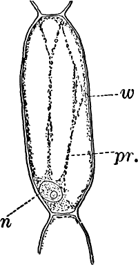
Fig. 1.—A single cell from a hair on the stamen of the
common spiderwort (Tradescantia), × 150. pr. protoplasm; w, cell
wall; n, nucleus.
In order to study the structure of the cell more exactly we will
select such as may be examined without cutting them. A good example is
furnished by the common spiderwort (Fig. 1). Attached to the base of
the stamens (Fig. 85, B) are delicate hairs composed of chains of
cells, which may be examined alive by carefully removing a stamen and
placing it in a drop of water under a cover glass. Each cell (Fig. 1)
is an oblong sac, with a delicate colorless wall which chemical tests
show to be composed of cellulose, a substance closely resembling
starch. Within this sac, and forming a lining to it, is a thin layer
of colorless matter containing many fine granules. Bands and threads
of the same substance traverse the cavity of the cell, which is filled
with a deep purple homogeneous fluid. This fluid, which in most cells
is colorless, is called the cell sap, and is composed mainly of water.
Imbedded in the granular lining of the sac is a roundish body (n),
which itself has a definite membrane, and usually shows one or more
roundish bodies within, besides an indistinctly granular appearance.
This body is called the nucleus of the cell, and the small one within
it, the nucleolus.The membrane surrounding the cell is known as the cell wall, and in
young plant cells is always composed of cellulose.The granular substance lining the cell wall (Fig. 1, pr.) is called
“protoplasm,” and with the nucleus constitutes the living part of the
cell. If sufficiently magnified, the granules within the protoplasm
will be seen to be in active streaming motion. This movement, which is
very evident here, is not often so conspicuous, but still may often be
detected without difficulty.
The cell may be regarded as the unit of organic structure, and of
cells are built up all of the complicated structures of which the
bodies of the highest plants and animals are composed. We shall find
that the cells may become very much modified for various purposes, but
at first they are almost identical in structure, and essentially the
same as the one we have just considered.
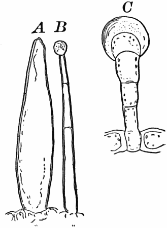
Fig. 3.—Hairs from the leaf stalk of a wild geranium.
A, single-celled hair. B and C, hairs consisting of a row of
cells. The terminal rounded cell secretes a peculiar scented oil that
gives the plant its characteristic odor. B, × 50; C, × 150.
Very many of the lower forms of life consist of but a single cell
which may occasionally be destitute of a cell wall. Such a form is
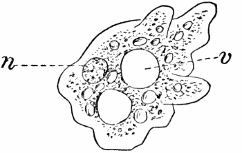
shown in Figure 2. Here we have a mass of protoplasm with a nucleus
(n) and cavities (vacuoles, v) filled with cell sap, but no cell
wall. The protoplasm is in constant movement, and by extensions of a
portion of the mass and contraction of other parts, the whole creeps
slowly along. Other naked cells (Fig. 12, B; Fig. 16, C) are
provided with delicate thread-like processes of protoplasm called
“cilia” (sing. cilium), which are in active vibration, and propel
the cell through the water.
Fig. 4.—A, cross section. B, longitudinal section
of the leaf stalk of wild geranium, showing its cellular structure.
Ep. epidermis. h, a hair, × 50. C, a cell from the prothallium
(young plant) of a fern, × 150. The contents of the cell contracted
by the action of a solution of sugar.
On placing a cell into a fluid denser than the cell sap (e.g. a
ten-per-cent solution of sugar in water), a portion of the water will
be extracted from the cell, and we shall then see the protoplasm
receding from the wall (Fig. 4, C), showing that it is normally in a
state of tension due to pressure from within of the cell sap. The cell
wall shows the same thing though in a less degree, owing to its being
much more rigid than the protoplasmic lining. It is owing to the
partial collapsing of the cells, consequent on loss of water, that
plants wither when the supply of water is cut off.
As cells grow, new ones are formed in various ways. If the new cells
remain together, cell aggregates, called tissues, are produced, and
of these tissues are built up the various organs of the higher plants.
The simplest tissues are rows of cells, such as form the hairs
covering the surface of the organs of many flowering plants (Fig. 3),
and are due to a division of the cells in a single direction. If the
divisions take place in three planes, masses of cells, such as make up
the stems, etc., of the higher plants, result (Fig. 4, A, B).
CHAPTER III.
CLASSIFICATION OF PLANTS.—PROTOPHYTES.
For the sake of convenience it is desirable to collect into groups
such plants as are evidently related; but as our knowledge of many
forms is still very imperfect, any classification we may adopt must be
to a great extent only provisional, and subject to change at any time,
as new forms are discovered or others become better understood.
The following general divisions are usually accepted: I. Sub-kingdom
(or Branch); II. Class; III. Order; IV. Family; V. Genus; VI. Species.
To illustrate: The white pine belongs to the highest great division
(sub-kingdom) of the plant kingdom. The plants of this division all
produce seeds, and hence are called “spermaphytes” (“seed plants”).
They may be divided into two groups (classes), distinguished by
certain peculiarities in the flowers and seeds. These are named
respectively “gymnosperms” and “angiosperms,” and to the first our
plant belongs. The gymnosperms may be further divided into several
subordinate groups (orders), one of which, the conifers, or
cone-bearing evergreens, includes our plant. This order includes
several families, among them the fir family (Abietineæ), including
the pines and firs. Of the sub-divisions (genera, sing. genus) of
the fir family, one of the most familiar is the genus Pinus, which
embraces all the true pines. Comparing different kinds of pines, we
find that they differ in the form of the cones, arrangement of the
leaves, and other minor particulars. The form we have selected differs
from all other native forms in its cones, and also in having the
leaves in fives, instead of twos or threes, as in most other kinds.
Therefore to distinguish the white pine from all other pines, it is
given a “specific” name, strobus.
The following table will show more plainly what is meant:
Sub-kingdom,
Spermaphyta.
Includes all spermaphytes, or seed plants.
Class,
Gymnospermæ.
All naked-seeded plants.
Order,
Coniferæ.
All cone-bearing evergreens.
Family,
Abietineæ.
Firs, Pines, etc.
Genus,
Pinus.
Pines.
Species,
Strobus.
White Pine.
SUB-KINGDOM I.
Protophytes.
The name Protophytes (Protophyta) has been applied to a large number
of simple plants, which differ a good deal among themselves. Some of
them differ strikingly from the higher plants, and resemble so
remarkably certain low forms of animal life as to be quite
indistinguishable from them, at least in certain stages. Indeed, there
are certain forms that are quite as much animal as vegetable in their
attributes, and must be regarded as connecting the two kingdoms. Such
forms are the slime moulds (Fig. 5), Euglena (Fig. 9), Volvox
(Fig. 10), and others.
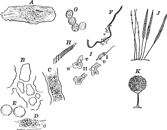
Fig. 5.—A, a portion of a slime mould growing on a
bit of rotten wood, × 3. B, outline of a part of the same, × 25.
C, a small portion showing the densely granular character of the
protoplasm, × 150. D, a group of spore cases of a slime mould
(Trichia), of about the natural size. E, two spore cases, × 5. The
one at the right has begun to open. F, a thread (capillitium) and
spores of Trichia, × 50. G, spores. H, end of the thread, × 300.
I, zoöspores of Trichia, × 300. i, ciliated form; ii, amœboid
forms. n, nucleus. v, contractile vacuole. J, K, sporangia of
two common slime moulds. J, Stemonitis, × 2. K, Arcyria, × 4.
Other protophytes, while evidently enough of vegetable nature, are
nevertheless very different in some respects from the higher plants.
The protophytes may be divided into three classes: I. The slime moulds
(Myxomycetes); II. The Schizophytes; III. The green monads
(Volvocineæ).
Class I.—The Slime Moulds.
These curious organisms are among the most puzzling forms with which
the botanist has to do, as they are so much like some of the lowest
forms of animal life as to be scarcely distinguishable from them, and
indeed they are sometimes regarded as animals rather than plants. At
certain stages they consist of naked masses of protoplasm of very
considerable size, not infrequently several centimetres in diameter.
These are met with on decaying logs in damp woods, on rotting leaves,
and other decaying vegetable matter. The commonest ones are bright
yellow or whitish, and form soft, slimy coverings over the substratum
(Fig. 5, A), penetrating into its crevices and showing sensitiveness
toward light. The plasmodium, as the mass of protoplasm is called, may
be made to creep upon a slide in the following way: A tumbler is
filled with water and placed in a saucer filled with sand. A strip of
blotting paper about the width of the slide is now placed with one end
in the water, the other hanging over the edge of the glass and against
one side of a slide, which is thus held upright, but must not be
allowed to touch the side of the tumbler. The strip of blotting paper
sucks up the water, which flows slowly down the surface of the slide
in contact with the blotting paper. If now a bit of the substance upon
which the plasmodium is growing is placed against the bottom of the
slide on the side where the stream of water is, the protoplasm will
creep up against the current of water and spread over the slide,
forming delicate threads in which most active streaming movements of
the central granular protoplasm may be seen under the microscope, and
the ends of the branches may be seen to push forward much as we saw in
the amœba. In order that the experiment may be successful, the whole
apparatus should be carefully protected from the light, and allowed to
stand for several hours. This power of movement, as well as the power
to take in solid food, are eminently animal characteristics, though
the former is common to many plants as well.
After a longer or shorter time the mass of protoplasm contracts and
gathers into little heaps, each of which develops into a structure
that has no resemblance to any animal, but would be at once placed
with plants. In one common form (Trichia) these are round or
pear-shaped bodies of a yellow color, and about as big as a pin head
(Fig. 5, D), occurring in groups on rotten logs in damp woods.
Others are stalked (Arcyria, Stemonitis) (Fig. 5, J, K), and
of various colors,—red, brown, etc. The outer part of the structure
is a more or less firm wall, which breaks when ripe, discharging a
powdery mass, mixed in most forms with very fine fibres.
When strongly magnified the fine dust is found to be made up of
innumerable small cells with thick walls, marked with ridges or
processes which differ much in different species. The fibres also
differ much in different genera. Sometimes they are simple, hair-like
threads; in others they are hollow tubes with spiral thickenings,
often very regularly placed, running around their walls.The spores may sometimes be made to germinate by placing them in a
drop of water, and allowing them to remain in a warm place for about
twenty-four hours. If the experiment has been successful, at the end
of this time the spore membrane will have burst, and the contents
escaped in the form of a naked mass of protoplasm (Zoöspore) with a
nucleus, and often showing a vacuole (Fig. 5, v), that alternately
becomes much distended, and then disappears entirely. On first
escaping it is usually provided with a long, whip-like filament of
protoplasm, which is in active movement, and by means of which the
cell swims actively through the water (Fig. 5, I i). Sometimes such
a cell will be seen to divide into two, the process taking but a short
time, so that the numbers of these cells under favorable conditions
may become very large. After a time the lash is withdrawn, and the
cell assumes much the form of a small amœba (I ii).
The succeeding stages are difficult to follow. After repeatedly
dividing, a large number of these amœba-like cells run together,
coalescing when they come in contact, and forming a mass of protoplasm
that grows, and finally assumes the form from which it started.
Of the common forms of slime moulds the species of Trichia (Figs.
D, I) and Physarum are, perhaps, the best for studying the
germination, as the spores are larger than in most other forms, and
germinate more readily. The experiment is apt to be most successful if
the spores are sown in a drop of water in which has been infused some
vegetable matter, such as a bit of rotten wood, boiling thoroughly to
kill all germs. A drop of this fluid should be placed on a perfectly
clean cover glass, which it is well to pass once or twice through a
flame, and the spores transferred to this drop with a needle
previously heated. By these precautions foreign germs will be avoided,
which otherwise may interfere seriously with the growth of the young
slime moulds. After sowing the spores in the drop of culture fluid,
the whole should be inverted over a so-called “moist chamber.” This is
simply a square of thick blotting paper, in which an opening is cut
small enough to be entirely covered by the cover glass, but large
enough so that the drop in the centre of the cover glass will not
touch the sides of the chamber, but will hang suspended clear in it.
The blotting paper should be soaked thoroughly in pure water
(distilled water is preferable), and then placed on a slide, covering
carefully with the cover glass with the suspended drop of fluid
containing the spores. The whole should be kept under cover so as to
prevent loss of water by evaporation. By this method the spores may be
examined conveniently without disturbing them, and the whole may be
kept as long as desired, so long as the blotting paper is kept wet, so
as to prevent the suspended drop from drying up.
Class II.—Schizophytes.
The Schizophytes are very small plants, though not infrequently
occurring in masses of considerable size. They are among the commonest
of all plants, and are found everywhere. They multiply almost entirely
by simple transverse division, or splitting of the cells, whence their
name. There are two pretty well-marked orders,—the blue-green slimes
(Cyanophyceæ) and the bacteria (Schizomycetes). They are
distinguished, primarily, by the first (with a very few exceptions)
containing chlorophyll (leaf-green), which is entirely absent from
nearly all of the latter.
The blue-green slimes: These are, with few exceptions, green plants of
simple structure, but possessing, in addition to the ordinary green
pigment (chlorophyll, or leaf-green), another coloring matter, soluble
in water, and usually blue in color, though sometimes yellowish or
red.
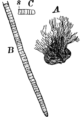
Fig. 6.—Blue-green slime (Oscillaria). A, mass of
filaments of the natural size. B, single filament, × 300. C, a
piece of a filament that has become separated. s, sheath, × 300.
As a representative of the group, we will select one of the commonest
forms (Oscillaria), known sometimes as green slime, from forming a
dark blue-green or blackish slimy coat over the mud at the bottom of
stagnant or sluggish water, in watering troughs, on damp rocks, or
even on moist earth. A search in the places mentioned can hardly fail
to secure plenty of specimens for study. If a bit of the slimy mass is
transferred to a china dish, or placed with considerable water on a
piece of stiff paper, after a short time the edge of the mass will
show numerous extremely fine filaments of a dark blue-green color,
radiating in all directions from the mass (Fig. 6, a). The filaments
are the individual plants, and possess considerable power of motion,
as is shown by letting the mass remain undisturbed for a day or two,
at the end of which time they will have formed a thin film over the
surface of the vessel in which they are kept; and the radiating
arrangement of the filaments can then be plainly seen.
If the mass is allowed to dry on the paper, it often leaves a bright
blue stain, due to the blue pigment in the cells of the filament. This
blue color can also be extracted by pulverizing a quantity of the
dried plants, and pouring water over them, the water soon becoming
tinged with a decided blue. If now the water containing the blue
pigment is filtered, and the residue treated with alcohol, the latter
will extract the chlorophyll, becoming colored of a yellow-green.
The microscope shows that the filaments of which the mass is composed
(Fig. 6, B) are single rows of short cylindrical cells of uniform
diameter, except at the end of the filament, where they usually become
somewhat smaller, so that the tip is more or less distinctly pointed.
The protoplasm of the cells has a few small granules scattered through
it, and is colored uniformly of a pale blue-green. No nucleus can be
seen.If the filament is broken, there may generally be detected a delicate,
colorless sheath that surrounds it, and extends beyond the end cells
(Fig. 6, c). The filament increases in length by the individual
cells undergoing division, this always taking place at right angles to
the axis of the filament. New filaments are produced simply by the
older ones breaking into a number of pieces, each of which rapidly
grows to full size.
The name “oscillaria” arises from the peculiar oscillating or swinging
movements that the plant exhibits. The most marked movement is a
swaying from side to side, combined with a rotary motion of the free
ends of the filaments, which are often twisted together like the
strands of a rope. If the filaments are entirely free, they may often
be observed to move forward with a slow, creeping movement. Just how
these movements are caused is still a matter of controversy.
The lowest of the Cyanophyceæ are strictly single-celled, separating
as soon as formed, but cohering usually in masses or colonies by means
of a thick mucilaginous substance that surrounds them (Fig. 7, D).
The higher ones are filaments, in which there may be considerable
differentiation. These often occur in masses of considerable size,
forming jelly-like lumps, which may be soft or quite firm (Fig. 7,
A, B). They are sometimes found on damp ground, but more commonly
attached to plants, stones, etc., in water. The masses vary in color
from light brown to deep blackish green, and in size from that of a
pin head to several centimetres in diameter.
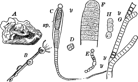
Fig. 7.—Forms of Cyanophyceæ. A, Nostoc. B,
Glœotrichia, × 1. C, individual of Glœotrichia. D,
Chroöcoccus. E, Nostoc. F, Oscillaria. G, H, Tolypothrix.
All × 300. y, heterocyst. sp. spore.
In the higher forms special cells called heterocysts are found. They
are colorless, or light yellowish, regularly disposed; but their
function is not known. Besides these, certain cells become
thick-walled, and form resting cells (spores) for the propagation of
the plant (Fig. 7, C. sp.). In species where the sheath of the
filament is well marked (Fig. 7, H), groups of cells slip out of the
sheath, and develop a new one, thus giving rise to a new plant.
The bacteria (Schizomycetes), although among the commonest of
organisms, owing to their excessive minuteness, are difficult to
study, especially for the beginner. They resemble, in their general
structure and methods of reproduction, the blue-green slimes, but are,
with very few exceptions, destitute of chlorophyll, although often
possessing bright pigments,—blue, violet, red, etc. It is one of
these that sometimes forms blood-red spots in flour paste or bits of
bread that have been kept very moist and warm. They are universally
present where decomposition is going on, and are themselves the
principal agents of decay, which is the result of their feeding upon
the substance, as, like all plants without chlorophyll, they require
organic matter for food. Most of the species are very tenacious of
life, and may be completely dried up for a long time without dying,
and on being placed in water will quickly revive. Being so extremely
small, they are readily carried about in the air in their dried-up
condition, and thus fall upon exposed bodies, setting up decomposition
if the conditions are favorable.
A simple experiment to show this may be performed by taking two test
tubes and partly filling them with an infusion of almost any organic
substance (dried leaves or hay, or a bit of meat will answer). The
fluid should now be boiled so as to kill any germs that may be in it;
and while hot, one of the vessels should be securely stopped up with a
plug of cotton wool, and the other left open. The cotton prevents
access of all solid particles, but allows the air to enter. If proper
care has been taken, the infusion in the closed vessel will remain
unchanged indefinitely; but the other will soon become turbid, and a
disagreeable odor will be given off. Microscopic examination shows the
first to be free from germs of any kind, while the second is swarming
with various forms of bacteria.
These little organisms have of late years attracted the attention of
very many scientists, from the fact that to them is due many, if not
all, contagious diseases. The germs of many such diseases have been
isolated, and experiments prove beyond doubt that these are alone the
causes of the diseases in question.
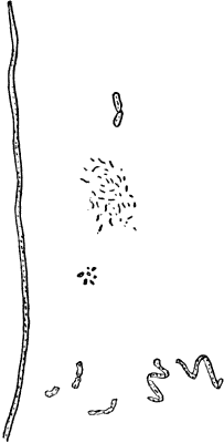
If a drop of water containing bacteria is examined, we find them to be
excessively small, many of them barely visible with the strongest
lenses. The larger ones (Fig. 8) recall quite strongly the smaller
species of oscillaria, and exhibit similar movements. Others are so
small as to appear as mere lines and dots, even with the strongest
lenses. Among the common forms are small, nearly globular cells;
oblong, rod-shaped or thread-shaped filaments, either straight or
curved, or even spirally twisted. Frequently they show a quick
movement which is probably in all cases due to cilia, which are,
however, too small to be seen in most cases.
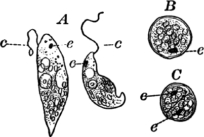
Fig. 9.—Euglena. A, individual in the active
condition. E, the red “eye-spot.” c, flagellum. n, nucleus. B,
resting stage. C, individual dividing, × 300.
Reproduction is for the most part by simple transverse division, as in
oscillaria; but occasionally spores are produced also.
Class III.—Green Monads (Volvocineæ).
This group of the protophytes is unquestionably closely related to
certain low animals (Monads or Flagellata), with which they are
sometimes united. They are characterized by being actively motile, and
are either strictly unicellular, or the cells are united by a
gelatinous envelope into a colony of definite form.
Of the first group, Euglena (Fig. 9), may be selected as a type.
This organism is found frequently among other algæ, and occasionally
forms a green film on stagnant water. It is sometimes regarded as a
plant, sometimes as an animal, and is an elongated, somewhat worm-like
cell without a definite cell wall, so that it can change its form to
some extent. The protoplasm contains oval masses, which are bright
green in color; but the forward pointed end of the cell is colorless,
and has a little depression. At this end there is a long vibratile
protoplasmic filament (c), by means of which the cell moves. There
is also to be seen near this end a red speck (e) which is probably
sensitive to light. A nucleus can usually be seen if the cell is first
killed with an iodine solution, which often will render the flagellum
(c) more evident, this being invisible while the cell is in motion.
The cells multiply by division. Previous to this the flagellum is
withdrawn, and a firm cell wall is formed about the cell (Fig. 9,
B). The contents then divide into two or more parts, which
afterwards escape as new individuals.
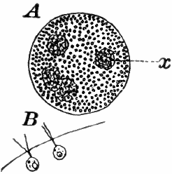
Fig. 10.—Volvox. A, mature colony, containing
several smaller ones (x), × 50. B, Two cells showing the cilia,
× 300.
Of the forms that are united in colonies[2] one of the best known is
Volvox (Fig. 10). This plant is sometimes found in quiet water,
where it floats on or near the surface as a dark green ball, just
large enough to be seen with the naked eye. They may be kept for some
time in aquaria, and will sometimes multiply rapidly, but are very
susceptible to extremes of temperature, especially of heat.
The colony (Fig. 10, A) is a hollow sphere, the numerous green cells
of which it is composed forming a single layer on the outside. By
killing with iodine, and using a strong lens, each cell is seen to be
somewhat pear-shaped (Fig. B), with the pointed end out. Attached to
this end are two vibratile filaments (cilia or flagella), and the
united movements of these cause the rolling motion of the whole
colony. Usually a number of young colonies (Fig. x) are found within
the mother colony. These arise by the repeated bipartition of a single
cell, and escape finally, forming independent colonies.Another (sexual) form of reproduction occurs, similar to that found in
many higher plants; but as it only occurs at certain seasons, it is
not likely to be met with by the student.
Other forms related to Volvox, and sometimes met with, are
Gonium, in which there are sixteen cells, forming a flat square;
Pandorina and Eudorina, with sixteen cells, forming an oval or
globular colony like Volvox, but much smaller. In all of these the
structure of the cells is essentially as in Volvox.
CHAPTER IV.
SUB-KINGDOM II.
Algæ.[3]
In the second sub-kingdom of plants is embraced an enormous assemblage
of plants, differing widely in size and complexity, and yet showing a
sufficiently complete gradation from the lowest to the highest as to
make it impracticable to make more than one sub-kingdom to include
them. They are nearly all aquatic forms, although many of them will
survive long periods of drying, such forms occurring on moist earth,
rocks, or the trunks of trees, but only growing when there is a
plentiful supply of water.
All of them possess chlorophyll, which, however, in many forms, is
hidden by the presence of a brown or red pigment. They are ordinarily
divided into three classes—I. The Green Algæ (Chlorophyceæ);
II. Brown Algæ (Phæophyceæ); III. Red Algæ (Rhodophyceæ).
Class I.—Green Algæ.
The green algæ are to be found almost everywhere where there is
moisture, but are especially abundant in sluggish or stagnant fresh
water, being much less common in salt water. They are for the most
part plants of simple structure, many being unicellular, and very few
of them plants of large size.
We may recognize five well-marked orders of the green algæ—I. Green
slimes (Protococcaceæ); II. Confervaceæ; III. Pond scums
(Conjugatæ); IV. Siphoneæ; V. Stone-worts (Characeæ).
Order I.—Protococcaceæ.
The members of this order are minute unicellular plants, growing
either in water or on the damp surfaces of stones, tree trunks, etc.
The plants sometimes grow isolated, but usually the cells are united
more or less regularly into colonies.
A common representative of the order is the common green slime,
Protococcus (Fig. 11, A, C), which forms a dark green slimy
coating over stones, tree trunks, flower pots, etc. Owing to their
minute size the structure can only be made out with the microscope.
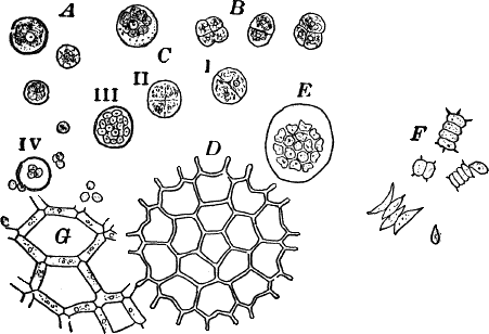
Fig. 11.—Protococcaceæ. A, C, Protococcus. A,
single cells. B, cells dividing by fission. C, successive steps in
the process of internal cell division. In C iv, the young cells have
mostly become free. D, a full-grown colony of Pediastrum. E, a
young colony still surrounded by the membrane of the mother cell. F,
Scenedesmus. All, × 300. G, small portion of a young colony of the
water net (Hydrodictyon), × 150.
Scraping off a little of the material mentioned into a drop of water
upon a slide, and carefully separating it with needles, a cover glass
may be placed over the preparation, and it is ready for examination.
When magnified, the green film is found to be composed of minute
globular cells of varying size, which may in places be found to be
united into groups. With a higher power, each cell (Fig. 11, A) is
seen to have a distinct cell wall, within which is colorless
protoplasm. Careful examination shows that the chlorophyll is confined
to several roundish bodies that are not usually in immediate contact
with the wall of the cell. These green masses are called chlorophyll
bodies (chloroplasts). Toward the centre of the cell, especially if it
has first been treated with iodine, the nucleus may be found. The size
of the cells, as well as the number of chloroplasts, varies a good
deal.With a little hunting, specimens in various stages of division may be
found. The division takes place in two ways. In the first (Fig. 11,
B), known as fission, a wall is formed across the cell, dividing it
into two cells, which may separate immediately or may remain united
until they have undergone further division. In this case the original
cell wall remains as part of the wall of the daughter cells. Fission
is the commonest form of cell multiplication throughout the vegetable
kingdom.The second form of cell division or internal cell division is shown at
C. Here the protoplasm and nucleus repeatedly divide until a number
of small cells are formed within the old one. These develop cell
walls, and escape by the breaking of the old cell wall, which is left
behind, and takes no part in the process. The cells thus formed are
sometimes provided with two cilia, and are capable of active movement.Internal cell division, as we shall see, is found in most plants, but
only at special times.Closely resembling Protococcus, and answering quite as well for
study, are numerous aquatic forms, such as Chlorococcum (Fig. 12).
These are for the most part destitute of a firm cell wall, but are
imbedded in masses of gelatinous substance like many Cyanophyceæ.
The chloroplasts are smaller and less distinct than in Protococcus.
The cells are here oval rather than round, and often show a clear
space at one end.
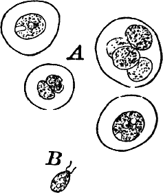
Fig. 12.—Chlorococcum, a plant related to
Protococcus, but the naked cells are surrounded by a colorless
gelatinous envelope. A, motionless cells. B, a cell that has
escaped from its envelope and is ciliated, × 300.
Owing to the absence of a definite membrane, a distinction between
fission and internal cell division can scarcely be made here. Often
the cells escape from the gelatinous envelope, and swim actively by
means of two cilia at the colorless end (Fig. 12, B). In this stage
they closely resemble the individuals of a Volvox colony, or other
green Flagellata, to which there is little doubt that they are
related.There are a number of curious forms common in fresh water that are
probably related to Protococcus, but differ in having the cells
united in colonies of definite form. Among the most striking are the
different species of Pediastrum (Fig. 11, D, E), often met with
in company with other algæ, and growing readily in aquaria when once
established. They are of very elegant shapes, and the number of cells
some multiple of four, usually sixteen.The cells form a flat disc, the outer ones being generally provided
with a pair of spines.New individuals arise by internal division of the cells, the contents
of each forming as many parts as there are cells in the whole colony.
The young cells now escape through a cleft in the wall of the mother
cell, but are still surrounded by a delicate membrane (Fig. 11, E).
Within this membrane the young cells arrange themselves in the form of
the original colony, and grow together, forming a new colony.A much larger but rarer form is the water net (Fig. 11, G), in which
the colony has the form of a hollow net, the spaces being surrounded
by long cylindrical cells placed end to end. Other common forms belong
to the genus Scenedesmus (Fig. 11, F), of which there are many
species.
Order II.—Confervaceæ.
Under this head are included a number of forms of which the simplest
ones approach closely, especially in their younger stages, the
Protococcaceæ. Indeed, some of the so-called Protococcaceæ are
known to be only the early stages of these plants.
A common member of this order is Cladophora, a coarse-branching
alga, growing commonly in running water, where it forms tufts,
sometimes a metre or more in length. By floating out a little of it in
a saucer, it is easy to see that it is made up of branching filaments.
The microscope shows (Fig. 13, A) that these filaments are rows of
cylindrical cells with thick walls showing evident stratification. At
intervals branches are given off, which may in turn branch, giving
rise to a complicated branching system. These branches begin as little
protuberances of the cell wall at the top of the cell. They increase
rapidly in length, and becoming slightly contracted at the base, a
wall is formed across at this point, shutting it off from the mother
cell.The protoplasm lines the wall of the cell, and extends in the form of
thin plates across the cavity of the cell, dividing it up into a
number of irregular chambers. Imbedded in the protoplasm are numerous
flattened chloroplasts, which are so close together as to make the
protoplasm appear almost uniformly green. Within the chloroplasts are
globular, glistening bodies, called “pyrenoids.” The cell has several
nuclei, but they are scarcely evident in the living cell. By placing
the cells for a few hours in a one per cent watery solution of chromic
acid, then washing thoroughly and staining with borax carmine, the
nuclei will be made very evident (Fig. 13, B). Such preparations may
be kept permanently in dilute glycerine.
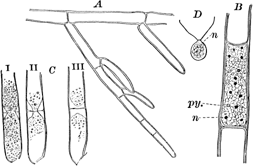
Fig. 13.—Cladophora. A, a fragment of a plant,
× 50. B, a single cell treated with chromic acid, and stained with
alum cochineal. n, nucleus. py. pyrenoid, × 150. C, three stages
in the division of a cell. i, 1.45 p.m.; ii, 2.55 p.m.; iii,
4.15 p.m., × 150. D, a zoöspore × 350.
If a mass of actively growing filaments is examined, some of the cells
will probably be found in process of fission. The process is very
simple, and may be easily followed (Fig. 13, C). A ridge of
cellulose is formed around the cell wall, projecting inward, and
pushing in the protoplasm as it grows. The process is continued until
the ring closes in the middle, cutting the protoplasmic body
completely in two, and forms a firm membrane across the middle of the
cell. The protoplasm at this stage (C iii.) is somewhat contracted,
but soon becomes closely applied to the new wall. The whole process
lasts, at ordinary temperatures (20°-25° C.), from three to four
hours.At certain times, but unfortunately not often to be met with, the
contents of some of the cells form, by internal division, a large
number of small, naked cells (zoöspores) (Fig. 13, D), which escape
and swim about actively for a time, and afterwards become invested
with a cell wall, and grow into a new filament. These cells are called
zoöspores, from their animal-like movements. They are provided with
two cilia, closely resembling the motile cells of the Protococcaceæ
and Volvocineæ.
There are very many examples of these simple Confervaceæ, some like
Conferva being simple rows of cells, others like Stigeoclonium
(Fig. 14, A), Chætophora and Draparnaldia (Fig. 14, B, C),
very much branched. The two latter forms are surrounded by masses of
transparent jelly, which sometimes reach a length of several
centimetres.
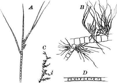
Fig. 14.—Confervaceæ. A, Stigeoclonium. B,
Draparnaldia, × 50. C, a piece of Draparnaldia, × 2. D, part
of a filament of Conferva, × 300.
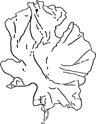
Among the marine forms related to these may be mentioned the sea
lettuce (Ulva), shown in Figure 15. The thin, bright-green,
leaf-like fronds of this plant are familiar to every seaside student.
Somewhat higher than Cladophora and its allies, especially in the
differentiation of the reproductive parts, are the various species of
Œdogonium and its relatives. There are numerous species of
Œdogonium not uncommon in stagnant water growing in company with
other algæ, but seldom forming masses by themselves of sufficient size
to be recognizable to the naked eye.
The plant is in structure much like Cladophora, except that it is
unbranched, and the cells have but a single nucleus (Fig. 16, E).
Even when not fruiting the filaments may usually be recognized by
peculiar cap-shaped structures at the top of some of the cells. These
arise as the result of certain peculiarities in the process of cell
division, which are too complicated to be explained here.There are two forms of reproduction, non-sexual and sexual. In the
first the contents of certain cells escape in the form of large
zoöspores (Fig. 16, C), of oval form, having the smaller end
colorless and surrounded by a crown of cilia. After a short period of
active motion, the zoöspore comes to rest, secretes a cell wall about
itself, and the transparent end becomes flattened out into a disc
(E, d), by which it fastens itself to some object in the water.
The upper part now rapidly elongates, and dividing repeatedly by cross
walls, develops into a filament like the original one. In many species
special zoöspores are formed, smaller than the ordinary ones, that
attach themselves to the filaments bearing the female reproductive
organ (oögonium), and grow into small plants bearing the male organ
(antheridium), (Fig. 16, B).
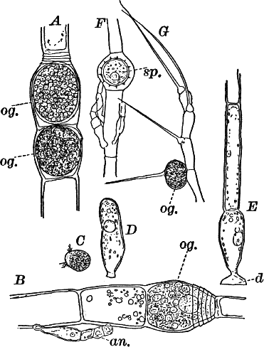
Fig. 16.—A, portion of a filament of Œdogonium,
with two oögonia (og.). The lower one shows the opening. B, a
similar filament, to which is attached a small male plant with an
antheridium (an.). C, a zoöspore of Œdogonium. D, a similar
spore germinating. E, base of a filament showing the disc (d) by
which it is attached. F, another species of Œdogonium with a ripe
spore (sp.). G, part of a plant of Bulbochæte. C, D, × 300;
the others × 150.
The sexual reproduction takes place as follows: Certain cells of a
filament become distinguished by their denser contents and by an
increase in size, becoming oval or nearly globular in form (Fig. 16,
A, B). When fully grown, the contents contract and form a naked
cell, which sometimes shows a clear area at one point on the surface.
This globular mass of protoplasm is the egg cell, or female cell, and
the cell containing it is called the “oögonium.” When the egg cell is
ripe, the oögonium opens by means of a little pore at one side
(Fig. 16, A).In other cells, either of the same filament or else of the small male
plants already mentioned, small motile cells, called spermatozoids,
are formed. These are much smaller than the egg cell, and resemble the
zoöspores in form, but are much smaller, and without chlorophyll. When
ripe they are discharged from the cells in which they were formed, and
enter the oögonium. By careful observation the student may possibly be
able to follow the spermatozoid into the oögonium, where it enters the
egg cell at the clear spot on its surface. As a result of the entrance
of the spermatozoid (fertilization), the egg cell becomes surrounded
by a thick brown wall, and becomes a resting spore. The spore loses
its green color, and the wall becomes dark colored and differentiated
into several layers, the outer one often provided with spines
(Fig. 16, F). As these spores do not germinate for a long time, the
process is only known in a comparatively small number of species, and
can hardly be followed by the ordinary student.
Much like Œdogonium, but differing in being branched, is the genus
Bulbochæte, characterized also by hairs swollen at the base, and
prolonged into a delicate filament (Fig. 16, G).
The highest members of the Confervaceæ are those of the genus
Coleochæte (Fig. 17), of which there are several species found in
the United States. These show some striking resemblances to the red
seaweeds, and possibly form a transition from the green algæ to the
red. The commonest species form bright-green discs, adhering firmly
to the stems and floating leaves of water lilies and other aquatics.
In aquaria they sometimes attach themselves in large numbers to the
glass sides of the vessel.
Growing from the upper surface are numerous hairs, consisting of a
short, sheath-like base, including a very long and delicate filament
(Fig. 17, B). In their methods of reproduction they resemble
Œdogonium, but the reproductive organs are more specialized.
CHAPTER V.
Green Algæ—Continued.
Order III.—Pond Scums (Conjugatæ).
The Conjugatæ, while in some respects approaching the Confervaceæ
in structure, yet differ from them to such an extent in some respects
that their close relationship is doubtful. They are very common and
familiar plants, some of them forming great floating masses upon the
surface of every stagnant pond and ditch, being commonly known as
“pond scum.” The commonest of these pond scums belong to the genus
Spirogyra, and one of these will illustrate the characteristics of
the order. When in active growth these masses are of a vivid green,
and owing to the presence of a gelatinous coating feel slimy, slipping
through the hands when one attempts to lift them from the water.
Spread out in water, the masses are seen to be composed of slender
threads, often many centimetres in length, and showing no sign of
branching.
Fig. 18.—A, a filament of a common pond scum
(Spirogyra) separating into two parts. B, a cell undergoing
division. The cell is seen in optical section, and the chlorophyll
bands are omitted, n, nʹ, the two nuclei. C, a complete cell.
n, nucleus. py. pyrenoid. D, E, successive stages in the
process of conjugation. G, a ripe spore. H, a form in which
conjugation takes place between the cells of the same filament. All
× 150.
For microscopical examination the larger species are preferable. When
one of these is magnified (Fig. 18, A, C), the unbranched filament
is shown to be made up of perfectly cylindrical cells, with rather
delicate walls. The protoplasm is confined to a thin layer lining the
walls, except for numerous fine filaments that radiate from the
centrally placed nucleus (n), which thus appears suspended in the
middle of the cell. The nucleus is large and distinct in the larger
species, and has a noticeably large and conspicuous nucleolus. The
most noticeable thing about the cell is the green spiral bands running
around it. These are the chloroplasts, which in all the Conjugatæ
are of very peculiar forms. The number of these bands varies much in
different species of Spirogyra, but is commonly two or three. These
chloroplasts, like those of other plants, are not noticeably different
in structure from the ordinary protoplasm, as is shown by extracting
the chlorophyll, which may be done by placing the plants in alcohol
for a short time. This extracts the chlorophyll, but a microscopic
examination of the decolored cells shows that the bands remain
unchanged, except for the absence of color. These bands are flattened,
with irregularly scalloped margins, and at intervals have rounded
bodies (pyrenoids) imbedded in them (Fig. 18, C, py.). The
pyrenoids, especially when the plant has been exposed to the light for
some time, are surrounded by a circle of small granules, which become
bluish when iodine is applied, showing them to be starch. (To show the
effect of iodine on starch on a large scale, mix a little flour, which
is nearly all starch, with water, and add a little iodine. The starch
will immediately become colored blue, varying in intensity with the
amount of iodine.) The cells divide much as in Cladophora, but the
nucleus here takes part in the process. The division naturally occurs
only at night, but by reducing the temperature at night to near the
freezing point (4° C., or a little lower), the process may be checked.
The experiment is most conveniently made when the temperature out of
doors approaches the freezing point. Then it is only necessary to
keep the plants in a warm room until about 10 p.m., when they may be
put out of doors for the night. On bringing them in in the morning,
the division will begin almost at once, and may be easily studied. The
nucleus divides into two parts, which remain for a time connected by
delicate threads (Fig. 18, B), that finally disappear. At first no
nucleoli are present in the daughter nuclei, but they appear before
the division is complete.New filaments are formed by the breaking up of the old ones, this
sometimes being very rapid. As the cells break apart, the free ends
bulge strongly, showing the pressure exerted upon the cell wall by the
contents (Fig. 18, A).
Spores like those of Œdogonium are formed, but the process is
somewhat different. It occurs in most species late in the spring, but
may sometimes be met with at other times. The masses of fruiting
plants usually appear brownish colored. If spores have been formed
they can, in the larger species at least, be seen with a hand lens,
appearing as rows of dark-colored specks.
Two filaments lying side by side send out protuberances of the cell
wall that grow toward each other until they touch (Fig. 18, D). At
the point of contact, the wall is absorbed, forming a continuous
channel from one cell to the other. This process usually takes place
in all the cells of the two filaments, so that the two filaments,
connected by tubes at regular intervals, have the form of a ladder.In some species adjoining cells of the same filament become connected,
the tubes being formed at the end of the cells (Fig. 18, H), and the
cell in which the spore is formed enlarges.Soon after the channel is completed, the contents of one cell flow
slowly through it into the neighboring cell, and the protoplasm of the
two fuses into one mass. (The union of the nuclei has also been
observed.) The young spore thus formed contracts somewhat, becoming
oval in form, and soon secretes a thick wall, colorless at first, but
afterwards becoming brown and more or less opaque. The chlorophyll
bands, although much crowded, are at first distinguishable, but later
lose the chlorophyll, and become unrecognizable. Like the resting
spores of Œdogonium these require a long period of rest before
germinating.
There are various genera of the pond scums, differing in the form of
the chloroplasts and also in the position of the spores. Of these may
be mentioned Zygnema (Fig. 19, A), with two star-shaped
chloroplasts in each cell, and Mesocarpus (Fig. 19, B, D), in
which the single chloroplast has the form of a thin median plate. (B
shows the appearance from in front, C from the side, showing the
thickness of the plate.) Mesocarpus and the allied genera have the
spore formed between the filaments, the contents of both the uniting
cells leaving them.
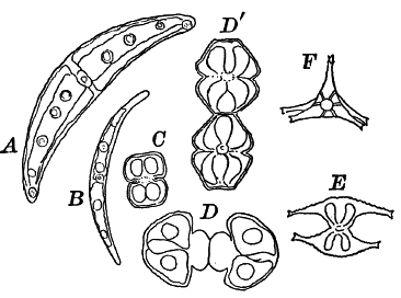
Fig. 20.—Forms of Desmids. A, B, Closterium.
C, D, Dʹ, Cosmarium. D, and Dʹ show the process of
division. E, F, Staurastrum; E seen from the side, F from
the end.
Evidently related to the pond scums, but differing in being for the
most part strictly unicellular, are the desmids (Fig. 20). They are
confined to fresh water, and seldom occur in masses of sufficient size
to be seen with the naked eye, usually being found associated with
pond scums or other filamentous forms. Many of the most beautiful
forms may be obtained by examining the matter adhering to the leaves
and stems of many floating water plants, especially the bladder weed
(Utricularia) and other fine-leaved aquatics.
The desmids include the most beautiful examples of unicellular plants
to be met with, the cells having extremely elegant outlines. The cell
shows a division into two parts, and is often constricted in the
middle, each division having a single large chloroplast of peculiar
form. The central part of the cell in which the nucleus lies is
colorless.Among the commonest forms, often growing with Spirogyra, are various
species of Closterium (Fig. 20, A, B), recognizable at once by
their crescent shape. The cell appears bright green, except at the
ends and in the middle. The large chloroplast in each half is composed
of six longitudinal plates, united at the axis of the cell. Several
large pyrenoids are always found, often forming a regular line through
the central axis. At each end of the cell is a vacuole containing
small granules that show an active dancing movement.
The desmids often have the power of movement, swimming or creeping
slowly over the slide as we examine them, but the mechanism of these
movements is still doubtful.
In their reproduction they closely resemble the pond scums.
Order IV.—Siphoneæ.
The Siphoneæ are algæ occurring both in fresh and salt water, and
are distinguished from other algæ by having the form of a tube,
undivided by partition walls, except when reproduction occurs. The
only common representatives of the order in fresh water are those
belonging to the genus Vaucheria, but these are to be had almost
everywhere. They usually occur in shallow ditches and ponds, growing
on the bottom, or not infrequently becoming free, and floating where
the water is deeper. They form large, dark green, felted masses, and
are sometimes known as “green felts.” Some species grow also on the
wet ground about springs. An examination of one of the masses shows it
to be made up of closely matted, hair-like threads, each of which is
an individual plant.
In transferring the plants to the slide for microscopic examination,
they must be handled very carefully, as they are very easily injured.
Each thread is a long tube, branching sometimes, but not divided into
cells as in Spirogyra or Cladophora. If we follow it to the tip,
the contents here will be found to be denser, this being the growing
point. By careful focusing it is easy to show that the protoplasm is
confined to a thin layer lining the wall, the central cavity of the
tube being filled with cell sap. In the protoplasm are numerous
elongated chloroplasts (cl.). and a larger or smaller number of
small, shining, globular bodies (ol.). These latter are drops of
oil, and, when the filaments are injured, sometimes run together, and
form drops of large size. No nucleus can be seen in the living plant,
but by treatment with chromic acid and staining, numerous very small
nuclei may be demonstrated.
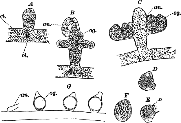
Fig. 21.—A, C, successive stages in the
development of the sexual organs of a green felt (Vaucheria). an.
antheridium. og. oögonium. D, a ripe oögonium. E, the same after
it has opened. o, the egg cell. F, a ripe spore. G, a species in
which the sexual organs are borne separately on the main filament.
A, F, × 150. G, × 50. cl. chloroplasts. ol. oil.
When the filaments are growing upon the ground, or at the bottom of
shallow water, the lower end is colorless, and forms a more or less
branching root-like structure, fastening it to the earth. These
rootlets, like the rest of the filament, are undivided by walls.One of the commonest and at the same time most characteristic species
is Vaucheria racemosa (Fig. 21, A, F). The plant multiplies
non-sexually by branches pinched off by a constriction at the point
where they join the main filament, or by the filament itself becoming
constricted and separating into several parts, each one constituting a
new individual.The sexual organs are formed on special branches, and their
arrangement is such as to make the species instantly recognizable.The first sign of their development is the formation of a short branch
(Fig. 21, A) growing out at right angles to the main filament. This
branch becomes club-shaped, and the end somewhat pointed and more
slender, and curves over. This slender, curved portion is almost
colorless, and is soon shut off from the rest of the branch. It is
called an “antheridium,” and within are produced, by internal
division, numerous excessively small spermatozoids.As the branch grows, its contents become very dense, the oil drops
especially increasing in number and size. About the time that the
antheridium becomes shut off, a circle of buds appears about its base
(Fig. 21, B, og.). These are the young oögonia, which rapidly
increase in size, assuming an oval form, and become separated by walls
from the main branch (C). Unlike the antheridium, the oögonia
contain a great deal of chlorophyll, appearing deep green.When ripe, the antheridium opens at the end and discharges the
spermatozoids, which are, however, so very small as scarcely to be
visible except with the strongest lenses. They are little oval bodies
with two cilia, which may sometimes be rendered visible by staining
with iodine.
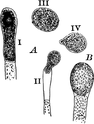
Fig. 22.—A, non-sexual reproduction in Vaucheria
sessilis. B, non-sexual spore of V. geminata, × 50.
The oögonia, which at first are uniformly colored, just before
maturity show a colorless space at the top, from which the
chloroplasts and oil drops have disappeared (D), and at the same
time this portion pushes out in the form of a short beak. Soon after
the wall is absorbed at this point, and a portion of the contents is
forced out, leaving an opening, and at the same time the remaining
contents contract to form a round mass, the germ or egg cell (Fig. 21,
E, o). Almost as soon as the oögonium opens, the spermatozoids
collect about it and enter; but, on account of their minuteness, it is
almost impossible to follow them into the egg cell, or to determine
whether several or only one enter. The fertilized egg cell becomes
almost at once surrounded by a wall, which rapidly thickens, and forms
a resting spore. As the spore ripens, it loses its green color,
becoming colorless, with a few reddish brown specks scattered through
it (F).In some species the sexual organs are borne directly on the filament
(Fig. 21, G).Large zoöspores are formed in some of the green felts (Fig. 22, A),
and are produced singly in the ends of branches that become swollen,
dark green, and filled with very dense protoplasm. This end becomes
separated by a wall from the rest of the branch, the end opens, and
the contents escape as a very large zoöspore, covered with numerous
short cilia (A ii). After a short period of activity, this loses its
cilia, develops a wall, and begins to grow (III, IV). Other species
(B) produce similar spores, which, however, are not motile, and
remain within the mother cell until they are set free by the decay of
its wall.
Order V.—Characeæ.
The Characeæ, or stone-worts, as some of them are called, are so
very different from the other green algæ that it is highly probable
that they should be separated from them.
The type of the order is the genus Chara (Fig. 23), called
stone-worts from the coating of carbonate of lime found in most of
them, giving them a harsh, stony texture. Several species are common
growing upon the bottom of ponds and slow streams, and range in size
from a few centimetres to a metre or more in height.
The plant (Fig. 23, A) consists of a central jointed axis with
circles of leaves at each joint or node. The distance between the
nodes (internodes) may in the larger species reach a length of several
centimetres. The leaves are slender, cylindrical structures, and like
the stem divided into nodes and internodes, and have at the nodes
delicate leaflets.
At each joint of the leaf, in fruiting specimens, attached to the
inner side, are borne two small, roundish bodies, in the commoner
species of a reddish color (Fig. 23, A, r). The lower of the two
is globular, and bright scarlet in color; the other, more oval and
duller.
Examined with a lens the main axis presents a striated appearance. The
whole plant is harsh to the touch and brittle, owing to the limy
coating. It is fastened to the ground by fine, colorless hairs, or
rootlets.
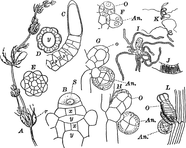
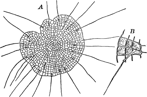
Fig. 23.—A, plant of a stone-wort (Chara),
one-half natural size. r, reproductive organs. B, longitudinal
section through the apex. S, apical cell. x, nodes. y,
internodes. C, a young leaf. D, cross section of an internode.
E, of a node of a somewhat older leaf. F, G, young sexual organs
seen in optical section. o, oögonium. An. antheridium. H,
superficial view. G, I, group of filaments containing
spermatozoids. J, a small portion of one of these more magnified,
showing a spermatozoid in each cell. K, free spermatozoids. L, a
piece of a leaf with ripe oögonium (o), and antheridium (An.).
B, H, × 150. J, K, × 300. I, × 50. L, × 25.
By making a series of longitudinal sections with a sharp razor through
the top of the plant, and magnifying sufficiently, it is found to end
in a single, nearly hemispherical cell (Fig. 23, B, S). This from
its position is called the “apical cell,” and from it are derived all
the tissues of the plant. Segments are cut off from its base, and
these divide again into two by a wall parallel to the first. Of the
two cells thus formed one undergoes no further division and forms the
central cell of an internode (y); the other divides repeatedly,
forming a node or joint (x).As the arrangement of these cells is essentially the same in the
leaves and stem, we will examine it in the former, as by cutting
several cross-sections of the whole bunch of young leaves near the top
of the plant, we shall pretty certainly get some sections through a
joint. The arrangement is shown in Figure 23, E.As the stem grows, a covering is formed over the large internodal cell
(y) by the growth of cells from the nodes. These grow both from
above and below, meeting in the middle of the internode and completely
hiding the long axial cell. A section across the internode shows the
large axial cell (y) surrounded by the regularly arranged cells of
the covering or cortex (Fig. 23, D).All the cells contain a layer of protoplasm next the wall with
numerous oval chloroplasts. If the cells are uninjured, they often
show a very marked movement of the protoplasm. These movements are
best seen, however, in forms like Nitella, where the long internodal
cells are not covered with a cortex. In Chara they are most evident
in the root hairs that fasten the plant to the ground.The growth of the leaves is almost identical with that of the stem,
but the apical growth is limited, and the apical cell becomes finally
very long and pointed (Fig. 23, C). In some species the chloroplasts
are reddish in the young cells, assuming their green color as the
cells approach maturity.
The plant multiplies non-sexually by means of special branches that
may become detached, but there are no non-sexual spores formed.
The sexual organs have already been noticed arising in pairs at the
joints of the leaves. The oögonium is formed above, the antheridium
below.The young oögonium (F, O) consists of a central cell, below which
is a smaller one surrounded by a circle of five others, which do not
at first project above the central cell, but later completely envelop
it (G). Each of these five cells early becomes divided into an upper
and a lower one, the latter becoming twisted as it elongates, and the
central cell later has a small cell cut off from its base by an
oblique wall. The central cell forms the egg cell, which in the ripe
oögonium (L, O) is surrounded by five, spirally twisted cells, and
crowned by a circle of five smaller ones, which become of a yellowish
color when full grown. They separate at the time of fertilization to
allow the spermatozoids to enter the oögonium.The antheridium consists at first of a basal cell and a terminal one.
The latter, which is nearly globular, divides into eight nearly
similar cells by walls passing through the centre. In each of these
eight cells two walls are next formed parallel to the outer surface,
so that the antheridium (apart from the basal cell) contains
twenty-four cells arranged in three concentric series (G, an.).
These cells, especially the outer ones, develop a great amount of a
red pigment, giving the antheridium its characteristic color.The diameter of the antheridium now increases rapidly, and the central
cells separate, leaving a large space within. Of the inner cells, the
second series, while not increasing in diameter, elongate, assuming an
oblong form, and from the innermost are developed long filaments (I,
J) composed of a single row of cells, in each of which is formed a
spermatozoid.The eight outer cells are nearly triangular in outline, fitting
together by deeply indented margins, and having the oblong cells with
the attached filaments upon their inner faces.If a ripe antheridium is crushed in a drop of water, after lying a few
minutes the spermatozoids will escape through small openings in the
side of the cells. They are much larger than any we have met with.
Each is a colorless, spiral thread with about three coils, one end
being somewhat dilated with a few granules; the other more pointed,
and bearing two extremely long and delicate cilia (K). To see the
cilia it is necessary to kill the spermatozoids with iodine or some
other reagent.After fertilization the outer cells of the oögonium become very hard,
and the whole falls off, germinating after a sufficient period of
rest.
According to the accounts of Pringsheim and others, the young plant
consists at first of a row of elongated cells, upon which a bud is
formed that develops into the perfect plant.
There are two families of the Characeæ, the Chareæ, of which
Chara is the type, and the Nitelleæ, represented by various
species of Nitella and Tolypella. The second family have the
internodes without any cortex—that is, consisting of a single long
cell; and the crown at the top of the oögonium is composed of ten
cells instead of five. They are also destitute of the limy coating of
the Chareæ.
Both as regards the structure of the plant itself, as well as the
reproductive organs, especially the very complex antheridium, the
Characeæ are very widely separated from any other group of plants,
either above or below them.
CHAPTER VI.
THE BROWN ALGÆ (Phæophyceæ).
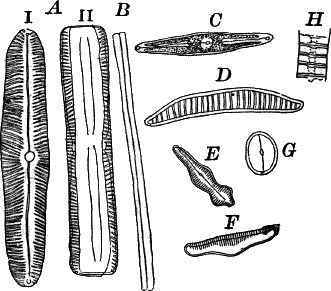
Fig. 24.—Forms of diatoms. A, Pinnularia. i, seen
from above; ii, from the side. B, Fragillaria (?). C,
Navicula. D, F, Eunotia. E, Gomphonema. G, Cocconeis.
H, Diatoma. All × 300.
These plants are all characterized by the presence of a brown pigment,
in addition to the chlorophyll, which almost entirely conceals the
latter, giving the plants a brownish color, ranging from a light
yellowish brown to nearly black. One order of plants that possibly
belongs here (Diatomaceæ) are single celled, but the others are for
the most part large seaweeds. The diatoms, which are placed in this
class simply on account of the color, are probably not closely related
to the other brown algæ, but just where they should be placed is
difficult to say. In some respects they approach quite closely the
desmids, and are not infrequently regarded as related to them. They
are among the commonest of organisms occurring everywhere in stagnant
and running water, both fresh and salt, forming usually, slimy,
yellowish coatings on stones, mud, aquatic plants, etc. Like the
desmids they may be single or united into filaments, and not
infrequently are attached by means of a delicate gelatinous stalk
(Fig. 25).
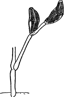
They are at once distinguished from the desmids by their color, which
is always some shade of yellowish or reddish brown. The commonest
forms, e.g. Navicula (Fig. 24, C), are boat-shaped when seen
from above, but there is great variety in this respect. The cell wall
is always impregnated with large amounts of flint, so that after the
cell dies its shape is perfectly preserved, the flint making a perfect
cast of it, looking like glass. These flinty shells exhibit
wonderfully beautiful and delicate markings which are sometimes so
fine as to test the best lenses to make them out.This shell is composed of two parts, one shutting over the other like
a pill box and its cover. This arrangement is best seen in such large
forms as Pinnularia (Fig. 24, A ii).
Most of the diatoms show movements, swimming slowly or gliding over
solid substances; but like the movements of Oscillaria and the
desmids, the movements are not satisfactorily understood, although
several explanations have been offered.
They resemble somewhat the desmids in their reproduction.
The True Brown Algæ.
These are all marine forms, many of great size, reaching a length in
some cases of a hundred metres or more, and showing a good deal of
differentiation in their tissues and organs.
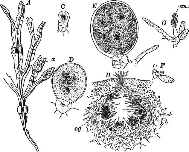
Fig. 26.—A, a branch of common rock weed (Fucus),
one-half natural size. x, end of a branch bearing conceptacles. B,
section through a conceptacle containing oögonia (og.), × 25. C,
E, successive stages in the development of the oögonium, × 150. F,
G, antheridia. In G, one of the antheridia has discharged the mass
of spermatozoids (an.), × 150.
One of the commonest forms is the ordinary rock weed (Fucus), which
covers the rocks of our northeastern coast with a heavy drapery for
several feet above low-water mark, so that the plants are completely
exposed as the tide recedes. The commonest species, F. vesiculosus
(Fig. 26, A), is distinguished by the air sacs with which the stems
are provided. The plant is attached to the rock by means of a sort of
disc or root from which springs a stem of tough, leathery texture, and
forking regularly at intervals, so that the ultimate branches are very
numerous, and the plant may reach a length of a metre or more. The
branches are flattened and leaf-like, the centre traversed by a
thickened midrib. The end of the growing branches is occupied by a
transversely elongated pit or depression. The growing point is at the
bottom of this pit, and by a regular forking of the growing point the
symmetrical branching of the plant is brought about. Scattered over
the surface are little circular pits through whose openings protrude
bunches of fine hairs. When wet the plant is flexible and leathery,
but it may become quite dry and hard without suffering, as may be seen
when the plants are exposed to the sun at low tide.
The air bladders are placed in pairs, for the most part, and buoy up
the plant, bringing it up to the surface when covered with water.
The interior of the plant is very soft and gelatinous, while the outer
part forms a sort of tough rind of much firmer consistence. The ends
of some of the branches (Fig. 26, A, x) are usually much swollen,
and the surface covered with little elevations from which may often be
seen protruding clusters of hairs like those arising from the other
parts of the plant. A section through one of these enlarged ends shows
that each elevation corresponds to a cavity situated below it. On some
of the plants these cavities are filled with an orange-yellow mass; in
others there are a number of roundish olive-brown bodies large enough
to be easily seen. The yellow masses are masses of antheridia; the
round bodies, the oögonia.
If the plants are gathered while wet, and packed so as to prevent
evaporation of the water, they will keep perfectly for several days,
and may readily be shipped for long distances. If they are to be
studied away from the seashore, sections for microscopic examination
should be mounted in salt water (about 3 parts in weight of common
salt to 100 of water). If fresh material is not to be had, dried
specimens or alcoholic material will answer pretty well.
To study the minute structure of the plant, make a thin cross-section,
and mount in salt water. The inner part or pith is composed of loosely
arranged, elongated cells, placed end to end, and forming an irregular
network, the large spaces between filled with the mucilaginous
substance derived from the altered outer walls of these cells. This
mucilage is hard when dry, but swells up enormously in water,
especially fresh water. The cells grow smaller and more compact toward
the outside of the section, until there are no spaces of any size
between those of the outside or rind. The cells contain small
chloroplasts like those of the higher plants, but owing to the
presence of the brown pigment found in all of the class, in addition
to the chlorophyll, they appear golden brown instead of green.No non-sexual reproductive bodies are known in the rock weeds, beyond
small branches that occur in clusters on the margins of the main
branches, and probably become detached, forming new plants. In some of
the lower forms, however, e.g. Ectocarpus and Laminaria
(Fig. 28, A, C), zoöspores are formed.The sexual organs of the rock weed, as we have already seen, are borne
in special cavities (conceptacles) in the enlarged ends of some of the
branches. In the species here figured, F. vesiculosus, the
antheridia and oögonia are borne on separate plants; but in others,
e.g. F. platycarpus, they are both in the same conceptacle.The walls of the conceptacle (Fig. 26, B) are composed of closely
interwoven filaments, from which grow inward numerous hairs, filling
up the space within, and often extending out through the opening at
the top.The reproductive bodies arise from the base of these hairs. The
oögonia (Fig. 26, C, E) arise as nearly colorless cells, that
early become divided into two cells, a short basal cell or stalk and a
larger terminal one, the oögonium proper. The latter enlarges rapidly,
and its contents divide into eight parts. The division is at first
indicated by a division of the central portion, which includes the
nucleus, and is colored brown, into two, four, and finally eight
parts, after which walls are formed between these. The brown color
spreads until the whole oögonium is of a nearly uniform olive-brown
tint.When ripe, the upper part of the oögonium dissolves, allowing the
eight cells, still enclosed in a delicate membrane, to escape
(Fig. 27, H). Finally, the walls separating the inner cells of the
oögonium become also absorbed, as well as the surrounding membrane,
and the eight egg cells escape into the water (Fig. 27, I) as naked
balls of protoplasm, in which a central nucleus may be dimly seen.The antheridia (Fig. 26, F, G) are small oblong cells, at first
colorless, but when ripe containing numerous glistening, reddish brown
dots, each of which is part of a spermatozoid. When ripe, the contents
of the antheridium are forced out into the water (G), leaving the
empty outer wall behind, but still surrounded by a thin membrane.
After a few minutes this membrane is dissolved, and the spermatozoids
are set free. These (Fig. 27, K) are oval in form, with two long
cilia attached to the side where the brown speck, seen while still
within the antheridium, is conspicuous.The act of fertilization may be easily observed by laying fresh
antheridia into a drop of water containing recently discharged egg
cells. To obtain these, all that is necessary is to allow freshly
gathered plants to remain in the air until they are somewhat dry, when
the ripe sexual cells will be discharged from the openings of the
conceptacles, exuding as little drops, those with antheridia being
orange-yellow; the masses of oögonia, olive. Within a few minutes
after putting the oögonia into water, the egg cells may be seen to
escape into the water, when some of the antheridia may be added. The
spermatozoids will be quickly discharged, and collect immediately in
great numbers about the egg cells, to which they apply themselves
closely, often setting them in rotation by the movements of their
cilia, and presenting a most extraordinary spectacle (J). Owing to
the small size of the spermatozoids, and the opacity of the eggs, it
is impossible to see whether more than one spermatozoid penetrates it;
but from what is known in other cases it is not likely. The egg now
secretes a wall about itself, and within a short time begins to grow.
It becomes pear-shaped, the narrow portion becoming attached to the
parent plant or to some other object by means of rootlets, and the
upper part grows into the body of the young plant (Fig. 27, M).
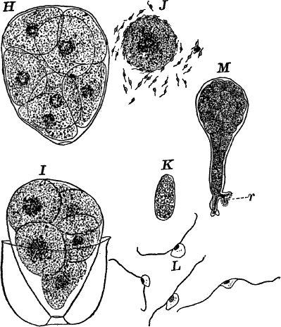
Fig. 27.—H, the eight egg cells still surrounded by
the inner membrane of the oögonium. I, the egg cells escaping into
the water. J, a single egg cell surrounded by spermatozoids. K,
mass of spermatozoids surrounded by the inner membrane of the
antheridium. L, spermatozoids. M, young plant. r, the roots.
K, × 300; L, × 600; the others, × 150.
The simpler brown seaweeds, so far as known, multiply only by means of
zoöspores, which may grow directly into new plants, or, as has been
observed in some species, two zoöspores will first unite. A few, like
Ectocarpus (Fig. 28, A), are simple, branched filaments, but most
are large plants with complex tissues. Of the latter, a familiar
example is the common kelp, “devil’s apron” (Laminaria), often three
to four metres in length, with a stout stalk, provided with root-like
organs, by which it is firmly fastened. Above, it expands into a
broad, leaf-like frond, which in some species is divided into strips.
Related to the kelps is the giant kelp of the Pacific (Macrocystis),
which is said sometimes to reach a length of three hundred metres.
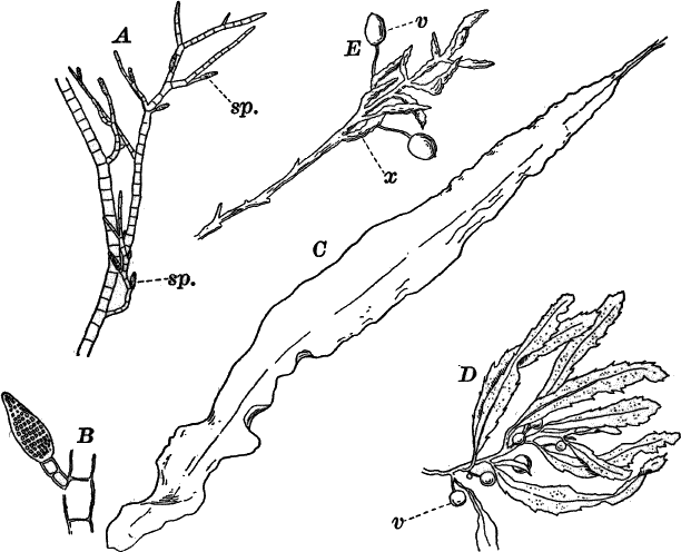
Fig. 28.—Forms of brown seaweeds. A, Ectocarpus,
× 50. Sporangia (sp.). B, a single sporangium, × 150. C, kelp
(Laminaria), × ⅛. D, E, gulf weed (Sargassum). D, one-half
natural size. E, natural size. v, air bladders. x, conceptacle
bearing branches.
The highest of the class are the gulf weeds (Sargassum), plants of
the warmer seas, but one species of which is found from Cape Cod
southward (Fig. 28, D, E). These plants possess distinct stems and
leaves, and there are stalked air bladders, looking like berries,
giving the plant a striking resemblance to the higher land plants.
CHAPTER VII.
Class III.—The Red Algæ (Rhodophyceæ).
These are among the most beautiful and interesting members of the
plant kingdom, both on account of their beautiful colors and the
exquisitely graceful forms exhibited by many of them. Unfortunately
for inland students they are, with few exceptions, confined to salt
water, and consequently fresh material is not available. Nevertheless,
enough can be done with dried material to get a good idea of their
general appearance, and the fruiting plants can be readily preserved
in strong alcohol. Specimens, simply dried, may be kept for an
indefinite period, and on being placed in water will assume perfectly
the appearance of the living plants. Prolonged exposure, however, to
the action of fresh water extracts the red pigment that gives them
their characteristic color. This pigment is found in the chlorophyll
bodies, and usually quite conceals the chlorophyll, which, however,
becomes evident so soon as the red pigment is removed.
The red seaweeds differ much in the complexity of the plant body, but
all agree in the presence of the red pigment, and, at least in the
main, in their reproduction. The simpler ones consist of rows of
cells, usually branching like Cladophora; others form cell plates
comparable to Ulva (Fig. 30, C, D); while others, among which is
the well-known Irish moss (Chondrus), form plants of considerable
size, with pretty well differentiated tissues. In such forms the outer
cells are smaller and firmer, constituting a sort of rind; while the
inner portions are made up of larger and looser cells, and may be
called the pith. Between these extremes are all intermediate forms.
They usually grow attached to rocks, shells, wood, or other plants,
such as the kelps and even the larger red seaweeds. They are most
abundant in the warmer seas, but still a considerable number may be
found in all parts of the ocean, even extending into the Arctic
regions.
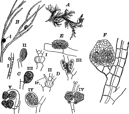
Fig. 29.—A, a red seaweed (Callithamnion), of the
natural size. B, a piece of the same, × 50. t, tetraspores. C
i–v, successive stages in the development of the tetraspores, × 150.
D I, II young procarps. tr. trichogyne. iii, young; iv, ripe spore
fruit. I, III, × 150. iv, × 50. E, an antheridium, × 150. F, spore
fruit of Polysiphonia. The spores are here surrounded by a case,
× 50.
The methods of reproduction may be best illustrated by a specific
example, and preferably one of the simpler ones, as these are most
readily studied microscopically.
The form here illustrated (Callithamnion) grows attached to wharves,
etc., below low-water mark, and is extremely delicate, collapsing
completely when removed from the water. The color is a bright rosy
red, and with its graceful form and extreme delicacy it makes one of
the most beautiful of the group.
If alcoholic material is used, it may be mounted for examination
either in water or very dilute glycerine.
The plant is composed of much-branched, slender filaments, closely
resembling Cladophora in structure, but with smaller cells (Fig. 29,
B). The non-sexual reproduction is by means of special spores, which
from being formed in groups of four, are known as tetraspores. In the
species under consideration the mother cell of the tetraspores arises
as a small bud near the upper end of one of the ordinary cells
(Fig. 29, C i). This bud rapidly increases in size, assuming an oval
form, and becoming cut off from the cell of the stem (Fig. 29, C
ii). The contents now divide into four equal parts, arranged like the
quadrants of a sphere. When ripe, the wall of the mother cell gives
way, and the four spores escape into the water and give rise to new
plants. These spores, it will be noticed, differ in one important
particular from corresponding spores in most algæ, in being unprovided
with cilia, and incapable of spontaneous movement.Occasionally in the same plant that bears tetraspores, but more
commonly in special ones, there are produced the sexual organs, and
subsequently the sporocarps, or fruits, developed from them. The
plants that bear them are usually stouter that the non-sexual ones,
and the masses of ripe carpospores are large enough to be readily seen
with the naked eye.If a plant bearing ripe spores is selected, the young stages of the
female organ (procarp) may generally be found by examining the younger
parts of the plant. The procarp arises from a single cell of the
filament. This cell undergoes division by a series of longitudinal
walls into a central cell and about four peripheral ones (Fig. 29, D
i). One of the latter divides next into an upper and a lower cell, the
former growing out into a long, colorless appendage known as a
trichogyne (Fig. 29, D, tr.).The antheridia (Fig. 29, E) are hemispherical masses of closely set
colorless cells, each of which develops a single spermatozoid which,
like the tetraspores, is destitute of cilia, and is dependent upon the
movement of the water to convey it to the neighborhood of the procarp.
Occasionally one of these spermatozoids may be found attached to the
trichogyne, and in this way fertilization is effected. Curiously
enough, neither the cell which is immediately fertilized, nor the one
beneath it, undergo any further change; but two of the other
peripheral cells on opposite sides of the filament grow rapidly and
develop into large, irregular masses of spores (Fig. 29, D III, IV).
While the plant here described may be taken as a type of the group,
it must be borne in mind that many of them differ widely, not only in
the structure of the plant body, but in the complexity of the sexual
organs and spores as well. The tetraspores are often imbedded in the
tissues of the plant, or may be in special receptacles, nor are they
always arranged in the same way as here described, and the same is
true of the carpospores. These latter are in some of the higher forms,
e.g. Polysiphonia (Fig. 29, F), contained in urn-shaped
receptacles, or they may be buried within the tissues of the plant.
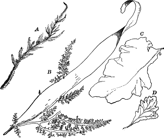
Fig. 30.—Marine red seaweeds. A, Dasya. B,
Rhodymenia (with smaller algæ attached). C, Grinnellia. D,
Delesseria. A, B, natural size; the others reduced one-half.
The fresh-water forms are not common, but may occasionally be met with
in mill streams and other running water, attached to stones and
woodwork, but are much inferior in size and beauty to the marine
species. The red color is not so pronounced, and they are, as a rule,
somewhat dull colored.
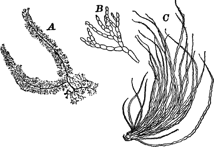
Fig. 31.—Fresh-water red algæ. A, Batrachospermum,
× about 12. B, a branch of the same, × 150. C, Lemanea, natural
size.
The commonest genera are Batrachospermum and Lemanea (Fig. 31).
CHAPTER VIII.
SUB-KINGDOM III.
Fungi.
The name “Fungi” has been given to a vast assemblage of plants,
varying much among themselves, but on the whole of about the same
structural rank as the algæ. Unlike the algæ, however, they are
entirely destitute of chlorophyll, and in consequence are dependent
upon organic matter for food, some being parasites (growing upon
living organisms), others saprophytes (feeding on dead matter). Some
of them show close resemblances in structure to certain algæ, and
there is reason to believe that they are descended from forms that
originally had chlorophyll; others are very different from any green
plants, though more or less evidently related among themselves.
Recognizing then these distinctions, we may make two divisions of the
sub-kingdom: I. The Alga-Fungi (Phycomycetes), and
II. The True Fungi (Mycomycetes).
Class I.—Phycomycetes.
These are fungi consisting of long, undivided, often branching tubular
filaments, resembling quite closely those of Vaucheria or other
Siphoneæ, but always destitute of any trace of chlorophyll. The
simplest of these include the common moulds (Mucorini), one of which
will serve to illustrate the characteristics of the order.
If a bit of fresh bread, slightly moistened, is kept under a bell jar
or tumbler in a warm room, in the course of twenty-four hours or so it
will be covered with a film of fine white threads, and a little later
will produce a crop of little globular bodies mounted on upright
stalks. These are at first white, but soon become black, and the
filaments bearing them also grow dark-colored.
These are moulds, and have grown from spores that are in the
atmosphere falling on the bread, which offers the proper conditions
for their growth and multiplication.
One of the commonest moulds is the one here figured (Fig. 32), and
named Mucor stolonifer, from the runners, or “stolons,” by which it
spreads from one point to another. As it grows it sends out these
runners along the surface of the bread, or even along the inner
surface of the glass covering it. They fasten themselves at intervals
to the substratum, and send up from these points clusters of short
filaments, each one tipped with a spore case, or “sporangium.”
For microscopical study they are best mounted in dilute glycerine
(about one-quarter glycerine to three-quarters pure water). After
carefully spreading out the specimens in this mixture, allow a drop of
alcohol to fall upon the preparation, and then put on the cover glass.
The alcohol drives out the air, which otherwise interferes badly with
the examination.The whole plant consists of a very long, much-branched, but undivided
tubular filament. Where it is in contact with the substratum,
root-like outgrowths are formed, not unlike those observed in
Vaucheria. At first the walls are colorless, but later become dark
smoky brown in color. A layer of colorless granular protoplasm lines
the wall, becoming more abundant toward the growing tips of the
branches. The spore cases, “sporangia,” arise at the ends of upright
branches (Fig. 32, C), which at first are cylindrical (a), but
later enlarge at the end (b), and become cut off by a convex wall
(c). This wall pushes up into the young sporangium, forming a
structure called the “columella.” When fully grown, the sporangium is
globular, and appears quite opaque, owing to the numerous granules in
the protoplasm filling the space between the columella and its outer
wall. This protoplasm now divides into a great number of small oval
cells (spores), which rapidly darken, owing to a thick, black wall
formed about each one, and at the same time the columella and the
stalk of the sporangium become dark-colored.When ripe, the wall of the sporangium dissolves, and the spores
(Fig. 32, E) are set free. The columella remains unchanged, and
some of the spores often remain sticking to it (Fig. 32, D).
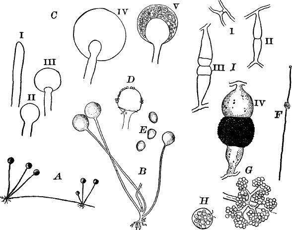
Fig. 32.—A, common black mould (Mucor), × 5. B,
three nearly ripe spore cases, × 25. C, development of the spore
cases, i–iv, × 150; v, × 50. D, spore case which has discharged its
spores. E, spores, × 300. F, a form of Mucor mucedo, with small
accessory spore cases, × 5. G, the spore cases, × 50. H, a single
spore case, × 300. I, development of the zygospore of a black mould,
× 45 (after De Bary).
Spores formed in a manner strongly recalling those of the pond scums
are also known, but only occur after the plants have grown for a long
time, and hence are rarely met with (Fig. 32, I).
Another common mould (M. mucedo), often growing in company with the
one described, differs from it mainly in the longer stalk of the
sporangium, which is also smaller, and in not forming runners. This
species sometimes bears clusters of very small sporangia attached to
the middle of the ordinary sporangial filament (Fig. 32, F, H).
These small sporangia have no columella.
Other moulds are sometimes met with, parasitic upon the larger species
of Mucor.
Related to the black moulds are the insect moulds (Entomopthoreæ),
which attack and destroy insects. The commonest of these attacks the
house flies in autumn, when the flies, thus infested, may often be
found sticking to window panes, and surrounded by a whitish halo of
the spores that have been thrown off by the fungus.
Order II.—White Rusts and Mildews (Peronosporeæ)
These are exclusively parasitic fungi, and grow within the tissues of
various flowering plants, sometimes entirely destroying them.
As a type of this group we will select a very common one (Cystopus
bliti), that is always to be found in late summer and autumn growing
on pig weed (Amarantus). It forms whitish, blister-like blotches
about the size of a pin head on the leaves and stems, being commonest
on the under side of the leaves (Fig. 33, A). In the earlier stages
the leaf does not appear much affected, but later becomes brown and
withered about the blotches caused by the fungus.
If a thin vertical section of the leaf is made through one of these
blotches, and mounted as described for Mucor, the latter is found to
be composed of a mass of spores that have been produced below the
epidermis of the leaf, and have pushed it up by their growth. If the
section is a very thin one, we may be able to make out the structure
of the fungus, and then find it to be composed of irregular, tubular,
much-branched filaments, which, however, are not divided by
cross-walls. These filaments run through the intercellular spaces of
the leaf, and send into the cells little globular suckers, by means of
which the fungus feeds.The spores already mentioned are formed at the ends of crowded
filaments, that push up, and finally rupture the epidermis (Fig. 33,
B). They are formed by the ends of the filaments swelling up and
becoming constricted, so as to form an oval spore, which is then cut
off by a wall. The portion of the filament immediately below acts in
the same way, and the process is repeated until a chain of half a
dozen or more may be produced, the lowest one being always the last
formed. When ripe, the spores are separated by a thin neck, and
become very easily broken off.In order to follow their germination it is only necessary to place a
few leaves with fresh patches of the fungus under a bell jar or
tumbler, inverted over a dish full of water, so as to keep the air
within saturated with moisture, but taking care to keep the leaves out
of the water. After about twenty-four hours, if some of the spores are
scraped off and mounted in water, they will germinate in the course of
an hour or so. The contents divide into about eight parts, which
escape from the top of the spore, which at this time projects as a
little papilla. On escaping, each mass of protoplasm swims away as a
zoöspore, with two extremely delicate cilia. After a short time it
comes to rest, and, after developing a thin cell wall, germinates by
sending out one or two filaments (Fig. 33, C, E).
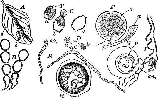
Fig. 33.—A, leaf of pig-weed (Amarantus), with
spots of white rust (c), one-half natural size. B, non-sexual
spores (conidia). C, the same germinating. D, zoöspores. E,
germinating zoöspores. sp. the spore. F, young. G, mature sexual
organs. In G, the tube may be seen connecting the antheridium
(an.), with the egg cell (o). H, a ripe resting spore still
surrounded by the wall of the oögonium. I, a part of a filament of
the fungus, showing its irregular form. All × 300.
Under normal conditions the spores probably germinate when the leaves
are wet, and the filaments enter the plant through the breathing pores
on the lower surface of the leaves, and spread rapidly through the
intercellular spaces.Later on, spores of a very different kind are produced. Unlike those
already studied, they are formed some distance below the epidermis,
and in order to study them satisfactorily, the fungus must be freed
from the host plant. In order to do this, small pieces of the leaf
should be boiled for about a minute in strong caustic potash, and then
treated with acetic or hydrochloric acid. By this means the tissues of
the leaf become so soft as to be readily removed, while the fungus is
but little affected. The preparation should now be washed and mounted
in dilute glycerine.The spores (oöspores) are much larger than those first formed, and
possess an outer coat of a dark brown color (Fig. 33, H). Each spore
is contained in a large cell, which arises as a swelling of one of the
filaments, and becomes shut off by a wall. At first (Fig. 33, F) its
contents are granular, and fill it completely, but later contract to
form a globular mass of protoplasm (G. o), the germ cell or egg
cell. The whole is an oögonium, and differs in no essential respect
from that of Vaucheria.Frequently a smaller cell (antheridium), arising from a neighboring
filament, and in close contact with the oögonium, may be detected
(Fig. 33, F, G, an.), and in exceptionally favorable cases a
tube is to be seen connecting it with the germ cell, and by means of
which fertilization is effected.After being fertilized, the germ cell secretes a wall, at first thin
and colorless, but later becoming thick and dark-colored on the
outside, and showing a division into several layers, the outermost of
which is dark brown, and covered with irregular reticulate markings.
These spores do not germinate at once, but remain over winter
unchanged.
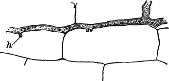
Fig. 34.—Fragment of a filament of the white rust of
the shepherd’s-purse, showing the suckers (h), × 300.
It is by no means impossible that sometimes the germ cell may develop
into a spore without being fertilized, as is the case in many of the
water moulds.
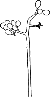
Closely related to the species above described is another one
(C. candidus), which attacks shepherd’s-purse, radish, and others of
the mustard family, upon which it forms chalky white blotches, and
distorts the diseased parts of the plant very greatly.
Very similar to the white rusts in most respects, but differing in the
arrangement of the non-sexual spores, are the mildews (Peronospora,
Phytophthora). These plants form mouldy-looking patches on the
leaves and stems of many plants, and are often very destructive. Among
them are the vine mildew (Peronospora viticola) (Fig. 35), the
potato fungus (Phytophthora infestans), and many others.
Order III.—Saprolegniaceæ (Water Moulds).
These plants resemble quite closely the white rusts, and are probably
related to them. They grow on decaying organic matter in water, or
sometimes on living water animals, fish, crustaceans, etc. They may
usually be had for study by throwing into water taken from a stagnant
pond or aquarium, a dead fly or some other insect. After a few days it
will probably be found covered with a dense growth of fine, white
filaments, standing out from it in all directions (Fig. 36, A).
Somewhat later, if carefully examined with a lens, little round, white
bodies may be seen scattered among the filaments.
Fig. 36.—A, an insect that has decayed in water, and
become attacked by a water mould (Saprolegnia), natural size. B, a
ripe zoösporangium, × 100. C, the same discharging the spores. D,
active. E, germinating zoöspores, × 300. F, a second sporangium
forming below the empty one. G i–iv, development of the oögonium,
× 100. H, ripe oögonium filled with resting spores, × 100.
On carefully removing a bit of the younger growth and examining it
microscopically, it is found to consist of long filaments much like
those of Vaucheria, but entirely destitute of chlorophyll. In places
these filaments are filled with densely granular protoplasm, which
when highly magnified exhibits streaming movements. The protoplasm
contains a large amount of oil in the form of small, shining drops.In the early stages of its growth the plant multiplies by zoöspores,
produced in great numbers in sporangia at the ends of the branches.
The protoplasm collects here much as we saw in V. sessilis, the end
of the filament becoming club-shaped and ending in a short
protuberance (Fig. 36, B). This end becomes separated by a wall, and
the contents divide into numerous small cells that sometimes are
naked, and sometimes have a delicate membrane about them. The first
sign of division is the appearance in the protoplasm of delicate lines
dividing it into numerous polygonal areas which soon become more
distinct, and are seen to be distinct cells whose outlines remain more
or less angular on account of the mutual pressure. When ripe, the end
of the sporangium opens, and the contained cells are discharged
(Fig. 36, C). In case they have no membrane, they swim away at once,
each being provided with two cilia, and resembling almost exactly the
zoöspores of the white rust (Fig. 36, D, E). When the cells are
surrounded by a membrane they remain for some time at rest, but
finally the contents escape as a zoöspore, like those already
described. By killing the zoöspores with a little iodine the granular
nature of the protoplasm is made more evident, and the cilia may be
seen. They soon come to rest, and germinate in the same way as those
of the white rusts and mildews.As soon as the sporangium is emptied, a new one is formed, either by
the filament growing up through it (Fig. 36, F) and the end being
again cut off, or else by a branch budding out just below the base of
the empty sporangium, and growing up by the side of it.Besides zoöspores there are also resting spores developed. Oögonia
like those of Vaucheria or the Peronosporeæ are formed usually
after the formation of zoöspores has ceased; but in many cases,
perhaps all, these develop without being fertilized. Antheridia are
often wanting, and even when they are present, it is very doubtful
whether fertilization takes place.[5]The oögonia (Fig. 36, G, H) arise at the end of the main
filaments, or of short side branches, very much as do the sporangia,
from which they differ at this stage in being of globular form. The
contents contract to form one or several egg cells, naked at first,
but later becoming thick-walled resting spores (H).
CHAPTER IX.
THE TRUE FUNGI (Mycomycetes).
The great majority of the plants ordinarily known as fungi are
embraced under this head. While some of the lower forms show
affinities with the Phycomycetes, and through them with the algæ,
the greater number differ very strongly from all green plants both in
their habits and in their structure and reproduction. It is a
much-disputed point whether sexual reproduction occurs in any of them,
and it is highly probable that in the great majority, at any rate, the
reproduction is purely non-sexual.
Probably to be reckoned with the Mycomycetes, but of doubtful
affinities, are the small unicellular fungi that are the main causes
of alcoholic fermentation; these are the yeast fungi
(Saccharomycetes). They cause the fermentation of beer and wine, as
well as the incipient fermentation in bread, causing it to “rise” by
the giving off of bubbles of carbonic acid gas during the process.
If a little common yeast is put into water containing starch or sugar,
and kept in a warm place, in a short time bubbles of gas will make
their appearance, and after a little longer time alcohol may be
detected by proper tests; in short, alcoholic fermentation is taking
place in the solution.
If a little of the fermenting liquid is examined microscopically, it
will be found to contain great numbers of very small, oval cells, with
thin cell walls and colorless contents. A careful examination with a
strong lens (magnifying from 500–1000 diameters) shows that the
protoplasm, in which are granules of varying size, does not fill the
cell completely, but that there are one or more large vacuoles or
spaces filled with colorless cell sap. No nucleus is visible in the
living cell, but it has been shown that a nucleus is present.If growth is active, many of the cells will be seen dividing. The
process is somewhat different from ordinary fission and is called
budding (Fig. 37, B). A small protuberance appears at the bud or at
the side of the cell, and enlarges rapidly, assuming the form of the
mother cell, from which it becomes completely separated by the
constriction of the base, and may fall off at once, or, as is more
frequently the case, may remain attached for a time, giving rise
itself to other buds, so that not infrequently groups of half a dozen
or more cells are met with (Fig. 37, B, C).
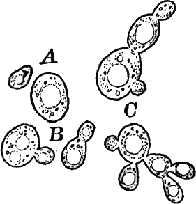
That the yeast cells are the principal agents of alcoholic
fermentation may be shown in much the same way that bacteria are shown
to cause ordinary decomposition. Liquids from which they are excluded
will remain unfermented for an indefinite time.
There has been much controversy as to the systematic position of the
yeast fungi, which has not yet been satisfactorily settled, the
question being whether they are to be regarded as independent plants
or only one stage in the life history of some higher fungi (possibly
the Smuts), which through cultivation have lost the power of
developing further.
Class I.—The Smuts (Ustillagineæ).
The smuts are common and often very destructive parasitic fungi,
living entirely within the tissues of the higher plants. Owing to
this, as well as to the excessively small spores and difficulty in
germinating them, the plants are very difficult of study, except in a
general way, and we will content ourselves with a glance at one of the
common forms, the corn smut (Ustillago maydis). This familiar fungus
attacks Indian corn, forming its spores in enormous quantities in
various parts of the diseased plant, but particularly in the flowers
(“tassel” and young ear).
The filaments, which resemble somewhat those of the white rusts,
penetrate all parts of the plant, and as the time approaches for the
formation of the spores, these branch extensively, and at the same
time become soft and mucilaginous (Fig. 38, B). The ends of these
short branches enlarge rapidly and become shut off by partitions, and
in each a globular spore (Fig. 38, C) is produced. The outer wall is
very dark-colored and provided with short spines. To study the
filaments and spore formation, very thin sections should be made
through the young kernels or other parts in the vicinity, before they
are noticeably distorted by the growth of the spore-bearing filaments.
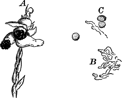
Fig. 38.—A, “tassel” of corn attacked by smut
(Ustillago). B, filaments of the fungus from a thin section of a
diseased grain, showing the beginning of the formation of the spores,
× 300. C, ripe spores, × 300.
As the spores are forming, an abnormal growth is set up in the cells
of the part attacked, which in consequence becomes enormously enlarged
(Fig. 38, A), single grains sometimes growing as large as a walnut.
As the spores ripen, the affected parts, which are at first white,
become a livid gray, due to the black spores shining through the
overlying white tissues. Finally the masses of spores burst through
the overlying cells, appearing like masses of soot, whence the popular
name for the plant.
The remaining Mycomycetes are pretty readily divisible into two
great classes, based upon the arrangement of the spores. The first of
these is known as the Ascomycetes (Sac fungi), the other the
Basidiomycetes (mushrooms, puff-balls, etc.).
Class II.—Ascomycetes (Sac Fungi).
This class includes a very great number of common plants, all
resembling each other in producing spores in sacs (asci, sing.
ascus) that are usually oblong in shape, and each containing eight
spores, although the number is not always the same. Besides the spores
formed in these sacs (ascospores), there are other forms produced in
various ways.
There are two main divisions of the class, the first including only a
few forms, most of which are not likely to be met with by the student.
In these the spore sacs are borne directly upon the filaments without
any protective covering. The only form that is at all common is a
parasitic fungus (Exoascus) that attacks peach-trees, causing the
disease of the leaves known as “curl.”
All of the common Ascomycetes belong to the second division, and
have the spore sacs contained in special structures called spore
fruits, that may reach a diameter of several centimetres in a few
cases, though ordinarily much smaller.
Among the simpler members of this group are the mildews
(Perisporiaceæ), mostly parasitic forms, living upon the leaves and
stems of flowering plants, sometimes causing serious injury by their
depredations. They form white or grayish downy films on the surface of
the plant, in certain stages looking like hoar-frost. Being very
common, they may be readily obtained, and are easily studied. One of
the best species for study (Podosphæra) grows abundantly on the
leaves of the dandelion, especially when the plants are growing under
unfavorable conditions. The same species is also found on other plants
of the same family. It may be found at almost any time during the
summer; but for studying, the spore fruits material should be
collected in late summer or early autumn. It at first appears as
white, frost-like patches, growing dingier as it becomes older, and
careful scrutiny of the older specimens will show numerous brown or
blackish specks scattered over the patches. These are the spore
fruits.
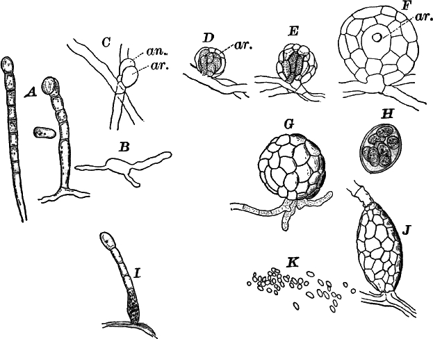
Fig. 39.—A, spore-bearing filaments of the dandelion
mildew (Podosphæra), × 150. B, a germinating spore, × 150. C–F,
development of the spore fruit, × 300. ar. archicarp. G, a ripe
spore fruit, × 150. H, the spore sac removed from the spore fruit,
× 150. I, spore-bearing filament attacked by another fungus
(Cicinnobulus), causing the enlargement of the basal cell, × 150.
J, a more advanced stage, × 300. K, spores, × 300.
For microscopical study, fresh material may be used, or, if necessary,
dried specimens. The latter, before mounting, should be soaked for a
short time in water, to which has been added a few drops of
caustic-potash solution. This will remove the brittleness, and swell
up the dried filaments to their original proportions. A portion of the
plant should be carefully scraped off the leaf on which it is growing,
thoroughly washed in pure water, and transferred to a drop of water or
very dilute glycerine, in which it should be carefully spread out with
needles. If air bubbles interfere with the examination, they may be
driven off with alcohol, and then the cover glass put on. If the
specimen is mounted in glycerine, it will keep indefinitely, if care
is taken to seal it up. The plant consists of much-interlaced
filaments, divided at intervals by cross-walls.[6] They are nearly
colorless, and the contents are not conspicuous. These filaments send
up vertical branches (Fig. 39, A), that become divided into a series
of short cells by means of cross-walls. The cells thus formed are at
first cylindrical, but later bulge out at the sides, becoming broadly
oval, and finally become detached as spores (conidia). It is these
spores that give the frosty appearance to the early stages of the
fungus when seen with the naked eye. The spores fall off very easily
when ripe, and germinate quickly in water, sending out two or more
tubes that grow into filaments like those of the parent plant
(Fig. 39, B).
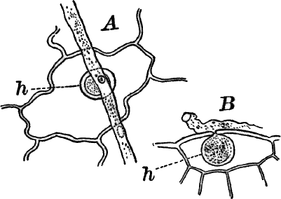
Fig. 40.—Chrysanthemum mildew (Erysiphe), showing
the suckers (h) by which the filaments are attached to the leaf.
A, surface view. B, vertical section of the leaf, × 300.
The spore fruits, as already observed, are formed toward the end of
the season, and, in the species under consideration at least, appear
to be the result of a sexual process. The sexual organs (if they are
really such) are extremely simple, and, owing to their very small
size, are not easily found. They arise as short branches at a point
where two filaments cross; one of them (Fig. 39, C, ar.), the
female cell, or “archicarp,” is somewhat larger than the other and
nearly oval in form, and soon becomes separated by a partition from
the filament that bears it. The other branch (antheridium) grows up in
close contact with the archicarp, and like it is shut off by a
partition from its filament. It is more slender than the archicarp,
but otherwise differs little from it. No actual communication can be
shown to be present between the two cells, and it is therefore still
doubtful whether fertilization really takes place. Shortly after these
organs are full-grown, several short branches grow up about them, and
soon completely envelop them (D, E). These branches soon grow
together, and cross-walls are formed in them, so that the young spore
fruit appears surrounded by a single layer of cells, sufficiently
transparent, however, to allow a view of the interior.The antheridium undergoes no further change, but the archicarp soon
divides into two cells,—a small basal one and a larger upper cell.
There next grow from the inner surface of the covering cells, short
filaments, that almost completely fill the space between the
archicarp and the wall. An optical section of such a stage (Fig. 39,
F) shows a double wall and the two cells of the archicarp. The spore
fruit now enlarges rapidly, and the outer cells become first yellow
and then dark brown, the walls becoming thicker and harder as they
change color. Sometimes special filaments or appendages grow out from
their outer surfaces, and these are also dark-colored. Shortly before
the fruit is ripe, the upper cell of the archicarp, which has
increased many times in size, shows a division of its contents into
eight parts, each of which develops a wall and becomes an oval spore.
By crushing the ripe spore fruit, these spores still enclosed in the
mother cell (ascus) may be forced out (Fig. 39, H). These spores do
not germinate at once, but remain dormant until the next year.
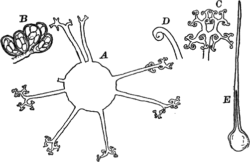
Fig. 41.—Forms of mildews (Erysiphe). A,
Microsphæra, a spore fruit, × 150. B, cluster of spore sacs of the
same, × 150. C, a single appendage, × 300. D, end of an appendage
of Uncinula, × 300. E, appendage of Phyllactinia, × 150.
Frequently other structures, resembling somewhat the spore fruits, are
found associated with them (Fig. 39, I, K), and were for a long
time supposed to be a special form of reproductive organ; but they are
now known to belong to another fungus (Cicinnobulus), parasitic upon
the mildew. They usually appear at the base of the chains of conidia,
causing the basal cell to enlarge to many times its original size, and
finally kill the young conidia, which shrivel up. A careful
examination reveals the presence of very fine filaments within those
of the mildew, which may be traced up to the base of the conidial
branch, where the receptacle of the parasite is forming. The spores
contained in these receptacles are very small (Fig. 39, K), and when
ripe exude in long, worm-shaped masses, if the receptacle is placed in
water.
The mildews may be divided into two genera: Podosphæra, with a
single ascus in the spore fruit; and Erysiphe, with two or more. In
the latter the archicarp branches, each branch bearing a spore sac
(Fig. 41, B).
The appendages growing out from the wall of the spore fruit are often
very beautiful in form, and the two genera given above are often
subdivided according to the form of these appendages.
A common mould closely allied to the mildews is found on various
articles of food when allowed to remain damp, and is also very common
on botanical specimens that have been poorly dried, and hence is often
called “herbarium mould” (Eurotium herbariorum).
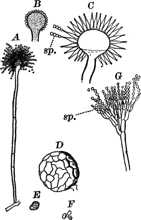
Fig. 42.—A, spore bearing filament of the herbarium
mould (Eurotium), × 150. B, C, another species showing the way
in which the spores are borne—optical section—× 150. D, spore
fruit of the herbarium mould, × 150. E, spore sac. F, spores,
× 300. G, spore-bearing filament of the common blue mould
(Penicillium), × 300. sp. the spores.
The conidia are of a greenish color, and produced on the ends of
upright branches which are enlarged at the end, and from which grow
out little prominences, which give rise to the conidia in the same way
as we have seen in the mildews (Fig. 42, A).Spore fruits much like those of the mildews are formed later, and are
visible to the naked eye as little yellow grains (Fig. 42, D). These
contain numerous very small spore sacs (E), each with eight spores.
There are numerous common species of Eurotium, differing in color
and size, some being yellow or black, and larger than the ordinary
green form.
Another form, common everywhere on mouldy food of all kinds, as well
as in other situations, is the blue mould (Penicillium). This, in
general appearance, resembles almost exactly the herbarium mould, but
is immediately distinguishable by a microscopic examination (Fig. 42,
G).
In studying all of these forms, they may be mounted, as directed for
the black moulds, in dilute glycerine; but must be handled with great
care, as the spores become shaken off with the slightest jar.
Of the larger Ascomycetes, the cup fungi (Discomycetes) may be
taken as types. The spore fruit in these forms is often of
considerable size, and, as their name indicates, is open, having the
form of a flat disc or cup. A brief description of a common one will
suffice to give an idea of their structure and development.
Ascobolus (Fig. 43) is a small, disc-shaped fungus, growing on horse
dung. By keeping some of this covered with a bell jar for a week or
two, so as to retain the moisture, at the end of this time a large
crop of the fungus will probably have made its appearance. The part
visible is the spore fruit (Fig. 43, A), of a light brownish color,
and about as big as a pin-head.
Its development may be readily followed by teasing out in water the
youngest specimens that can be found, taking care to take up a little
of the substratum with it, as the earliest stages are too small to be
visible to the naked eye. The spore fruits arise from filaments not
unlike those of the mildews, and are preceded by the formation of an
archicarp composed of several cells, and readily seen through the
walls of the young fruit (Fig. 43, B). In the study of the early
stages, a potash solution will be found useful in rendering them
transparent.The young fruit has much the same structure as that of the mildews,
but the spore sacs are much more numerous, and there are special
sterile filaments developed between them. If the young spore fruit is
treated with chlor-iodide of zinc, it is rendered quite transparent,
and the young spore sacs colored a beautiful blue, so that they are
readily distinguishable.
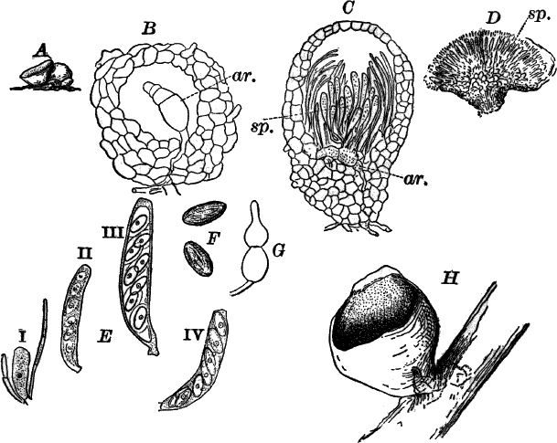
Fig. 43.—A, a small cup fungus (Ascobolus), × 5.
B, young spore fruit, × 300. ar. archicarp. C, an older one,
× 150. ar. archicarp. sp. young spore sacs. D, section through a
full-grown spore fruit (partly diagrammatic), × 25. sp. spore sacs.
E, development of spore sacs and spores: i–iii, × 300; iv, × 150.
F, ripe spores. G, a sterile filament (paraphysis), × 300. H,
large scarlet cup fungus (Peziza), natural size.
The development of the spore sacs may be traced by carefully crushing
the young spore fruits in water. The young spore sacs (Fig. 43, E i)
are colorless, with granular protoplasm, in which a nucleus can often
be easily seen. The nucleus subsequently divides repeatedly, until
there are eight nuclei, about which the protoplasm collects to form as
many oval masses, each of which develops a wall and becomes a spore
(Figs. ii–iv). These are imbedded in protoplasm, which is at first
granular, but afterwards becomes almost transparent. As the spores
ripen, the wall acquires a beautiful violet-purple color, changing
later to a dark purple-brown, and marked with irregular longitudinal
ridges (Fig. 43, F). The full-grown spore sacs (Fig. 43, E, W)
are oblong in shape, and attached by a short stalk. The sterile
filaments between them often become curiously enlarged at the end
(G). As the spore fruit ripens, it opens at the top, and spreads out
so as to expose the spore sacs as they discharge their contents
(Fig. 43, D).
Of the larger cup fungi, those belonging to the genus Peziza
(Fig. 43, H) are common, growing on bits of rotten wood on the
ground in woods. They are sometimes bright scarlet or orange-red, and
very showy. Another curious form is the morel (Morchella), common in
the spring in dry woods. It is stalked like a mushroom, but the
surface of the conical cap is honeycombed with shallow depressions,
lined with the spore sacs.
Order Lichenes.
Under the name of lichens are comprised a large number of fungi,
differing a good deal in structure, but most of them not unlike the
cup fungi. They are, with few exceptions, parasitic upon various forms
of algæ, with which they are so intimately associated as to form
apparently a single plant. They grow everywhere on exposed rocks, on
the ground, trunks of trees, fences, etc., and are found pretty much
the world over. Among the commonest of plants are the lichens of the
genus Parmelia (Fig. 44, A), growing everywhere on tree trunks,
wooden fences, etc., forming gray, flattened expansions, with much
indented and curled margins. When dry, the plant is quite brittle, but
on moistening becomes flexible, and at the same time more or less
decidedly green in color. The lower surface is white or brown, and
often develops root-like processes by which it is fastened to the
substratum. Sometimes small fragments of the plant become detached in
such numbers as to form a grayish powder over certain portions of it.
These, when supplied with sufficient moisture, will quickly produce
new individuals.
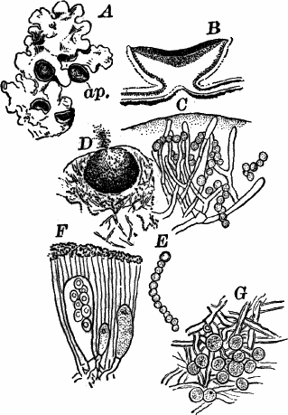
Fig. 44.—A, a common lichen (Parmelia), of the
natural size. ap. spore fruit. B, section through one of the spore
fruits, × 5. C, section through the body of a gelatinous lichen
(Collema), showing the Nostoc individuals surrounded by the fungus
filaments, × 300. D, a spermagonium of Collema, × 25. E, a
single Nostoc thread. F, spore sacs and paraphyses of Usnea,
× 300. G, Protococcus cells and fungus filaments of Usnea.
Not infrequently the spore fruits are to be met with flat discs of a
reddish brown color, two or three millimetres in diameter, and closely
resembling a small cup fungus. They are at first almost closed, but
expand as they mature (Fig. 44, A, ap.).
If a thin vertical section of the plant is made and sufficiently
magnified, it is found to be made up of somewhat irregular,
thick-walled, colorless filaments, divided by cross-walls as in the
other sac-fungi. In the central parts of the plant these are rather
loose, but toward the outside become very closely interwoven and often
grown together, so as to form a tough rind. Among the filaments of the
outer portion are numerous small green cells, that closer examination
shows to be individuals of Protococcus, or some similar green algæ,
upon which the lichen is parasitic. These are sufficiently abundant to
form a green line just inside the rind if the section is examined with
a simple lens (Fig. 44, B).The spore fruits of the lichens resemble in all essential respects
those of the cup fungi, and the spore sacs (Fig. 44, F) are much the
same, usually, though not always, containing eight spores, which are
sometimes two-celled. The sterile filaments between the spore sacs
usually have thickened ends, which are dark-colored, and give the
color to the inner surface of the spore fruit.In Figure 45, H, is shown one of the so-called “Soredia,”[7] a
group of the algæ, upon which the lichen is parasitic, surrounded by
some of the filaments, the whole separating spontaneously from the
plant and giving rise to a new one.
Owing to the toughness of the filaments, the finer structure of the
lichens is often difficult to study, and free use of caustic potash is
necessary to soften and make them manageable.
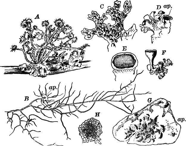
Fig. 45.—Forms of lichens. A, a branch with lichens
growing upon it, one-half natural size. B, Usnea, natural size.
ap. spore fruit. C, Sticta, one-half natural size. D,
Peltigera, one-half natural size. ap. spore fruit. E, a single
spore fruit, × 2. F, Cladonia, natural size. G, a piece of bark
from a beech, with a crustaceous lichen (Graphis) growing upon it,
× 2. ap. spore fruit. H, Soredium of a lichen, × 300.
According to their form, lichens are sometimes divided into the bushy
(fruticose), leafy (frondose), incrusting (crustaceous), and
gelatinous. Of the first, the long gray Usnea (Fig. 45, A, B),
which drapes the branches of trees in swamps, is a familiar example;
of the second, Parmelia, Sticta (Fig. 45, C) and Peltigera
(D) are types; of the third, Graphis (G), common on the trunks
of beech-trees, to which it closely adheres; and of the last,
Collema (Fig. 44, C, D, E), a dark greenish, gelatinous form,
growing on mossy tree trunks, and looking like a colony of Nostoc,
which indeed it is, but differing from an ordinary colony in being
penetrated everywhere by the filaments of the fungus growing upon it.
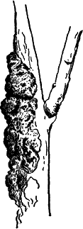
Not infrequently in this form, as well as in other lichens, special
cavities, known as spermogonia (Fig. 44, D), are found, in which
excessively small spores are produced, which have been claimed to be
male reproductive cells, but the latest investigations do not support
this theory.
The last group of the Ascomycetes are the “black fungi,”
Pyrenomycetes, represented by the black knot of cherry and plum
trees, shown in Figure 46. They are mainly distinguished from the cup
fungi by producing their spore sacs in closed cavities. Some are
parasites; others live on dead wood, leaves, etc., forming very hard
masses, generally black in color, giving them their common name. Owing
to the hardness of the masses, they are very difficult to manipulate;
and, as the structure is not essentially different from that of the
Discomycetes, the details will not be entered into here.
Of the parasitic forms, one of the best known is the “ergot” of rye,
more or less used in medicine. Other forms are known that attack
insects, particularly caterpillars, which are killed by their attacks.
CHAPTER X.
Fungi—Continued.
Class Basidiomycetes.
The Basidiomycetes include the largest and most highly developed of
the fungi, among which are many familiar forms, such as the mushrooms,
toadstools, puff-balls, etc. Besides these large and familiar forms,
there are other simpler and smaller ones that, according to the latest
investigations, are probably related to them, though formerly regarded
as constituting a distinct group. The most generally known of these
lower Basidiomycetes are the so-called rusts. The larger
Basidiomycetes are for the most part saprophytes, living in decaying
vegetable matter, but a few are true parasites upon trees and others
of the flowering plants.
All of the group are characterized by the production of spores at the
top of special cells known as basidia,[8] the number produced upon a
single basidium varying from a single one to several.
Of the lower Basidiomycetes, the rusts (Uredineæ) offer common and
easily procurable forms for study. They are exclusively parasitic in
their habits, growing within the tissues of the higher land plants,
which they often injure seriously. They receive their popular name
from the reddish color of the masses of spores that, when ripe, burst
through the epidermis of the host plant. Like many other fungi, the
rusts have several kinds of spores, which are often produced on
different hosts; thus one kind of wheat rust lives during part of its
life within the leaves of the barberry, where it produces spores quite
different from those upon the wheat; the cedar rust, in the same way,
is found at one time attacking the leaves of the wild crab-apple and
thorn.
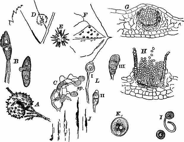
Fig. 47.—A, a branch of red cedar attacked by a rust
(Gymnosporangium), causing a so-called “cedar apple,” × ½. B,
spores of the same, one beginning to germinate, × 300. C, a spore
that has germinated, each cell producing a short, divided filament
(basidium), which in turn gives rise to secondary spores (sp.),
× 300. D, part of the leaf of a hawthorn attacked by the cluster cup
stage of the same fungus, upper side showing spermogonia, natural
size. E, cluster cups (Roestelia) of the same fungus, natural
size. F, tip of a leaf of the Indian turnip (Arisæma), bearing the
cluster cup (Æcidium) stage of a rust, × 2. G, vertical section
through a young cluster cup. H, similar section through a mature
one, × 50. I, germinating spores of H, × 300. J, part of a corn
leaf, with black rust, natural size. K, red rust spore of the wheat
rust (Puccinia graminis), × 300. L, forms of black-rust spores: i,
Uromyces; ii, Puccinia; iii, Phragmidium.
The first form met with in most rusts is sometimes called the
“cluster-cup” stage, and in many species is the only stage known. In
Figure 47, F, is shown a bit of the leaf of the Indian turnip
(Arisæma) affected by one of these “cluster-cup” forms. To the naked
eye, or when slightly magnified, the masses of spores appear as bright
orange spots, mostly upon the lower surface. The affected leaves are
more or less checked in their growth, and the upper surface shows
lighter blotches, corresponding to the areas below that bear the
cluster cups. These at first appear as little elevations of a
yellowish color, and covered with the epidermis; but as the spores
ripen they break through the epidermis, which is turned back around
the opening, the whole forming a little cup filled with a bright
orange red powder, composed of the loose masses of spores.
Putting a piece of the affected leaf between two pieces of pith so as
to hold it firmly, with a little care thin vertical sections of the
leaf, including one of the cups, may be made, and mounted, either in
water or glycerine, removing the air with alcohol. We find that the
leaf is thickened at this point owing to a diseased growth of the
cells of the leaf, induced by the action of the fungus. The mass of
spores (Fig. 47, G) is surrounded by a closely woven mass of
filaments, forming a nearly globular cavity. Occupying the bottom of
the cup are closely set, upright filaments, each bearing a row of
spores, arranged like those of the white rusts, but so closely crowded
as to be flattened at the sides. The outer rows have thickened walls,
and are grown together so as to form the wall of the cup.The spores are filled with granular protoplasm, in which are numerous
drops of orange-yellow oil, to which is principally due their color.
As the spores grow, they finally break the overlying epidermis, and
then become rounded as the pressure from the sides is relieved. They
germinate within a few hours if placed in water, sending out a tube,
into which pass the contents of the spore (Fig. 47, I).
One of the most noticeable of the rusts is the cedar rust
(Gymnosporangium), forming the growths known as “cedar apples,”
often met with on the red cedar. These are rounded masses, sometimes
as large as a walnut, growing upon the small twigs of the cedar
(Fig. 47, A). This is a morbid growth of the same nature as those
produced by the white rusts and smuts. If one of these cedar apples is
examined in the late autumn or winter, it will be found to have the
surface dotted with little elevations covered by the epidermis, and on
removing this we find masses of forming spores. These rupture the
epidermis early in the spring, and appear then as little spikes of a
rusty red color. If they are kept wet for a few hours, they enlarge
rapidly by the absorption of water, and may reach a length of four or
five centimetres, becoming gelatinous in consistence, and sometimes
almost entirely hiding the surface of the “apple.” In this stage the
fungus is extremely conspicuous, and may frequently be met with after
rainy weather in the spring.
This orange jelly, as shown by the microscope, is made up of elongated
two-celled spores (teleuto spores), attached to long gelatinous stalks
(Fig. 47, B). They are thick-walled, and the contents resemble those
of the cluster-cup spores described above.To study the earlier stages of germination it is best to choose
specimens in which the masses of spores have not been moistened. By
thoroughly wetting these, and keeping moist, the process of
germination may be readily followed. Many usually begin to grow within
twenty-four hours or less. Each cell of the spore sends out a tube
(Fig. 47, C), through an opening in the outer wall, and this tube
rapidly elongates, the spore contents passing into it, until a short
filament (basidium) is formed, which then divides into several short
cells. Each cell develops next a short, pointed process, which swells
up at the end, gradually taking up all the contents of the cell, until
a large oval spore (sp.) is formed at the tip, containing all the
protoplasm of the cell.
Experiments have been made showing that these spores do not germinate
upon the cedar, but upon the hawthorn or crab-apple, where they
produce the cluster-cup stage often met with late in the summer. The
affected leaves show bright orange-yellow spots about a centimetre in
diameter (Fig. 47, D), and considerably thicker than the other parts
of the leaf. On the upper side of these spots may be seen little black
specks, which microscopic examination shows to be spermogonia,
resembling those of the lichens. Later, on the lower surface, appear
the cluster cups, whose walls are prolonged so that they form little
tubular processes of considerable length (Fig. 47, E).
In most rusts the teleuto spores are produced late in the summer or
autumn, and remain until the following spring before they germinate.
They are very thick-walled, the walls being dark-colored, so that in
mass they appear black, and constitute the “black-rust” stage
(Fig. 47, J). Associated with these, but formed earlier, and
germinating immediately, are often to be found large single-celled
spores, borne on long stalks. They are usually oval in form, rather
thin-walled, but the outer surface sometimes provided with little
points. The contents are reddish, so that in mass they appear of the
color of iron rust, and cause the “red rust” of wheat and other
plants, upon which they are growing.
The classification of the rusts is based mainly upon the size and
shape of the teleuto spores where they are known, as the cluster-cup
and red-rust stages are pretty much the same in all. Of the commoner
genera Melampsora, and Uromyces (Fig. 47, L i), have unicellular
teleuto spores; Puccinia (ii) and Gymnosporangium, two-celled
spores; Triphragmium, three-celled; and Phragmidium (iii), four or
more.
The rusts are so abundant that a little search can scarcely fail to
find some or all of the stages. The cluster-cup stages are best
examined fresh, or from alcoholic material; the teleuto spores may be
dried without affecting them.
Probably the best-known member of the group is the wheat rust
(Puccinia graminis), which causes so much damage to wheat and
sometimes to other grains. The red-rust stage may be found in early
summer; the black-rust spores in the stubble and dead leaves in the
autumn or spring, forming black lines rupturing the epidermis.
Probably to be associated with the lower Basidiomycetes are the
large fungi of which Tremella (Fig. 51, A) is an example. They are
jelly-like forms, horny and somewhat brittle when dry, but becoming
soft when moistened. They are common, growing on dead twigs, logs,
etc., and are usually brown or orange-yellow in color.
Of the higher Basidiomycetes, the toadstools, mushrooms, etc., are
the highest, and any common form will serve for study. One of the most
accessible and easily studied forms is Coprinus, of which there are
several species growing on the excrement of various herbivorous
animals. They not infrequently appear on horse manure that has been
kept covered with a glass for some time, as described for Ascobolus.
After two or three weeks some of these fungi are very likely to make
their appearance, and new ones continue to develop for a long time.
Fig. 48.—A, young. B, full-grown fruit of a
toadstool (Coprinus), × 2. C, under side of the cap, showing the
radiating “gills,” or spore-bearing plates. D, section across one of
the young gills, × 150. E, F, portions of gills from a nearly ripe
fruit, × 300. sp. spores. x, sterile cell. In F, a basidium is
shown, with the young spores just forming. G, H, young fruits,
× 50.
The first trace of the plant, visible to the naked eye, is a little
downy, white speck, just large enough to be seen. This rapidly
increases in size, becoming oblong in shape, and growing finally
somewhat darker in color; and by the time it reaches a height of a few
millimetres a short stalk becomes perceptible, and presently the whole
assumes the form of a closed umbrella. The top is covered with little
prominences, that diminish in number and size toward the bottom. After
the cap reaches its full size, the stalk begins to grow, slowly at
first, but finally with great rapidity, reaching a height of several
centimetres within a few hours. At the same time that the stalk is
elongating, the cap spreads out, radial clefts appearing on its upper
surface, which flatten out very much as the folds of an umbrella are
stretched as it opens, and the spaces between the clefts appear as
ridges, comparable to the ribs of the umbrella (Fig. 48, B). The
under side of the cap has a number of ridges running from the centre
to the margin, and of a black color, due to the innumerable spores
covering their surface (C). Almost as soon as the umbrella opens,
the spores are shed, and the whole structure shrivels up and
dissolves, leaving almost no trace behind.
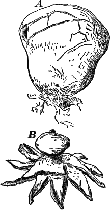
Fig. 49.—Basidiomycetes. A, common puff-ball
(Lycoperdon). B, earth star (Geaster). A, × ¼. B, one-half
natural size.
If we examine microscopically the youngest specimens procurable,
freeing from air with alcohol, and mounting in water or dilute
glycerine, we find it to be a little, nearly globular mass of
colorless filaments, with numerous cross-walls, the whole arising from
similar looser filaments imbedded in the substratum (Fig. 48, G). If
the specimen is not too young, a denser central portion can be made
out, and in still older ones (Fig. 48, H) this central mass has
assumed the form of a short, thick stalk, crowned by a flat cap, the
whole invested by a loose mass of filaments that merge more or less
gradually into the central portion. By the time the spore fruit (for
this structure corresponds to the spore fruit of the Ascomycetes)
reaches a height of two or three millimetres, and is plainly visible
to the naked eye, the cap grows downward at the margins, so as to
almost entirely conceal the stalk. A longitudinal section of such a
stage shows the stalk to be composed of a small-celled, close tissue
becoming looser in the cap, on whose inner surface the spore-bearing
ridges (“gills” or Lamellæ) have begun to develop. Some of these run
completely to the edge of the cap, others only part way. To study
their structure, make cross-sections of the cap of a nearly
full-grown, but unopened, specimen, and this will give numerous
sections of the young gills. We find them to be flat plates, composed
within of loosely interwoven filaments, whose ends stand out at right
angles to the surface of the gills, forming a layer of closely-set
upright cells (basidia) (Fig. 48, D). These are at first all alike,
but later some of them become club-shaped, and develop at the end
several (usually four) little points, at the end of which spores are
formed in exactly the same way as we saw in the germinating teleuto
spores of the cedar rust, all the protoplasm of the basidium passing
into the growing spores (Fig. 48, E, F). The ripe spores (E,
sp.) are oval, and possess a firm, dark outer wall. Occasionally
some of the basidia develop into very large sterile cells (E, x),
projecting far beyond the others, and often reaching the neighboring
gill.
Similar in structure and development to Coprinus are all the large
and common forms; but they differ much in the position of the
spore-bearing tissue, as well as in the form and size of the whole
spore fruit. They are sometimes divided, according to the position of
the spores, into three orders: the closed-fruited (Angiocarpous)
forms, the half-closed (Hemi-angiocarpous), and the open or
naked-fruited forms (Gymnocarpous).
Of the first, the puff-balls (Fig. 49) are common examples. One
species, the giant puff-ball (Lycoperdon giganteum), often reaches a
diameter of thirty to forty centimetres. The earth stars (Geaster)
have a double covering to the spore fruit, the outer one splitting at
maturity into strips (Fig. 49, B). Another pretty and common form is
the little birds’-nest fungus (Cyathus), growing on rotten wood or
soil containing much decaying vegetable matter (Fig. 50).
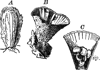
Fig. 50.—Birds’-nest fungus (Cyathus). A, young.
B, full grown. C, section through B, showing the “sporangia”
(sp.). All twice the natural size.
In the second order the spores are at first protected, as we have seen
in Coprinus, which belongs to this order, but finally become
exposed. Here belong the toadstools and mushrooms (Fig. 51, B), the
large shelf-shaped fungi (Polyporus), so common on tree trunks and
rotten logs (Fig. 51, C, D, E), and the prickly fungus
(Hydnum) (Fig. 51, G).
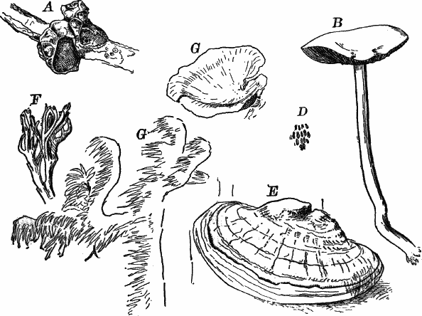
Fig. 51.—Forms of Basidiomycetes. A, Tremella,
one-half natural size. B, Agaricus, natural size. C, E,
Polyporus: C, × ½; E, × ¼. D, part of the under surface of
D, natural size. F, Clavaria, a small piece, natural size. G,
Hydnum, a piece of the natural size.
Of the last, or naked-fruited forms, the commonest belong to the
genus Clavaria (Fig. 51, F), smooth-branching forms, usually of a
brownish color, bearing the spores directly upon the surface of the
branches.
CHAPTER XI.
SUB-KINGDOM IV.
Bryophyta.
The Bryophytes, or mosses, are for the most part land plants, though a
few are aquatic, and with very few exceptions are richly supplied with
chlorophyll. They are for the most part small plants, few of them
being over a few centimetres in height; but, nevertheless, compared
with the plants that we have heretofore studied, quite complex in
their structure. The lowest members of the group are flattened,
creeping plants, or a few of them floating aquatics, without distinct
stem and leaves; but the higher ones have a pretty well-developed
central axis or stem, with simple leaves attached.
There are two classes—I. Liverworts (Hepaticæ), and II. Mosses
(Musci).
Class I.—The Liverworts.
One of the commonest of this class, and to be had at any time, is
named Madotheca. It is one of the highest of the class, having
distinct stem and leaves. It grows most commonly on the shady side of
tree trunks, being most luxuriant near the ground, where the supply of
moisture is most constant. It also occurs on stones and rocks in moist
places. It closely resembles a true moss in general appearance, and
from the scale-like arrangement of its leaves is sometimes called
“scale moss.”
The leaves (Fig. 52, A, B) are rounded in outline unequally,
two-lobed, and arranged in two rows on the upper side of the stem, so
closely overlapping as to conceal it entirely. On the under side are
similar but smaller leaves, less regularly disposed. The stems branch
at intervals, the branches spreading out laterally so that the whole
plant is decidedly flattened. On the under side are fine, whitish
hairs, that fasten it to the substratum. If we examine a number of
specimens, especially early in the spring, a difference will be
observed in the plants. Some of them will be found to bear peculiar
structures (Fig. 52, C, D), in which the spores are produced.
These are called “sporogonia.” They are at first globular, but when
ripe open by means of four valves, and discharge a greenish brown mass
of spores. An examination of the younger parts of the same plants will
probably show small buds (Fig. 54, H), which contain the female
reproductive organs, from which the sporogonia arise.
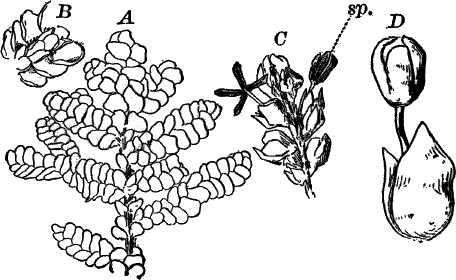
Fig. 52.—A, part of a plant of a leafy liverwort
(Madotheca), × 2. B, part of the same, seen from below, × 4. C,
a branch with two open sporogonia (sp.), × 4. D, a single
sporogonium, × 8.
On other plants may be found numerous short side branches (Fig. 53,
B), with very closely set leaves. If these are carefully separated,
the antheridia can just be seen as minute whitish globules, barely
visible to the naked eye. Plants that, like this one, have the male
and female reproductive organs on distinct plants, are said to be
“diœcious.”
A microscopical examination of the stem and leaves shows their
structure to be very simple. The former is cylindrical, and composed
of nearly uniform elongated cells, with straight cross-walls. The
leaves consist of a single layer of small, roundish cells, which, like
those of the stem, contain numerous rounded chloroplasts, to which is
due their dark green color.The tissues are developed from a single apical cell, but it is
difficult to obtain good sections through it.The antheridia are borne singly at the bases of the leaves on the
special branches already described (Fig. 53, A, an.). By carefully
dissecting with needles such a branch in a drop of water, some of the
antheridia will usually be detached uninjured, and may be readily
studied, the full-grown ones being just large enough to be seen with
the naked eye. They are globular bodies, attached by a stalk composed
of two rows of cells. The globular portion consists of a wall of
chlorophyll-bearing cells, composed of two layers below, but single
above (Fig. 53, C). Within is a mass of excessively small cells,
each of which contains a spermatozoid. In the young antheridium (A,
an.) the wall is single throughout, and the central cells few in
number. To study them in their natural position, thin longitudinal
sections of the antheridial branch should be made.
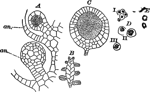
Fig. 53.—A, end of a branch from a male plant of
Madotheca. The small side branchlets bear the antheridia, × 2. B,
two young antheridia (an.), the upper one seen in optical section,
the lower one from without, × 150. C, a ripe antheridium, optical
section, × 50. D, sperm cells with young spermatozoids. E, ripe
spermatozoids, × 600.
When ripe, if brought into water, the antheridium bursts at the top
into a number of irregular lobes that curl back and allow the mass of
sperm cells to escape. The spermatozoids, which are derived
principally from the nucleus of the sperm cells (53, D) are so small
as to make a satisfactory examination possible only with very powerful
lenses. The ripe spermatozoid is coiled in a flat spiral (53, E),
and has two excessively delicate cilia, visible only under the most
favorable circumstances.The female organ in the bryophytes is called an “archegonium,” and
differs considerably from anything we have yet studied, but recalls
somewhat the structure of the oögonium of Chara. They are found in
groups, contained in little bud-like branches (54, H). In order to
study them, a plant should be chosen that has numbers of such buds,
and the smallest that can be found should be used. Those containing
the young archegonia are very small; but after one has been
fertilized, the leaves enclosing it grow much larger, and the bud
becomes quite conspicuous, being surrounded by two or three
comparatively large leaves. By dissecting the young buds, archegonia
in all stages of growth may be found.
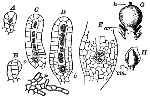
Fig. 54.—A–D, development of the archegonium of
Madotheca. B, surface view, the others in optical section. o,
egg cell, × 150. E, base of a fertilized archegonium, containing a
young embryo (em.), × 150. F, margin of one of the leaves
surrounding the archegonia. G, young sporogonium still surrounded by
the much enlarged base of the archegonium. h, neck of the
archegonium. ar. abortive archegonia, × 12. H, short branch
containing the young sporogonium, × 4.
When very young the archegonium is composed of an axial row of three
cells, surrounded by a single outer layer of cells, the upper ones
forming five or six regular rows, which are somewhat twisted (Fig. 54,
A, B). As it becomes older, the lower part enlarges slightly, the
whole looking something like a long-necked flask (C, D). The
centre of the neck is occupied by a single row of cells (canal cells),
with more granular contents than the outer cells, the lowest cell of
the row being somewhat larger than the others (Fig. 54, C, o).
When nearly ripe, the division walls of the canal cells are absorbed,
and the protoplasm of the lowest cell contracts and forms a globular
naked cell, the egg cell (D, o). If a ripe archegonium is placed
in water, it soon opens at the top, and the contents of the canal
cells are forced out, leaving a clear channel down to the egg cell. If
the latter is not fertilized, the inner walls of the neck cells turn
brown, and the egg cell dies; but if a spermatozoid penetrates to the
egg cell, the latter develops a wall and begins to grow, forming the
embryo or young sporogonium.
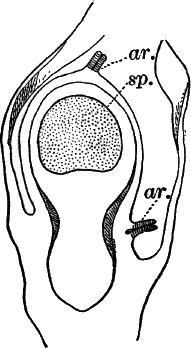
Fig. 55.—Longitudinal section of a nearly full-grown
sporogonium of Madotheca, which has not, however, broken through the
overlying cells, × 25. sp. cavity in which the spores are formed.
ar. abortive archegonium.
The first division wall to be formed in the embryo is transverse, and
is followed by vertical ones (Fig. 54, E, em.). As the embryo
enlarges, the walls of the basal part of the archegonium grow rapidly,
so that the embryo remains enclosed in the archegonium until it is
nearly full-grown (Fig. 55). As it increases in size, it becomes
differentiated into three parts: a wedge-shaped base or “foot”
penetrating downward into the upper part of the plant, and serving to
supply the embryo with nourishment; second, a stalk supporting the
third part, the capsule or spore-bearing portion of the fruit. The
capsule is further differentiated into a wall, which later becomes
dark colored, and a central cavity, in which are developed special
cells, some of which by further division into four parts produce the
spores, while the others, elongating enormously, give rise to special
cells, called elaters (Fig. 56, B).
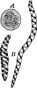
The ripe spores are nearly globular, contain chlorophyll and drops of
oil, and the outer wall is brown and covered with fine points
(Fig. 56, A). The elaters are long-pointed cells, having on the
inner surface of the wall a single or double dark brown spiral band.
These bands are susceptible to changes in moisture, and by their
movements probably assist in scattering the spores after the
sporogonium opens.
Just before the spores are ripe, the stalk of the sporogonium
elongates rapidly, carrying up the capsule, which breaks through the
archegonium wall, and finally splits into four valves, and discharges
the spores.
There are four orders of the liverworts represented in the United
States, three of which differ from the one we have studied in being
flattened plants, without distinct stems and leaves,—at least, the
leaves when present are reduced to little scales upon the lower
surface.
The first order (Ricciaceæ) are small aquatic forms, or grow on damp
ground or rotten logs. They are not common forms, and not likely to be
encountered by the student. One of the floating species is shown in
figure 57, A.
The second order, the horned liverworts (Anthoceroteæ), are
sometimes to be met with in late summer and autumn, forms growing
mostly on damp ground, and at once recognizable by their long-pointed
sporogonia, which open when ripe by two valves, like a bean pod
(Fig. 57, B).
The third order (Marchantiaceæ) includes the most conspicuous
members of the whole class. Some of them, like the common liverwort
(Marchantia), shown in Figure 57, F, K, and the giant liverwort
(Fig. 57, D), are large and common forms, growing on the ground in
shady places, the former being often found also in greenhouses. They
are fastened to the ground by numerous fine, silky hairs, and the
tissues are well differentiated, the upper surface of the plant having
a well-marked epidermis, with peculiar breathing pores, large enough
to be seen with the naked eye (Fig. 57, E, J, K) Each of these
is situated in the centre of a little area (Fig. 57, E), and beneath
it is a large air space, into which the chlorophyll-bearing cells
(cl.) of the plant project (J).
The sexual organs are often produced in these forms upon special
branches (G), or the antheridia may be sunk in discs on the upper
side of the stem (D, an.).
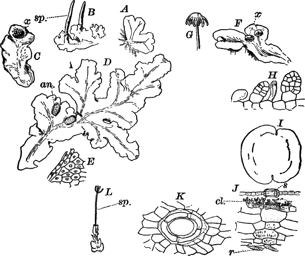
Fig. 57.—Forms of liverworts. A, Riccia, natural
size. B, Anthoceros (horned liverwort), natural size. sp.
sporogonia. C, Lunularia, natural size, x, buds. D, giant
liverwort (Conocephalus), natural size. an. antheridial disc. E,
small piece of the epidermis, showing the breathing pores, × 2. F,
common liverwort (Marchantia), × 2. x, cups containing buds. G,
archegonial branch of common liverwort, natural size. H, two young
buds from the common liverwort, × 150. I, a full-grown bud, × 25.
J, vertical section through the body of Marchantia, cutting
through a breathing pore (s), × 50. K, surface view of a breathing
pore, × 150. L, a leafy liverwort (Jungermannia). sp.
sporogonium, × 2.
Some forms, like Marchantia and Lunularia (Fig. 57, C), produce
little cups (x), circular in the first, semicircular in the second,
in which special buds (H, I) are formed that fall off and produce
new plants.
The highest of the liverworts (Jungermanniaceæ) are, for the most
part, leafy forms like Madotheca, and represented by a great many
common forms, growing usually on tree trunks, etc. They are much like
Madotheca in general appearance, but usually very small and
inconspicuous, so as to be easily overlooked, especially as their
color is apt to be brownish, and not unlike that of the bark on which
they grow (Fig. 57, L).
Class II.—The True Mosses.
The true mosses (Musci) resemble in many respects the higher
liverworts, such as Madotheca or Jungermannia, all of them having
well-marked stems and leaves. The spore fruit is more highly
developed than in the liverworts, but never contains elaters.
A good idea of the general structure of the higher mosses may be had
from a study of almost any common species. One of the most convenient,
as well as common, forms (Funaria) is to be had almost the year
round, and fruits at almost all seasons, except midwinter. It grows in
close patches on the ground in fields, at the bases of walls,
sometimes in the crevices between the bricks of sidewalks, etc. If
fruiting, it may be recognized by the nodding capsule on a long stalk,
that is often more or less twisted, being sensitive to changes in the
moisture of the atmosphere. The plant (Fig. 58, A, B) has a short
stem, thickly set with relatively large leaves. These are oblong and
pointed, and the centre is traversed by a delicate midrib. The base of
the stem is attached to the ground by numerous fine brown hairs.
The mature capsule is broadly oval in form (Fig. 58, C), and
provided with a lid that falls off when the spores are ripe. While the
capsule is young it is covered by a pointed membranous cap (B,
cal.) that finally falls off. When the lid is removed, a fine fringe
is seen surrounding the opening of the capsule, and serving the same
purpose as the elaters of the liverworts (Fig. 58, E).
Fig. 58.—A, fruiting plant of a moss (Funaria),
with young sporogonium (sp.), × 4. B, plant with ripe sporogonium.
cal. calyptra, × 2. C, sporogonium with calyptra removed. op.
lid, × 4. D, spores: i, ungerminated; ii–iv, germinating, × 300.
E, two teeth from the margin of the capsule, × 50. F, epidermal
cells and breathing pore from the surface of the sporogonium, × 150.
G, longitudinal section of a young sporogonium, × 12. sp. spore
mother cells. H, a small portion of G, magnified about 300 times.
sp. spore mother cells.
If the lower part of the stem is carefully examined with a lens, we
may detect a number of fine green filaments growing from it, looking
like the root hairs, except for their color. Sometimes the ground
about young patches of the moss is quite covered by a fine film of
such threads, and looking carefully over it probably very small moss
plants may be seen growing up here and there from it.
Fig. 59.—Longitudinal section through the summit of a
small male plant of Funaria. a, aʹ, antheridia. p, paraphysis.
L, section of a leaf, × 150.
This moss is diœcious. The male plants are smaller than the female,
and may be recognized by the bright red antheridia which are formed at
the end of the stem in considerable numbers, and surrounded by a
circle of leaves so that the whole looks something like a flower.
(This is still more evident in some other mosses. See Figure 65, E,
F.)
The leaves when magnified are seen to be composed of a single layer of
cells, except the midrib, which is made up of several thicknesses of
elongated cells. Where the leaf is one cell thick, the cells are
oblong in form, becoming narrower as they approach the midrib and the
margin. They contain numerous chloroplasts imbedded in the layer of
protoplasm that lines the wall. The nucleus (Fig. 63, C, n) may
usually be seen without difficulty, especially if the leaf is treated
with iodine. This plant is one of the best for studying the division
of the chloroplasts, which may usually be found in all stages of
division (Fig. 63, D). In the chloroplasts, especially if the plant
has been exposed to light for several hours, will be found numerous
small granules, that assume a bluish tint on the application of
iodine, showing them to be starch grains. If the plant is kept in the
dark for a day or two, these will be absent, having been used up; but
if exposed to the light again, new ones will be formed, showing that
they are formed only under the action of light.
Fig. 60.—A, B, young antheridia of Funaria,
optical section, × 150. C, two sperm cells of Atrichum. D,
spermatozoids of Sphagnum, × 600.
Starch is composed of carbon, hydrogen, and oxygen, and so far as is
known is only produced by chlorophyll-bearing cells, under the
influence of light. The carbon used in the manufacture of starch is
taken from the atmosphere in the form of carbonic acid, so that green
plants serve to purify the atmosphere by the removal of this
substance, which is deleterious to animal life, while at the same time
the carbon, an essential part of all living matter, is combined in
such form as to make it available for the food of other organisms.The marginal cells of the leaf are narrow, and some of them prolonged
into teeth.A cross-section of the stem (63, E) shows on the outside a single
row of epidermal cells, then larger chlorophyll-bearing cells, and in
the centre a group of very delicate, small, colorless cells, which in
longitudinal section are seen to be elongated, and similar to those
forming the midrib of the leaf. These cells probably serve for
conducting fluids, much as the similar but more perfectly developed
bundles of cells (fibro-vascular bundles) found in the stems and
leaves of the higher plants.The root hairs, fastening the plant to the ground, are rows of cells
with brown walls and oblique partitions. They often merge insensibly
into the green filaments (protonema) already noticed. These latter
have usually colorless walls, and more numerous chloroplasts, looking
very much like a delicate specimen of Cladophora or some similar
alga. If a sufficient number of these filaments is examined, some of
them will probably show young moss plants growing from them (Fig. 63,
A, k), and with a little patience the leafy plant can be traced
back to a little bud originating as a branch of the filament. Its
diameter is at first scarcely greater than that of the filament, but a
series of walls, close together, are formed, so placed as to cut off a
pyramidal cell at the top, forming the apical cell of the young moss
plant. This apical cell has the form of a three-sided pyramid with the
base upward. From it are developed three series of cells, cut off in
succession from the three sides, and from these cells are derived all
the tissues of the plant which soon becomes of sufficient size to be
easily recognizable.The protonemal filaments may be made to grow from almost any part of
the plant by keeping it moist, but grow most abundantly from the base
of the stem.The sexual organs are much like those of the liverworts and are borne
at the apex of the stems.The antheridia (Figs. 59, 60) are club-shaped bodies with a short
stalk. The upper part consists of a single layer of large
chlorophyll-bearing cells, enclosing a mass of very small, nearly
cubical, colorless, sperm cells each of which contains an excessively
small spermatozoid.The young antheridium has an apical cell giving rise to two series of
segments (Fig. 60, A), which in the earlier stages are very plainly
marked.When ripe the chlorophyll in the outer cells changes color, becoming
red, and if a few such antheridia from a plant that has been kept
rather dry for a day or two, are teased out in a drop of water, they
will quickly open at the apex, the whole mass of sperm cells being
discharged at once.Among the antheridia are borne peculiar hairs (Fig. 59, p) tipped by
a large globular cell.
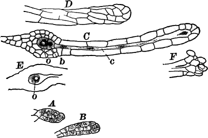
Fig. 61.—A, B, young; C, nearly ripe archegonium
of Funaria, optical section, × 150. D, upper part of the neck of
C, seen from without, showing how it is twisted. E, base of a ripe
archegonium. F, open apex of the same, × 150. o, egg cell. b,
ventral canal cell.
Owing to their small size the spermatozoids are difficult to see
satisfactorily and other mosses (e.g. peat mosses, Figure 64, the
hairy cap moss, Figure 65, I), are preferable where obtainable. The
spermatozoids of a peat moss are shown in Figure 60, D. Like all of
the bryophytes they have but two cilia.The archegonia (Fig. 61) should be looked for in the younger plants in
the neighborhood of those that bear capsules. Like the antheridia they
occur in groups. They closely resemble those of the liverworts, but
the neck is longer and twisted and the base more massive. Usually but
a single one of the group is fertilized.
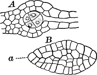
Fig. 62.—A, young embryo of Funaria, still
enclosed within the base of the archegonium, × 300. B, an older
embryo freed from the archegonium, × 150. a, the apical cell.
To study the first division of the embryo, it is usually necessary to
render the archegonium transparent, which may be done by using a
little caustic potash; or letting it lie for a few hours in dilute
glycerine will sometimes suffice. If potash is used it must be
thoroughly washed away, by drawing pure water under the cover glass
with a bit of blotting paper, until every trace of the potash is
removed. The first wall in the embryo is nearly at right angles to the
axis of the archegonium and divides the egg cell into nearly equal
parts. This is followed by nearly vertical walls in each cell
(Fig. 62, A). Very soon a two-sided apical cell (Fig. 62, B, a)
is formed in the upper half of the embryo, which persists until the
embryo has reached a considerable size. As in the liverworts the young
embryo is completely covered by the growing archegonium wall.The embryo may be readily removed from the archegonium by adding a
little potash to the water in which it is lying, allowing it to remain
for a few moments and pressing gently upon the cover glass with a
needle. In this way it can be easily forced out of the archegonium,
and then by thoroughly washing away the potash, neutralizing if
necessary with a little acetic acid, very beautiful preparations may
be made. If desired, these may be mounted permanently in glycerine
which, however, must be added very gradually to avoid shrinking the
cells.
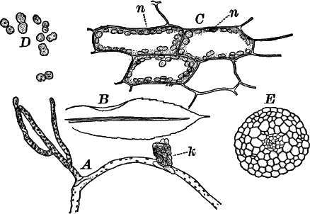
Fig. 63.—A, protonema of Funaria, with a bud
(k), × 50. B, outline of a leaf, showing also the thickened
midrib, × 12. C, cells of the leaf, × 300. n, nucleus. D,
chlorophyll granules undergoing division, × 300. E, cross-section of
the stem, × 50.
For some time the embryo has a nearly cylindrical form, but as it
approaches maturity the differentiation into stalk and capsule becomes
apparent. The latter increases rapidly in diameter, assuming gradually
the oval shape of the full-grown capsule. A longitudinal section of
the nearly ripe capsule (Fig. 58, G) shows two distinct portions; an
outer wall of two layers of cells, and an inner mass of cells in some
of which the spores are produced. This inner mass of cells is
continuous with the upper part of the capsule, but connected with the
side walls and bottom by means of slender, branching filaments of
chlorophyll-bearing cells.The spores arise from a single layer of cells near the outside of the
inner mass of cells (G, sp.). These cells (H, sp.) are filled
with glistening, granular protoplasm; have a large and distinct
nucleus, and no chlorophyll. They finally become entirely separated
and each one gives rise to four spores which closely resemble those of
the liverworts but are smaller.Near the base of the capsule, on the outside, are formed breathing
pores (Fig. 58, F) quite similar to those of the higher plants.If the spores are kept in water for a few days they will germinate,
bursting the outer brown coat, and the contents protruding through the
opening surrounded by the colorless inner spore membrane. The
protuberance grows rapidly in length and soon becomes separated from
the body of the spore by a wall, and lengthening, more and more, gives
rise to a green filament like those we found attached to the base of
the full-grown plant, and like those giving rise to buds that develop
into leafy plants.
Classification of the Mosses.
The mosses may be divided into four orders: I. The peat mosses
(Sphagnaceæ); II. Andreæaceæ; III. Phascaceæ; IV. The common
mosses (Bryaceæ).
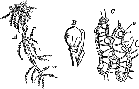
Fig. 64.—A, a peat moss (Sphagnum), × ½. B, a
sporogonium of the same, × 3. C, a portion of a leaf, × 150. The
narrow, chlorophyll-bearing cells form meshes, enclosing the large,
colorless empty cells, whose walls are marked with thickened bars, and
contain round openings (o).
The peat mosses (Fig. 64) are large pale-green mosses, growing often
in enormous masses, forming the foundation of peat-bogs. They are of a
peculiar spongy texture, very light when dry, and capable of absorbing
a great amount of water. They branch (Fig. 64, A), the branches
being closely crowded at the top, where the stems continue to grow,
dying away below.
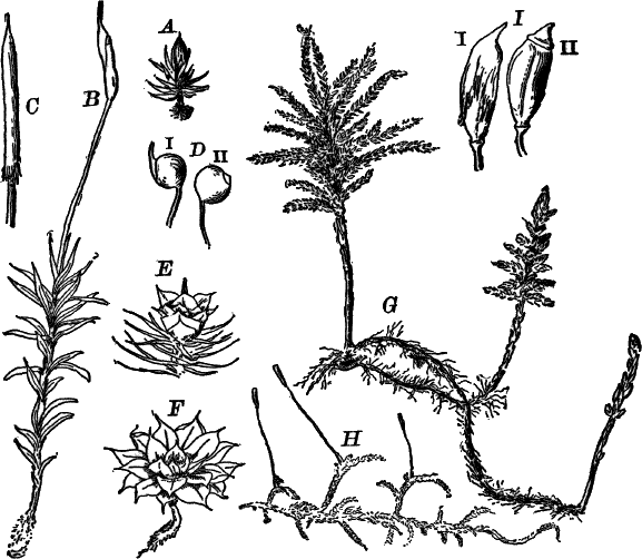
Fig. 65.—Forms of mosses. A, plant of Phascum,
× 3. B, fruiting plant of Atrichum, × 2. C, young capsule of
hairy-cap moss (Polytrichum), covered by the large, hairy calyptra.
D, capsules of Bartramia: i, with; ii, without the calyptra. E,
upper part of a male plant of Atrichum, showing the flower, × 2.
F, a male plant of Mnium, × 4. G, pine-tree moss (Clemacium),
× 1. H, Hypnum, × 1. I, ripe capsules of hairy-cap moss: i,
with; ii, without calyptra.
The sexual organs are rarely met with, but should be looked for late
in autumn or early spring. The antheridial branches are often
bright-colored, red or yellow, so as to be very conspicuous. The
capsules, which are not often found, are larger than in most of the
common mosses, and quite destitute of a stalk, the apparent stalk
being a prolongation of the axis of the plant in the top of which the
base of the sporogonium is imbedded. The capsule is nearly globular,
opening by a lid at the top (Fig. 64, B).
A microscopical examination of the leaves, which are quite destitute
of a midrib, shows them to be composed of a network of narrow
chlorophyll-bearing cells surrounding much larger empty ones whose
walls are marked with transverse thickenings, and perforated here and
there with large, round holes (Fig. 64, C). It is to the presence of
these empty cells that the plant owes its peculiar spongy texture, the
growing plants being fairly saturated with water.
The Andreæaceæ are very small, and not at all common. The capsule
splits into four valves, something like a liverwort.
The Phascaceæ are small mosses growing on the ground or low down on
the trunks of trees, etc. They differ principally from the common
mosses in having the capsule open irregularly and not by a lid. The
commonest forms belong to the genus Phascum (Fig. 65, A).
The vast majority of the mosses the student is likely to meet with
belong to the last order, and agree in the main with the one
described. Some of the commoner forms are shown in Figure 65.
CHAPTER XII.
SUB-KINGDOM V.
Pteridophytes.
If we compare the structure of the sporogonium of a moss or liverwort
with the plant bearing the sexual organs, we find that its tissues are
better differentiated, and that it is on the whole a more complex
structure than the plant that bears it. It, however, remains attached
to the parent plant, deriving its nourishment in part through the
“foot” by means of which it is attached to the plant.
In the Pteridophytes, however, we find that the sporogonium becomes
very much more developed, and finally becomes entirely detached from
the sexual plant, developing in most cases roots that fasten it to the
ground, after which it may live for many years, and reach a very large
size.
The sexual plant, which is here called the “prothallium,” is of very
simple structure, resembling the lower liverworts usually, and never
reaches more than about a centimetre in diameter, and is often much
smaller than this.
The common ferns are the types of the sub-kingdom, and a careful study
of any of these will illustrate the principal peculiarities of the
group. The whole plant, as we know it, is really nothing but the
sporogonium, originating from the egg cell in exactly the same way as
the moss sporogonium, and like it gives rise to spores which are
formed upon the leaves.
The spores may be collected by placing the spore-bearing leaves on
sheets of paper and letting them dry, when the ripe spores will be
discharged covering the paper as a fine, brown powder. If these are
sown on fine, rather closely packed earth, and kept moist and covered
with glass so as to prevent evaporation, within a week or two a fine,
green, moss-like growth will make its appearance, and by the end of
five or six weeks, if the weather is warm, little, flat, heart-shaped
plants of a dark-green color may be seen. These look like small
liverworts, and are the sexual plants (prothallia) of our ferns
(Fig. 66, F). Removing one of these carefully, we find on the lower
side numerous fine hairs like those on the lower surface of the
liverworts, which fasten it firmly to the ground. By and by, if our
culture has been successful, we may find attached to some of the
larger of these, little fern plants growing from the under side of the
prothallia, and attached to the ground by a delicate root. As the
little plant becomes larger the prothallium dies, leaving it attached
to the ground as an independent plant, which after a time bears the
spores.
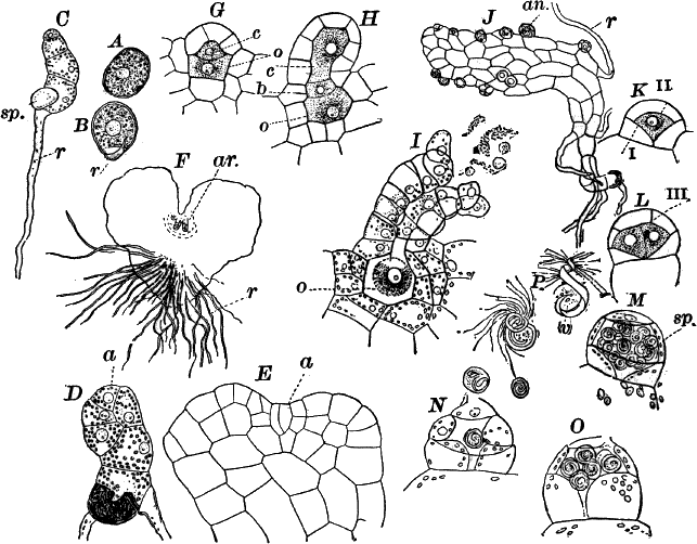
Fig. 66.—A, spore of the ostrich fern (Onoclea),
with the outer coat removed. B, germinating spore, × 150. C, young
prothallium, × 50. r, root hair. sp. spore membrane. D, E,
older prothallia. a, apical cell, × 150. F, a female prothallium,
seen from below, × 12. ar. archegonia. G, H, young archegonia,
in optical section, × 150. o, central cell. b, ventral canal cell.
c, upper canal cell. I, a ripe archegonium in the act of opening,
× 150. o, egg cell. J, a male prothallium, × 50. an. antheridia.
K, L, young antheridia, in optical section, × 300. M, ripe
antheridium, × 300. sp. sperm cells. N, O, antheridia that have
partially discharged their contents, × 300. P, spermatozoids, killed
with iodine, × 500. v, vesicle attached to the hinder end.
In choosing spores for germination it is best to select those of large
size and containing abundant chlorophyll, as they germinate more
readily. Especially favorable for this purpose are the spores of the
ostrich fern (Onoclea struthiopteris) (Fig. 70, I, J), or the
sensitive fern (O. sensibilis). Another common and readily grown
species is the lady fern (Asplenium filixfœmina) (Fig. 70, H). The
spores of most ferns retain their vitality for many months, and hence
can be kept dry until wanted.
The first stages of germination may be readily seen by sowing the
spores in water, where, under favorable circumstances, they will begin
to grow within three or four days. The outer, dry, brown coat of the
spore is first ruptured, and often completely thrown off by the
swelling of the spore contents. Below this is a second colorless
membrane which is also ruptured, but remains attached to the spore.
Through the orifice in the second coat, the inner delicate membrane
protrudes in the form of a nearly colorless papilla which rapidly
elongates and becomes separated from the body of the spore by a
partition, constituting the first root hair (Fig. 66, B, C, r).
The body of the spore containing most of the chlorophyll elongates
more slowly, and divides by a series of transverse walls so as to form
a short row of cells, resembling in structure some of the simpler algæ
(C).In order to follow the development further, spores must be sown upon
earth, as they do not develop normally in water beyond this stage.In studying plants grown on earth, they should be carefully removed
and washed in a drop of water so as to remove, as far as possible, any
adherent particles, and then may be mounted in water for microscopic
examination.In most cases, after three or four cross-walls are formed, two walls
arise in the end cell so inclined as to enclose a wedge-shaped cell
(a) from which are cut off two series of segments by walls directed
alternately right and left (Fig. 66, D, E, a), the apical cell
growing to its original dimensions after each pair of segments is cut
off. The segments divide by vertical walls in various directions so
that the young plant rapidly assumes the form of a flat plate of cells
attached to the ground by root hairs developed from the lower surfaces
of the cells, and sometimes from the marginal ones. As the division
walls are all vertical, the plant is nowhere more than one cell thick.
The marginal cells of the young segments divide more rapidly than the
inner ones, and soon project beyond the apical cell which thus comes
to lie at the bottom of a cleft in the front of the plant which in
consequence becomes heart-shaped (E, F). Sooner or later the
apical cell ceases to form regular segments and becomes
indistinguishable from the other cells.In the ostrich fern and lady fern the plants are diœcious. The male
plants (Fig. 66, J) are very small, often barely visible to the
naked eye, and when growing thickly form dense, moss-like patches.
They are variable in form, some irregularly shaped, others simple rows
of cells, and some have the heart shape of the larger plants.
The female plants (Fig. 66, F) are always comparatively large and
regularly heart-shaped, occasionally reaching a diameter of nearly or
quite one centimetre, so that they are easily recognizable without
microscopical examination.
All the cells of the plant except the root hairs contain large and
distinct chloroplasts much like those in the leaves of the moss, and
like them usually to be found in process of division.The archegonia arise from cells of the lower surface, just behind the
notch in front (Fig. 66, F, ar.). Previous to their formation the
cells at this point divide by walls parallel to the surface of the
plant, so as to form several layers of cells, and from the lowest
layer of cells the archegonia arise. They resemble those of the
liverworts but are shorter, and the lower part is completely sunk
within the tissues of the plant (Fig. 66, G, I). They arise as
single surface cells, this first dividing into three by walls parallel
to the outer surface. The lower cell undergoes one or two divisions,
but undergoes no further change; the second cell (C, o), becomes
the egg cell, and from it is cut off another cell (c), the canal
cell of the neck; the uppermost of the three becomes the neck. There
are four rows of neck cells, the two forward ones being longer than
the others, so that the neck is bent backward. In the full-grown
archegonium, there are two canal cells, the lower one (H, b)
called the ventral canal cell, being smaller than the other.Shortly before the archegonium opens, the canal cells become
disorganized in the same way as in the bryophytes, and the protoplasm
of the central cell contracts to form the egg cell which shows a
large, central nucleus, and in favorable cases, a clear space at the
top called the “receptive spot,” as it is here that the spermatozoid
enters. When ripe, if placed in water, the neck cells become very much
distended and finally open widely at the top, the upper ones not
infrequently being detached, and the remains of the neck cells are
forced out (Fig. 66, I).The antheridia (Fig. 66. J, M) arise as simple hemispherical
cells, in which two walls are formed (K I, II), the lower
funnel-shaped, the upper hemispherical and meeting the lower one so as
to enclose a central cell (shaded in the figure), from which the sperm
cells arise. Finally, a ring-shaped wall (L iii) is formed, cutting
off a sort of cap cell, so that the antheridium at this stage consists
of a central cell, surrounded by three other cells, the two lower
ring-shaped, the upper disc-shaped. The central cell, which contains
dense, glistening protoplasm, is destitute of chlorophyll, but the
outer cells have a few small chloroplasts. The former divides
repeatedly, until a mass of about thirty-two sperm cells is formed,
each giving rise to a large spirally-coiled spermatozoid. When ripe,
the mass of sperm cells crowds so upon the outer cells as to render
them almost invisible, and as they ripen they separate by a partial
dissolving of the division walls. When brought into water, the outer
cells of the antheridium swell strongly, and the matter derived from
the dissolved walls of the sperm cells also absorbs water, so that
finally the pressure becomes so great that the wall of the antheridium
breaks, and the sperm cells are forced out by the swelling up of the
wall cells (N, O). After lying a few moments in the water, the
wall of each sperm cell becomes completely dissolved, and the
spermatozoids are released, and swim rapidly away with a twisting
movement. They may be killed with a little iodine, when each is seen
to be a somewhat flattened band, coiled several times. At the forward
end, the coils are smaller, and there are numerous very long and
delicate cilia. At the hinder end may generally be seen a delicate sac
(P, v), containing a few small granules, some of which usually
show the reaction of starch, turning blue when iodine is applied.In studying the development of the antheridia, it is only necessary to
mount the plants in water and examine them directly; but the study of
the archegonia requires careful longitudinal sections of the
prothallium. To make these, the prothallium should be placed between
small pieces of pith, and the razor must be very sharp. It may be
necessary to use a little potash to make the sections transparent
enough to see the structure, but this must be used cautiously on
account of the great delicacy of the tissues.If a plant with ripe archegonia is placed in a drop of water, with the
lower surface uppermost, and at the same time male plants are put with
it, and the whole covered with a cover glass, the archegonia and
antheridia will open simultaneously; and, if examined with the
microscope, we shall see the spermatozoids collect about the open
archegonia, to which they are attracted by the substance forced out
when it opens. With a little patience, one or more may be seen to
enter the open neck through which it forces itself, by a slow twisting
movement, down to the egg cell. In order to make the experiment
successful, the plants should be allowed to become a little dry, care
being taken that no water is poured over them for a day or two
beforehand.The first divisions of the fertilized egg cell resemble those in the
moss embryo, except that the first wall is parallel with the
archegonium axis, instead of at right angles to it. Very soon,
however, the embryo becomes very different, four growing points being
established instead of the single one found in the moss embryo. The
two growing points on the side of the embryo nearest the archegonium
neck grow faster than the others, one of these outstripping the other,
and soon becoming recognizable as the first leaf of the embryo
(Fig. 67, A, L). The other (r) is peculiar, in having its
growing point covered by several layers of cells, cut off from its
outer face, a peculiarity which we shall find is characteristic of the
roots of all the higher plants, and, indeed, this is the first root of
the young fern. Of the other two growing points, the one next the leaf
grows slowly, forming a blunt cone (st.), and is the apex of the
stem. The other (f) has no definite form, and serves merely as an
organ of absorption, by means of which nourishment is supplied to the
embryo from the prothallium; it is known as the foot.
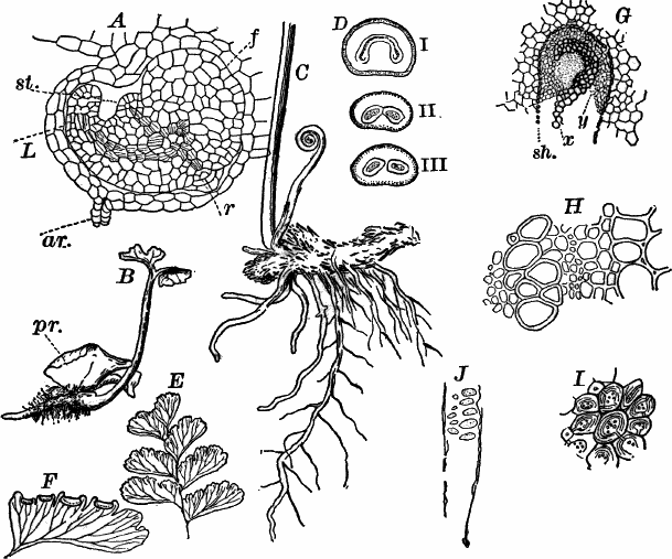
Fig. 67.—A, embryo of the ostrich fern just before
breaking through the prothallium, × 50. st. apex of stem. l, first
leaf. r, first root. ar. neck of the archegonium. B, young
plant, still attached to the prothallium (pr.). C, underground
stem of the maiden-hair fern (Adiantum), with one young leaf, and
the base of an older one, × 1. D, three cross-sections of a leaf
stalk: i, nearest the base; iii, nearest the blade of the leaf,
showing the division of the fibro-vascular bundle, × 5. E, part of
the blade of the leaf, × ½. F, a single spore-bearing leaflet,
showing the edge folded over to cover the sporangia, × 1. G, part of
the fibro-vascular bundle of the leaf stalk (cross-section), × 50.
x, woody part of the bundle. y, bast. sh. bundle sheath. H, a
small portion of the same bundle, × 150. I, stony tissue from the
underground stem, × 150. J, sieve tube from the underground stem,
× 300.
Up to this point, all the cells of the embryo are much alike, and the
embryo, like that of the bryophytes, is completely surrounded by the
enlarged base of the archegonium (compare Fig. 67, A, with Fig. 55);
but before the embryo breaks through the overlying cells a
differentiation of the tissues begins. In the axis of each of the four
divisions the cells divide lengthwise so as to form a cylindrical mass
of narrow cells, not unlike those in the stem of a moss. Here,
however, some of the cells undergo a further change; the walls thicken
in places, and the cells lose their contents, forming a peculiar
conducting tissue (tracheary tissue), found only in the two highest
sub-kingdoms. The whole central cylinder is called a “fibro-vascular
bundle,” and in its perfect form, at least, is found in no plants
below the ferns, which are also the first to develop true roots.
The young root and leaf now rapidly elongate, and burst through the
overlying cells, the former growing downward and becoming fastened in
the ground, the latter growing upward through the notch in the front
of the prothallium, and increasing rapidly in size (Fig. 67, B). The
leaf is more or less deeply cleft, and traversed by veins which are
continuations of the fibro-vascular bundle of the stalk, and
themselves fork once or twice. The surface of the leaf is covered with
a well-developed epidermis, and the cells occupying the space between
the veins contain numerous chloroplasts, so that the little plant is
now quite independent of the prothallium, which has hitherto supported
it. As soon as the fern is firmly established, the prothallium withers
away.
Comparing this now with the development of the sporogonium in the
bryophytes, it is evident that the young fern is the equivalent of the
sporogonium or spore fruit of the former, being, like it, the direct
product of the fertilized egg cell; and the prothallium represents the
moss or liverwort, upon which are borne the sexual organs. In the
fern, however, the sporogonium becomes entirely independent of the
sexual plant, and does not produce spores until it has reached a large
size, living many years. The sexual stage, on the other hand, is very
much reduced, as we have seen, being so small as to be ordinarily
completely overlooked; but its resemblance to the lower liverworts,
like Riccia, or the horned liverworts, is obvious. The terms
oöphyte (egg-bearing plant) and sporophyte (spore-bearing plant, or
sporogonium) are sometimes used to distinguish between the sexual
plant and the spore-bearing one produced from it.
The common maiden-hair fern (Adiantum pedatum) has been selected
here for studying the structure of the full-grown sporophyte, but
almost any other common fern will answer. The maiden-hair fern is
common in rich woods, and may be at once recognized by the form of its
leaves. These arise from a creeping, underground stem (Fig. 67, C),
which is covered with brownish scales, and each leaf consists of a
slender stalk, reddish brown or nearly black in color, which divides
into two equal branches at the top. Each of these main branches bears
a row of smaller ones on the outside, and these have a row of delicate
leaflets on each side (Fig. 67, E). The stem of the plant is
fastened to the ground by means of numerous stout roots. The youngest
of these, near the growing point of the stem, are unbranched, but the
older ones branch extensively (C).
On breaking the stem across, it is seen to be dark-colored, except in
the centre, which is traversed by a woody cylinder (fibro-vascular
bundle) of a lighter color. This is sometimes circular in sections,
sometimes horse-shoe shaped. Where the stem branches, the bundle of
the branch may be traced back to where it joins that of the main stem.
A thin cross-section of the stem shows, when magnified, three regions.
First, an outer row of cells, often absent in the older portions; this
is the epidermis. Second, within the epidermis are several rows of
cells similar to the epidermal cells, but somewhat larger, and like
them having dark-brown walls. These merge gradually into larger cells,
with thicker golden brown walls (Fig. 67, I). The latter, if
sufficiently magnified, show distinct striation of the walls, which
are often penetrated by deep narrow depressions or “pits.” This
thick-walled tissue is called “stony tissue” (schlerenchyma). All the
cells contain numerous granules, which the iodine test shows to be
starch. All of this second region lying between the epidermis and the
fibro-vascular bundle is known as the ground tissue. The third region
(fibro-vascular) is, as we have seen without the microscope, circular
or horse-shoe shaped. It is sharply separated from the ground tissue
by a row of small cells, called the “bundle sheath.” The cross-section
of the bundle of the leaf stalk resembles, almost exactly, that of the
stem; and, as it is much easier to cut, it is to be preferred in
studying the arrangement of the tissues of the bundle (Fig. 67, G).
Within the bundle sheath (sh.) there are two well-marked regions, a
central band (x) of large empty cells, with somewhat angular
outlines, and distinctly separated walls; and an outer portion (y)
filling up the space between these central cells and the bundle
sheath. The central tissue (x) is called the woody tissue (xylem);
the outer, the bast (phloem). The latter is composed of smaller cells
of variable form, and with softer walls than the wood cells.A longitudinal section of either the stem or leaf stalk shows that all
the cells are decidedly elongated, especially those of the
fibro-vascular bundle. The xylem (Fig. 68, C, x) is made up
principally of large empty cells, with pointed ends, whose walls are
marked with closely set, narrow, transverse pits, giving them the
appearance of little ladders, whence they are called “scalariform,” or
ladder-shaped markings. These empty cells are known as “tracheids,”
and tissue composed of such empty cells, “tracheary tissue.” Besides
the tracheids, there are a few small cells with oblique ends, and with
some granular contents.The phloem is composed of cells similar to the latter, but there may
also be found, especially in the stem, other larger ones (Fig. 67,
J), whose walls are marked with shallow depressions, whose bottoms
are finely pitted. These are the so-called “sieve tubes.”For microscopical examination, either fresh or alcoholic material may
be used, the sections being mounted in water. Potash will be found
useful in rendering opaque sections transparent.
The leaves, when young, are coiled up (Fig. 67, C), owing to growth
in the earlier stages being greater on the lower than on the upper
side. As the leaf unfolds, the stalk straightens, and the upper
portion (blade) becomes flat.
The general structure of the leaf stalk may be understood by making a
series of cross-sections at different heights, and examining them with
a hand lens. The arrangement is essentially the same as in the stem.
The epidermis and immediately underlying ground tissue are
dark-colored, but the inner ground tissue is light-colored, and much
softer than the corresponding part of the stem; and some of the outer
cells show a greenish color, due to the presence of chlorophyll.
The section of the fibro-vascular bundle differs at different heights.
Near the base of the stalk (Fig. D i) it is horseshoe-shaped; but,
if examined higher up, it is found to divide (II, III), one part going
to each of the main branches of the leaf. These secondary bundles
divide further, forming the veins of the leaflets.
The leaflets (E, F) are one-sided, the principal vein running
close to the lower edge, and the others branching from it, and forking
as they approach the upper margin, which is deeply lobed, the lobes
being again divided into teeth. The leaflets are very thin and
delicate, with extremely smooth surface, which sheds water perfectly.
If the plant is a large one, some of the leaves will probably bear
spores. The spore-bearing leaves are at once distinguished by having
the middle of each lobe of the leaflets folded over upon the lower
side (F). On lifting one of these flaps, numerous little rounded
bodies (spore cases) are seen, whitish when young, but becoming brown
as they ripen. If a leaf with ripe spore cases is placed upon a piece
of paper, as it dries the spores are discharged, covering the paper
with the spores, which look like fine brown powder.
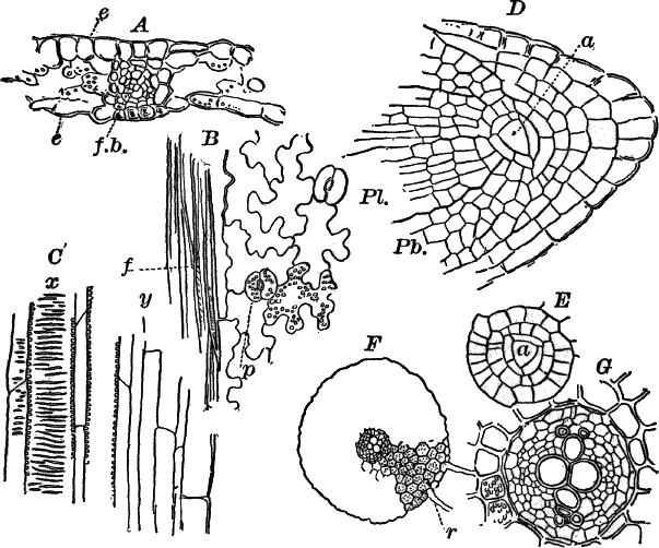
Fig. 68.—A, vertical section of the leaf of the
maiden-hair fern, which has cut across a vein (f.b.), × 150. B,
surface view of the epidermis from the lower surface of a leaf. f,
vein. p, breathing pore, × 150. C, longitudinal section of the
fibro-vascular bundle of the leaf stalk, showing tracheids with
ladder-shaped markings, × 150. D, longitudinal section through the
tip of a root, × 150. a, apical cell. Pl. young fibro-vascular
bundle. Pb. young ground tissue. E, cross-section of the root,
through the region of the apical cell (a), × 150. F, cross-section
through a full-grown root, × 25. r, root hairs. G, the
fibro-vascular bundle of the same, × 150.
A microscopical examination of the leaf stalk shows the tissues to be
almost exactly like those of the stem, except the inner ground
tissue, whose cells are thin-walled and colorless (soft tissue or
“parenchyma”) instead of stony tissue. The structure of the blade of
the leaf, however, shows a number of peculiarities. Stripping off a
little of the epidermis with a needle, or shaving off a thin slice
with a razor, it may be examined in water, removing the air if
necessary with alcohol. It is composed of a single layer of cells, of
very irregular outline, except where it overlies a vein (Fig. 68, B,
f). Here the cells are long and narrow, with heavy walls. The
epidermal cells contain numerous chloroplasts, and on the under
surface of the leaf breathing pores (stomata, sing. stoma), not
unlike those on the capsules of some of the bryophytes. Each breathing
pore consists of two special crescent-shaped epidermal cells (guard
cells), enclosing a central opening or pore communicating with an air
space below. They arise from cells of the young epidermis that divide
by a longitudinal wall, that separates in the middle, leaving the
space between.
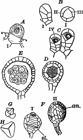
Fig. 69.—A, mother cell of the sporangium of the
maiden-hair fern, × 300. B, young sporangium, surface view, × 150:
i, from the side; ii, from above. C–E, successive stages in the
development of the sporangium seen in optical section, × 150. F,
nearly ripe sporangium, × 50: i, from in front; ii, from the side.
an. ring. st. point of opening. G, group of four spores, × 150.
H, a single spore, × 300.
By holding a leaflet between two pieces of pith, and using a very
sharp razor, cross-sections can be made. Such a section is shown in
Fig. 68, A. The epidermis (e) bounds the upper and lower surfaces,
and if a vein (f.b.) is cut across its structure is found to be like
that of the fibro-vascular bundle of the leaf stalk, but much
simplified.The ground tissue of the leaf is composed of very loose, thin-walled
cells, containing numerous chloroplasts. Between them are large and
numerous intercellular spaces, filled with air, and communicating with
the breathing pores. These are the principal assimilating cells of the
plant; i.e. they are principally concerned in the absorption and
decomposition of carbonic acid from the atmosphere, and the
manufacture of starch.The spore cases, or sporangia (Fig. 69), are at first little papillæ
(A), arising from the epidermal cells, from which they are early cut
off by a cross-wall. In the upper cell several walls next arise,
forming a short stalk, composed of three rows of cells, and an upper
nearly spherical cell—the sporangium proper. The latter now divides
by four walls (B, C, i–iv), into a central tetrahedral cell, and
four outer ones. The central cell, whose contents are much denser than
the outer ones, divides again by walls parallel to those first formed,
so that the young sporangium now consists of a central cell,
surrounded by two outer layers of cells. From the central cell a group
of cells is formed by further divisions (D), which finally become
entirely separated from each other. The outer cells of the spore case
divide only by walls, at right angles to their outer surface, so that
the wall is never more than two cells thick. Later, the inner of these
two layers becomes disorganized, so that the central mass of cells
floats free in the cavity of the sporangium, which is now surrounded
by but a single layer of cells (E).Each of the central cells divides into four spores, precisely as in
the bryophytes. The young spores (G, H) are nearly colorless and
are tetrahedral (like a three-sided pyramid) in form. As they ripen,
chlorophyll is formed in them, and some oil. The wall becomes
differentiated into three layers, the outer opaque and brown, the two
inner more delicate and colorless.Running around the outside of the ripe spore case is a single row of
cells (an.), differing from the others in shape, and having their
inner walls thickened. Near the bottom, two (sometimes four) of these
cells are wider than the others, and their walls are more strongly
thickened. It is at this place (st.) that the spore case opens. When
the ripe sporangium becomes dry, the ring of thickened cells (an.)
contracts more strongly than the others, and acts like a spring
pulling the sporangium open and shaking out the spores, which
germinate readily under favorable conditions, and form after a time
the sexual plants (prothallia).
The roots of the sporophyte arise in large numbers, the youngest being
always nearest the growing point of the stem or larger roots (Fig. 67,
C). The growing roots are pointed at the end which is also
light-colored, the older parts becoming dark brown. A cross-section of
the older portions shows a dark-brown ground tissue with a central,
light-colored, circular, fibro-vascular bundle (Fig. 68, F). Growing
from its outer surface are numerous brown root hairs (r).
When magnified the walls of all the outer cells (epidermis and ground
tissue) are found to be dark-colored but not very thick, and the cells
are usually filled with starch. There is a bundle sheath of
much-flattened cells separating the fibro-vascular bundle from the
ground tissue. The bundle (Fig. 68, G) shows a band of tracheary
tissue in the centre surrounded by colorless cells, all about alike.All of the organs of the fern grow from a definite apical cell, but it
is difficult to study except in the root.Selecting a fresh, pretty large root, a series of thin longitudinal
sections should be made either holding the root directly in the
fingers or placing it between pieces of pith. In order to avoid drying
of the sections, as is indeed true in cutting any delicate tissue, it
is a good plan to wet the blade of the razor. If the section has
passed through the apex, it will show the structure shown in
Figure 68, D. The apical cell (a) is large and distinct,
irregularly triangular in outline. It is really a triangular pyramid
(tetrahedron) with the base upward, which is shown by making a series
of cross-sections through the root tip, and comparing them with the
longitudinal sections. The cross-section of the apical cell (Fig. L)
appears also triangular, showing all its faces to be triangles.
Regular series of segments are cut off in succession from each of the
four faces of the apical cell. These segments undergo regular
divisions also, so that very early a differentiation of the tissues is
evident, and the three tissue systems (epidermal, ground, and
fibro-vascular) may be traced almost to the apex of the root (68,
D). From the outer series of segments is derived the peculiar
structure (root cap) covering the delicate growing point and
protecting it from injury.The apices of the stem and leaves, being otherwise protected, develop
segments only from the sides of the apical cell, the outer face never
having segments cut off from it.
CHAPTER XIII.
CLASSIFICATION OF THE PTERIDOPHYTES.
There are three well-marked classes of the Pteridophytes: the ferns
(Filicinæ); horse-tails (Equisetinæ); and the club mosses
(Lycopodinæ).
Class I.—Ferns (Filicinæ).
The ferns constitute by far the greater number of pteridophytes, and
their general structure corresponds with that of the maiden-hair fern
described. There are three orders, of which two, the true ferns
(Filices) and the adder-tongues (Ophioglossaceæ), are represented
in the United States. A third order, intermediate in some respects
between these two, and called the ringless ferns (Marattiaceæ), has
no representatives within our territory.
The classification is at present based largely upon the characters of
the sporophyte, the sexual plants being still very imperfectly known
in many forms.
The adder-tongues (Ophioglossaceæ) are mostly plants of rather small
size, ranging from about ten to fifty centimetres in height. There are
two genera in the United States, the true adder-tongues
(Ophioglossum) and the grape ferns (Botrychium). They send up but
one leaf each year, and this in fruiting specimens (Fig. 70, A) is
divided into two portions, the spore bearing (x) and the green
vegetative part. In Botrychium the leaves are more or less deeply
divided, and the sporangia distinct (Fig. 71, B). In Ophioglossum
the sterile division of the leaf is usually smooth and undivided, and
the spore-bearing division forms a sort of spike, and the sporangia
are much less distinct. The sporangia in both differ essentially from
those of the true ferns in not being derived from a single epidermal
cell, but are developed in part from the ground tissue of the leaf.
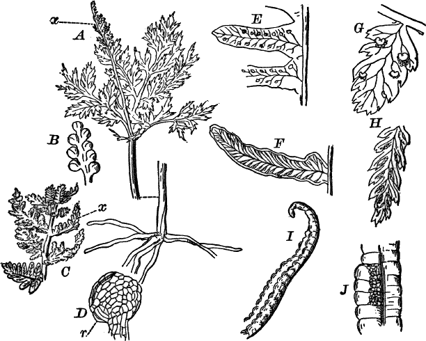
Fig. 70.—Forms of ferns. A, grape fern
(Botrychium), × ½. x, fertile part of the leaf. B, sporangia of
Botrychium, × 3. C, flowering fern (Osmunda). x, spore-bearing
leaflets, × ½. D, a sporangium of Osmunda, × 25. r, ring. E,
Polypodium, × 1. F, brake (Pteris), × 1. G, shield fern
(Aspidium), × 2. H, spleen-wort (Asplenium), × 2. I, ostrich
fern (Onoclea), × 1. J, the same, with the incurved edges of the
leaflet partially raised so as to show the masses of sporangia
beneath, × 2.
In the true ferns (Filices), the sporangia resemble those already
described, arising in all (unless possibly Osmunda) from a single
epidermal cell.
One group, the water ferns (Rhizocarpeæ), produce two kinds of
spores, large and small. The former produce male, the latter female
prothallia. In both cases the prothallium is small, and often scarcely
protrudes beyond the spore, and may be reduced to a single archegonium
or antheridium (Fig. 71, B, C) with only one or two cells
representing the vegetative cells of the prothallium (v). The water
ferns are all aquatic or semi-aquatic plants, few in number and scarce
or local in their distribution. The commonest are those of the genus
Marsilia (Fig. 71, A), looking like a four-leaved clover. Others
(Salvinia, Azolla) are floating forms (Fig. 71, D).
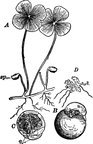
Fig. 71.—A, Marsilia, one of the Rhizocarpeæ
(after Underwood). sp. the “fruits” containing the sporangia. B, a
small spore of Pilularia, with the ripe antheridium protruding,
× 180. C, male prothallium removed from the spore, × 180. D,
Azolla (after Sprague), × 1.
Of the true ferns there are a number of families distinguished mainly
by the position of the sporangia, as well as by some differences in
their structure. Of our common ferns, those differing most widely from
the types are the flowering ferns (Osmunda), shown in Figure 70,
C, D. In these the sporangia are large and the ring (r)
rudimentary. The leaflets bearing the sporangia are more or less
contracted and covered completely with the sporangia, sometimes all
the leaflets of the spore-bearing leaf being thus changed, sometimes
only a few of them, as in the species figured.
Our other common ferns have the sporangia in groups (sori, sing.
sorus) on the backs of the leaves. These sori are of different shape
in different genera, and are usually protected by a delicate
membranous covering (indusium). Illustrations of some of the commonest
genera are shown in Figure 70, E, J.
Class II.—Horse-tails (Equisetinæ).
The second class of the pteridophytes includes the horse-tails
(Equisetinæ) of which all living forms belong to a single genus
(Equisetum). Formerly they were much more numerous than at present,
remains of many different forms being especially abundant in the coal
formations.
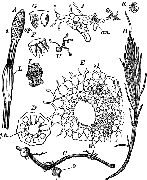
Fig. 72.—A, spore-bearing stem of the field
horse-tail (Equisetum), × 1. x, the spore-bearing cone. B,
sterile stem of the same, × ½. C, underground stem, with tubers
(o), × ½. D, cross-section of an aerial stem, × 5. f.b.
fibro-vascular bundle. E, a single fibro-vascular bundle, × 150.
tr. vessels. F, a single leaf from the cone, × 5. G, the same
cut lengthwise, through a spore sac (sp.), × 5. H, a spore, × 50.
I, the same, moistened so that the elaters are coiled up, × 150.
J, a male prothallium, × 50. an. an antheridium. K,
spermatozoids, × 300.
One of the commonest forms is the field horse-tail (Equisetum
arvense), a very abundant and widely distributed species. It grows in
low, moist ground, and is often found in great abundance growing in
the sand or gravel used as “ballast” for railway tracks.
The plant sends up branches of two kinds from a creeping underground
stem that may reach a length of a metre or more. This stem (Fig. 72,
C) is distinctly jointed, bearing at each joint a toothed sheath,
best seen in the younger portions, as they are apt to be destroyed in
the older parts. Sometimes attached to this are small tubers (o)
which are much-shortened branches and under favorable circumstances
give rise to new stems. They have a hard, brown rind, and are composed
within mainly of a firm, white tissue, filled with starch.
The surface of the stem is marked with furrows, and a section across
it shows that corresponding to these are as many large air spaces that
traverse the stem from joint to joint. From the joints numerous roots,
quite like those of the ferns, arise.
If the stem is dug up in the late fall or winter, numerous short
branches of a lighter color will be found growing from the joints.
These later grow up above ground into branches of two sorts. Those
produced first (Fig. 72, A), in April or May, are stouter than the
others, and nearly destitute of chlorophyll. They are usually twenty
to thirty centimetres in height, of a light reddish brown color, and,
like all the stems, distinctly jointed. The sheaths about the joints
(L) are much larger than in the others, and have from ten to twelve
large black teeth at the top. These sheaths are the leaves. At the top
of the branch the joints are very close together, and the leaves of
different form, and closely set so as to form a compact cone (x).
A cross-section of the stem (D) shows much the same structure as the
underground stem, but the number of air spaces is larger, and in
addition there is a large central cavity. The fibro-vascular bundles
(f.b.) are arranged in a circle, alternating with the air channels,
and each one has running through it a small air passage.
The cone at the top of the branch is made up of closely set,
shield-shaped leaves, which are mostly six-sided, on account of the
pressure. These leaves (F, G) have short stalks, and are arranged
in circles about the stem. Each one has a number of spore cases
hanging down from the edge, and opening by a cleft on the inner side
(G, sp.). They are filled with a mass of greenish spores that
shake out at the slightest jar when ripe.
The sterile branches (B) are more slender than the spore-bearing
ones, and the sheaths shorter. Surrounding the joints, apparently just
below the sheaths, but really breaking through their bases, are
circles of slender branches resembling the main branch, but more
slender. The sterile branches grow to a height of forty to fifty
centimetres, and from their bushy form the popular name of the plant,
“horse-tail,” is taken. The surface of the plant is hard and rough,
due to the presence of great quantities of flint in the epidermis,—a
peculiarity common to all the species.
The stem is mainly composed of large, thin-walled cells, becoming
smaller as they approach the epidermis. The outer cells of the ground
tissue in the green branches contain chlorophyll, and the walls of
some of them are thickened. The fibro-vascular bundles differ entirely
from those of the ferns. Each bundle is nearly triangular in section
(E), with the point inward, and the inner end occupied by a large
air space. The tracheary tissue is only slightly developed, being
represented by a few vessels[9] (tr.) at the outer angles of the
bundle, and one or two smaller ones close to the air channel. The rest
of the bundle is made up of nearly uniform, rather thin-walled,
colorless cells, some of which, however, are larger, and have
perforated cross-walls, representing the sieve tubes of the fern
bundle. There is no individual bundle sheath, but the whole circle of
bundles has a common outer sheath.The epidermis is composed of elongated cells whose walls present a
peculiar beaded appearance, due to the deposition of flint within
them. The breathing pores are arranged in vertical lines, and resemble
in general appearance those of the ferns, though differing in some
minor details. Like the other epidermal cells the guard cells have
heavy deposits of flint, which here are in the form of thick
transverse bars.The spore cases have thin walls whose cells, shortly before maturity,
develop thickenings upon their walls, which have to do with the
opening of the spore case. The spores (H, I) are round cells
containing much chlorophyll and provided with four peculiar
appendages called elaters. The elaters are extremely sensitive to
changes in moisture, coiling up tightly when moistened (I), but
quickly springing out again when dry (H). By dusting a few dry
spores upon a slide, and putting it under the microscope without any
water, the movement may be easily examined. Lightly breathing upon
them will cause the elaters to contract, but in a moment, as soon as
the moisture of the breath has evaporated, they will uncoil with a
quick jerk, causing the spores to move about considerably.The fresh spores begin to germinate within about twenty-four hours,
and the early stages, which closely resemble those of the ferns, may
be easily followed by sowing the spores in water. With care it is
possible to get the mature prothallia, which should be treated as
described for the fern prothallia. Under favorable conditions, the
first antheridia are ripe in about five weeks; the archegonia, which
are borne on separate plants, a few weeks later. The antheridia
(Fig. 72, J, an.) are larger than those of the ferns, and the
spermatozoids (K) are thicker and with fewer coils, but otherwise
much like fern spermatozoids.The archegonia have a shorter neck than those of the ferns, and the
neck is straight.Both male and female prothallia are much branched and very irregular
in shape.
There are a number of common species of Equisetum. Some of them,
like the common scouring rush (E. hiemale), are unbranched, and the
spores borne at the top of ordinary green branches; others have all
the stems branching like the sterile stems of the field horse-tail,
but produce a spore-bearing cone at the top of some of them.
Class III.—The Club Mosses (Lycopodinæ).
The last class of the pteridophytes includes the ground pines, club
mosses, etc., and among cultivated plants numerous species of the
smaller club mosses (Selaginella).
Two orders are generally recognized, although there is some doubt as
to the relationship of the members of the second order. The first
order, the larger club mosses (Lycopodiaceæ) is represented in the
northern states by a single genus (Lycopodium), of which the common
ground pine (L. dendroideum) (Fig. 73) is a familiar species. The
plant grows in the evergreen forests of the northern United States as
well as in the mountains further south, and in the larger northern
cities is often sold in large quantities at the holidays for
decorating. It sends up from a creeping, woody, subterranean stem,
numerous smaller stems which branch extensively, and are thickly set
with small moss-like leaves, the whole looking much like a little
tree. At the ends of some of the branches are small cones (A, x,
B) composed of closely overlapping, scale-like leaves, much as in a
fir cone. Near the base, on the inner surface of each of these scales,
is a kidney-shaped capsule (C, sp.) opening by a cleft along the
upper edge and filled with a mass of fine yellow powder. These
capsules are the spore cases.
The bases of the upright stems are almost bare, but become covered
with leaves higher up. The leaves are in shape like those of a moss,
but are thicker. The spore-bearing leaves are broader and when
slightly magnified show a toothed margin.
The stem is traversed by a central fibro-vascular cylinder that
separates easily from the surrounding tissue, owing to the rupture of
the cells of the bundle sheath, this being particularly frequent in
dried specimens. When slightly magnified the arrangement of the
tissues may be seen (Fig. 73, E). Within the epidermis is a mass of
ground tissue of firm, woody texture surrounding the central oval or
circular fibro-vascular cylinder. This shows a number of white bars
(xylem) surrounded by a more delicate tissue (phloem).
On magnifying the section more strongly, the cells of the ground
tissue (G) are seen to be oval in outline, with thick striated walls
and small intercellular spaces. Examined in longitudinal sections they
are long and pointed, belonging to the class of cells known as
“fibres.”
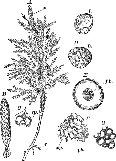
Fig. 73.—A, a club moss (Lycopodium), × ⅓. x,
cone. r, root. B, a cone, × 1. C, single scale with sporangium
(sp.). D, spores: i, from above; ii, from below, × 325. E, cross
section of stem, × 8. f.b. fibro-vascular bundle. F, portion of
the fibro-vascular bundle, × 150. G, cells of the ground tissue,
× 150.
The xylem (F, xy.) of the fibro-vascular bundle is composed of
tracheids, much like those of the ferns; the phloem is composed of
narrow cells, pretty much all alike.The spores (D) are destitute of chlorophyll and have upon the
outside a network of ridges, except on one side where three straight
lines converge, the spore being slightly flattened between them.Almost nothing is known of the prothallia of our native species.
The second order (Ligulatæ) is represented by two very distinct
families: the smaller club mosses (Selaginelleæ) and the quill-worts
(Isoeteæ). Of the former the majority are tropical, but are common
in greenhouses where they are prized for their delicate moss-like
foliage (Fig. 74, A).
Fig. 74.—A, one of the smaller club mosses
(Selaginella). sp. spore-bearing branch, × 2. B, part of a stem,
sending down naked rooting branches (r), × 1. C, longitudinal
section of a spike, with a single macrosporangium at the base; the
others, microsporangia, × 3. D, a scale and microsporangium, × 5.
E, young microsporangium, × 150. The shaded cells are the spore
mother cells. F, a young macrospore, × 150. G, section of the
stem, × 50. H, a single fibro-vascular bundle, × 150. I, vertical
section of the female prothallium of Selaginella, × 50. ar.
archegonium. J, section of an open archegonium, × 300. o, the egg
cell. K, microspore, with the contained male prothallium, × 300.
x, vegetative cell. sp. sperm cells. L, young plant, with the
attached macrospore, × 6. r, the first root. l, the first leaves.
The leaves in most species are like those of the larger club mosses,
but more delicate. They are arranged in four rows on the upper side of
the stem, two being larger than the others. The smaller branches grow
out sideways so that the whole branch appears flattened, reminding one
of the habit of the higher liverworts. Special leafless branches (B,
r) often grow downward from the lower side of the main branches, and
on touching the ground develop roots which fork regularly.
The sporangia are much like those of the ground pines, and produced
singly at the bases of scale leaves arranged in a spike or cone (A,
sp.), but two kinds of spores, large and small, are formed. In the
species figured the lower sporangium produces four large spores
(macrospores); the others, numerous small spores (microspores).
Even before the spores are ripe the development of the prothallium
begins, and this is significant, as it shows an undoubted
relationship between these plants and the lowest of the seed plants,
as we shall see when we study that group.
If ripe spores can be obtained by sowing them upon moist earth, the
young plants will appear in about a month. The microspore (Fig. 74,
K) produces a prothallium not unlike that of some of the water
ferns, there being a single vegetative cell (x), and the rest of the
prothallium forming a single antheridium. The spermatozoids are
excessively small, and resemble those of the bryophytes.The macrospore divides into two cells, a large lower one, and a
smaller upper one. The latter gives rise to a flat disc of cells
producing a number of small archegonia of simple structure (Fig. 74,
I, J). The lower cell produces later a tissue that serves to
nourish the young embryo.The development of the embryo recalls in some particulars that of the
seed plants, and this in connection with the peculiarities of the
sporangia warrants us in regarding the Ligulatæ as the highest of
existing pteridophytes, and to a certain extent connecting them with
the lowest of the spermaphytes.
Resembling the smaller club mosses in their development, but differing
in some important points, are the quill-worts (Isoeteæ). They are
mostly aquatic forms, growing partially or completely submerged, and
look like grasses or rushes. They vary from a few centimetres to half
a metre in height. The stem is very short, and the long cylindrical
leaves closely crowded together. The leaves which are narrow above are
widely expanded and overlapping at the base. The spores are of two
kinds, as in Selaginella, but the macrosporangia contain numerous
macrospores. The very large sporangia (M, sp.) are in cavities at
the bases of the leaves, and above each sporangium is a little pointed
outgrowth (ligula), which is also found in the leaves of
Selaginella. The quill-worts are not common plants, and owing to
their habits of growth and resemblance to other plants, are likely to
be overlooked unless careful search is made.
CHAPTER XIV.
SUB-KINGDOM VI.
Spermaphytes: Phænogams.
The last and highest great division of the vegetable kingdom has been
named Spermaphyta, “seed plants,” from the fact that the structures
known as seeds are peculiar to them. They are also commonly called
flowering plants, though this name might be also appropriately given
to certain of the higher pteridophytes.
In the seed plants the macrosporangia remain attached to the parent
plant, in nearly all cases, until the archegonia are fertilized and
the embryo plant formed. The outer walls of the sporangium now become
hard, and the whole falls off as a seed.
In the higher spermaphytes the spore-bearing leaves (sporophylls)
become much modified, and receive special names, those bearing the
microspores being commonly known as stamens; those bearing the
macrospores, carpels or carpophylls. The macrosporangia are also
ordinarily known as “ovules,” a name given before it was known that
these were the same as the macrosporangia of the higher pteridophytes.
In addition to the spore-bearing leaves, those surrounding them may be
much changed in form and brilliantly colored, forming, with the
enclosed sporophylls, the “flower” of the higher spermaphytes.
As might be expected, the tissues of the higher spermaphytes are the
most highly developed of all plants, though some of them are very
simple. The plants vary extremely in size, the smallest being little
floating plants, less than a millimetre in diameter, while others are
gigantic trees, a hundred metres and more in height.
There are two classes of the spermaphytes: I., the Gymnosperms, or
naked-seeded ones, in which the ovules (macrosporangia) are borne upon
open carpophylls; and II., Angiosperms, covered-seeded plants, in
which the carpophylls form a closed cavity (ovary) containing the
ovules.
Class I.—Gymnosperms (Gymnospermæ).
The most familiar of these plants are the common evergreen trees
(conifers), pines, spruces, cedars, etc. A careful study of one of
these will give a good idea of the most important characteristics of
the class, and one of the best for this purpose is the Scotch pine
(Pinus sylvestris), which, though a native of Europe, is not
infrequently met with in cultivation in America. If this species
cannot be had by the student, other pines, or indeed almost any other
conifer, will answer. The Scotch pine is a tree of moderate size,
symmetrical in growth when young, with a central main shaft, and
circles of branches at regular intervals; but as it grows older its
growth becomes irregular, and the crown is divided into several main
branches.[10] The trunk and branches are covered with a rough, scaly
bark of a reddish brown color, where it is exposed by the scaling off
of the outer layers. Covering the younger branches, but becoming
thinner on the older ones, are numerous needle-shaped leaves. These
are in pairs, and the base of each pair is surrounded by several dry,
blackish scales. Each pair of leaves is really attached to a very
short side branch, but this is so short as to make the leaves appear
to grow directly from the main branch. Each leaf is about ten
centimetres in length and two millimetres broad. Where the leaves are
in contact they are flattened, but the outer side is rounded, so that
a cross-section is nearly semicircular in outline. With a lens it is
seen that there are five longitudinal lines upon the surface of the
leaf, and careful examination shows rows of small dots corresponding
to these. These dots are the breathing pores. If a cross-section is
even slightly magnified it shows three distinct parts,—a whitish
outer border, a bright green zone, and a central oval, colorless area,
in which, with a little care, may be seen the sections of two
fibro-vascular bundles. In the green zone are sometimes to be seen
colorless spots, sections of resin ducts, containing the resin so
characteristic of the tissues of the conifers.
The general structure of the stem may be understood by making a
series of cross-sections through branches of different ages. In all,
three regions are distinguishable; viz., an outer region (bark or
cortex) (Fig. 76, A, c), composed in part of green cells, and, if
the section has been made with a sharp knife, showing a circle of
little openings, from each of which oozes a clear drop of resin. These
are large resin ducts (r). The centre is occupied by a soft white
tissue (pith), and the space between the pith and bark is filled by a
mass of woody tissue. Traversing the wood are numerous radiating
lines, some of which run from the bark to the pith, others only part
way. These are called the medullary rays. While in sections from
branches of any age these three regions are recognizable, their
relative size varies extremely. In a section of a twig of the present
year the bark and pith make up a considerable part of the section; but
as older branches are examined, we find a rapid increase in the
quantity of wood, while the thickness of the bark increases but
slowly, and the pith scarcely at all. In the wood, too, each year’s
growth is marked by a distinct ring (A i, ii). As the branches grow
in diameter the outer bark becomes split and irregular, and portions
die, becoming brown and hard.
The tree has a very perfect root system, but different from that of
any pteridophytes. The first root of the embryo persists as the main
or “tap” root of the full-grown tree, and from it branch off the
secondary roots, which in turn give rise to others.
The sporangia are borne on special scale-like leaves, and arranged
very much as in certain pteridophytes, notably the club mosses; but
instead of large and small spores being produced near together, the
two kinds are borne on special branches, or even on distinct trees
(e.g. red cedar). In the Scotch pine the microspores are ripe about
the end of May. The leaves bearing them are aggregated in small cones
(“flowers”), crowded about the base of a growing shoot terminating the
branches (Fig. 77, A ♂). The individual leaves (sporophylls) are
nearly triangular in shape, and attached by the smaller end. On the
lower side of each are borne two sporangia (pollen sacs) (C, sp.),
opening by a longitudinal slit, and filled with innumerable yellow
microspores (pollen spores), which fall out as a shower of yellow dust
if the branch is shaken.
The macrosporangia (ovules) are borne on similar leaves, known as
carpels, and, like the pollen sacs, borne in pairs, but on the upper
side of the sporophyll instead of the lower. The female flowers appear
when the pollen is ripe. The leaves of which they are composed are
thicker than those of the male flowers, and of a pinkish color. At the
base on the upper side are borne the two ovules (macrosporangia)
(Fig. 77, E, o), and running through the centre is a ridge that
ends in a little spine or point.
The ovule-bearing leaf has on the back a scale with fringed edge (F,
sc.), quite conspicuous when the flower is young, but scarcely to be
detected in the older cone. From the female flower is developed the
cone (Fig. 75, A), but the process is a slow one, occupying two
years. Shortly after the pollen is shed, the female flowers, which are
at first upright, bend downward, and assume a brownish color, growing
considerably in size for a short time, and then ceasing to grow for
several months.
Fig. 75.—Scotch pine (Pinus sylvestris). A, a ripe
cone, × ½. B, a year-old cone, × 1. C, longitudinal section of
B. D, a single scale of B, showing the sporangia (ovules) (o),
× 2. E, a scale from a ripe cone, with the seeds (s), × ½. F,
longitudinal section of a ripe seed, × 3. em. the embryo. G, a
germinating seed, × 2. r, the primary root. H, longitudinal
section through G, showing the first leaves of the young plant still
surrounded by the endosperm, × 4. I, an older plant with the leaves
(l) withdrawing from the seed coats, × 4. J, upper part of a young
plant, showing the circle of primary leaves (cotyledons), × 1. K,
section of the same, × 2. b, the terminal bud. L, cross-section of
the stem of the young plant, × 25. fb. a fibro-vascular bundle. M,
cross-section of the root, × 25. x, wood. ph. bast, of the
fibro-vascular bundle.
In Figure 75, B, is shown such a flower as it appears in the winter
and early spring following. The leaves are thick and fleshy, closely
pressed together, as is seen by dividing the flower lengthwise, and
each leaf ends in a long point (D). The ovules are still very small.
As the growth of the tree is resumed in the spring, the flower (cone)
increases rapidly in size and becomes decidedly green in color, the
ovules increasing also very much in size. If a scale from such a cone
is examined about the first of June, the ovules will probably be
nearly full-grown, oval, whitish bodies two to three millimetres in
length. A careful longitudinal section of the scale through the ovule
will show the general structure. Such a section is shown in Figure 77,
G. Comparing this with the sporangia of the pteridophytes, the first
difference that strikes us is the presence of an outer coat or
integument (in.), which is absent in the latter. The single
macrospore (sp.) is very large and does not lie free in the cavity
of the sporangium, but is in close contact with its wall. It is filled
with a colorless tissue, the prothallium, and if mature, with care it
is possible to see, even with a hand lens, two or more denser oval
bodies (ar.), the egg cells of the archegonia, which here are very
large. The integument is not entirely closed at the top, but leaves a
little opening through which the pollen spores entered when the flower
was first formed.
After the archegonia are fertilized the outer parts of the ovule
become hard and brown, and serve to protect the embryo plant, which
reaches a considerable size before the sporangium falls off. As the
walls of the ovule harden, the carpel or leaf bearing it undergoes a
similar change, becoming extremely hard and woody, and as each one
ends in a sharp spine, and they are tightly packed together, it is
almost impossible to separate them. The ripe cone (Fig. 75, A)
remains closed during the winter, but in the spring, about the time
the flowers are mature, the scales open spontaneously and discharge
the ripened ovules, now called seeds. Each seed (E, s) is
surrounded by a membranous envelope derived from the scale to which it
is attached, which becomes easily separated from the seed. The opening
of the cones is caused by drying, and if a number of ripe cones are
gathered in the winter or early spring, and allowed to dry in an
ordinary room, they will in a day or two open, often with a sharp,
crackling sound, and scatter the ripe seeds.
A section of a ripe seed (F) shows the embryo (em.) surrounded by
a dense, white, starch-bearing tissue derived from the prothallium
cells, and called the “endosperm.” This fills up the whole seed which
is surrounded by the hardened shell derived from the integument and
wall of the ovule. The embryo is elongated with a circle of small
leaves at the end away from the opening of the ovule toward which is
directed the root of the embryo.
The seed may remain unchanged for months, or even years, without
losing its vitality, but if the proper conditions are provided, the
embryo will develop into a new plant. To follow the further growth of
the embryo, the ripe seeds should be planted in good soil and kept
moderately warm and moist. At the end of a week or two some of the
seeds will probably have sprouted. The seed absorbs water, and the
protoplasm of the embryo renews its activity, beginning to feed upon
the nourishing substances in the cells of the endosperm. The embryo
rapidly increases in length, and the root pushes out of the seed
growing rapidly downward and fastening itself in the soil (G, r).
Cutting the seed lengthwise we find that the leaves have increased
much in length and become green (one of the few cases where
chlorophyll is formed in the absence of light). As these leaves
(called “cotyledons” or seed leaves) increase in length, they
gradually withdraw from the seed whose contents they have exhausted,
and the young plant enters upon an independent existence.
The young plant has a circle of leaves, about six in number,
surrounding a bud which is the growing point of the stem, and in many
conifers persists as long as the stem grows (Fig. 75, K, b). A
cross-section of the young stem shows about six separate
fibro-vascular bundles arranged in a circle (S, fb.). The root
shows a central fibro-vascular cylinder surrounded by a dark-colored
ground tissue. Growing from its surface are numerous root hairs
(Fig. 75, M).
For examining the microscopic structure of the pine, fresh material is
for most purposes to be preferred, but alcoholic material will answer,
and as the alcohol hardens the resin, it is for that reason
preferable.Cross-sections of the leaf, when sufficiently magnified, show that the
outer colorless border of the section is composed of two parts: the
epidermis of a single row of regular cells with very thick outer
walls, and irregular groups of cells lying below them. These latter
have thick walls appearing silvery and clearer than the epidermal
cells. They vary a good deal, in some leaves being reduced to a single
row, in others forming very conspicuous groups of some size. The green
tissue of the leaf is much more compact than in the fern we examined,
and the cells are more nearly round and the intercellular spaces
smaller. The chloroplasts are numerous and nearly round in shape.Scattered through the green tissue are several resin passages (r),
each surrounded by a circle of colorless, thick-walled cells, like
those under the epidermis. At intervals in the latter are
openings—breathing pores—(Fig. 76, J), below each of which is an
intercellular space (i). They are in structure like those of the
ferns, but the walls of the guard cells are much thickened like the
other epidermal cells.Each leaf is traversed by two fibro-vascular bundles of entirely
different structure from those of the ferns. Each is divided into two
nearly equal parts, the wood (x) lying toward the inner, flat side
of the leaf, the bast (T) toward the outer, convex side. This type
of bundle, called “collateral,” is the common form found in the stems
and leaves of seed plants. The cells of the wood or xylem are rather
larger than those of the bast or phloem, and have thicker walls than
any of the phloem cells, except the outermost ones which are
thick-walled fibres like those under the epidermis. Lying between the
bundles are comparatively large colorless cells, and surrounding the
whole central area is a single line of cells that separates it sharply
from the surrounding green tissue.In longitudinal sections, the cells, except of the mesophyll (green
tissue) are much elongated. The mesophyll cells, however, are short
and the intercellular spaces much more evident than in the
cross-section. The colorless cells have frequently rounded depressions
or pits upon their walls, and in the fibro-vascular bundle the
difference between the two portions becomes more obvious. The wood is
distinguished by the presence of vessels with close, spiral or
ring-shaped thickenings, while in the phloem are found sieve tubes,
not unlike those in the ferns.The fibro-vascular bundles of the stem of the seedling plant show a
structure quite similar to that of the leaf, but very soon a
difference is manifested. Between the two parts of the bundle the
cells continue to divide and add constantly to the size of the bundle,
and at the same time the bundles become connected by a line of similar
growing cells, so that very early we find a ring of growing cells
extending completely around the stem. As the cells in this ring
increase in number, owing to their rapid division, those on the
borders of the ring lose the power of dividing, and gradually assume
the character of the cells on which they border (Fig. 76, B,
cam.). The growth on the inside of the ring is more rapid than on
the outer border, and the ring continues comparatively near the
surface of the stem (Fig. 76, A, cam.). The spaces between the
bundles do not increase materially in breadth, and as the bundles
increase in size become in comparison very small, appearing in older
stems as mere lines between the solid masses of wood that make up the
inner portion of the bundles. These are the primary medullary rays,
and connect the pith in the centre of the stem with the bark. Later,
similar plates of cells are formed, often only a single cell thick,
and appearing when seen in cross-section as a single row of elongated
cells (C, m).As the stem increases in diameter the bundles become broader and
broader toward the outside, and taper to a point toward the centre,
appearing wedge-shaped, the inner ends projecting into the pith. The
outer limits of the bundles are not nearly so distinct, and it is not
easy to tell when the phloem of the bundles ends and the ground tissue
of the bark begins.A careful examination of a cross-section of the bark shows first, if
taken from a branch not more than two or three years old, the
epidermis composed of cells not unlike those of the leaf, but whose
walls are usually browner. Underneath are cells with brownish walls,
and often more or less dry and dead. These cells give the brown color
to the bark, and later both epidermis and outer ground tissue become
entirely dead and disappear. The bulk of the ground tissue is made up
of rather large, loose cells, the outer ones containing a good deal of
chlorophyll. Here and there are large resin ducts (Fig. 76, H),
appearing in cross-section as oval openings surrounded by several
concentric rows of cells, the innermost smaller and with denser
contents. These secrete the resin that fills the duct and oozes out
when the stem is cut. All of the cells of the bark contain more or
less starch.The phloem, when strongly magnified, is seen to be made up of cells
arranged in nearly regular radiating rows. Their walls are not very
thick and the cells are usually somewhat flattened in a radial
direction.Some of the cells are larger than the others, and these are found to
be, when examined in longitudinal section, sieve tubes (Fig. 76, E)
with numerous lateral sieve plates quite similar to those found in the
stems of ferns.
Fig. 76.—Scotch pine. A, cross-section of a
two-year-old branch, × 3. p, pith. c, bark. The radiating lines
are medullary rays. r, resin ducts. B, part of the same, × 150.
cam. cambium cells. x, tracheids. C, cross-section of a
two-year-old branch at the point where the two growth rings join: I,
the cells of the first year’s growth; II, those of the second year.
m, a medullary ray, × 150. D, longitudinal section of a branch,
showing the form of the tracheids and the bordered pits upon their
walls. m, medullary ray, × 150. E, part of a sieve tube, × 300.
F, cross-section of a tracheid passing through two of the pits in
the wall (p), × 300. G, longitudinal section of a branch, at right
angles to the medullary rays (m). At y, the section has passed
through the wall of a tracheid, bearing a row of pits, × 150. H,
cross-section of a resin duct, × 150. I, cross-section of a leaf,
× 20. fb. fibro-vascular bundle. r, resin duct. J, section of a
breathing pore, × 150. i, the air space below it.
The growing tissue (cambium), separating the phloem from the wood, is
made up of cells quite like those of the phloem, into which they
insensibly merge, except that their walls are much thinner, as is
always the case with rapidly growing cells. These cells (B, cam.)
are arranged in radial rows and divide, mainly by walls, at right
angles to the radii of the stem. If we examine the inner side of the
ring, the change the cells undergo is more marked. They become of
nearly equal diameter in all directions, and the walls become woody,
showing at the same time distinct stratification (B, x).On examining the xylem, where two growth rings are in contact, the
reason of the sharply marked line seen when the stem is examined with
the naked eye is obvious. On the inner side of this line (I), the
wood cells are comparatively small and much flattened, while the walls
are quite as heavy as those of the much larger cells (II) lying on
the outer side of the line. The small cells show the point where
growth ceased at the end of the season, the cells becoming smaller as
growth was feebler. The following year when growth commenced again,
the first wood cells formed by the cambium were much larger, as growth
is most vigorous at this time, and the wood formed of these larger
cells is softer and lighter colored than that formed of the smaller
cells of the autumn growth.The wood is mainly composed of tracheids, there being no vessels
formed except the first year. These tracheids are characterized by the
presence of peculiar pits upon their walls, best seen when thin
longitudinal sections are made in a radial direction. These pits
(Fig. 76, D, p) appear in this view as double circles, but if cut
across, as often happens in a cross-section of the stem, or in a
longitudinal section at right angles to the radius (tangential), they
are seen to be in shape something like an inverted saucer with a hole
through the bottom. They are formed in pairs, one on each side of the
wall of adjacent tracheids, and are separated by a very delicate
membrane (F, p, G, y). These “bordered” pits are very
characteristic of the wood of all conifers.The structure of the root is best studied in the seedling plant, or in
a rootlet of an older one. The general plan of the root is much like
that of the pteridophytes. The fibro-vascular bundle (Fig. 75, M,
fb.) is of the so-called radial type, there being three xylem masses
(x) alternating with as many phloem masses (ph.) in the root of
the seedling. This regularity becomes destroyed as the root grows
older by the formation of a cambium ring, something like that in the
stem.The development of the sporangia is on the whole much like that of the
club mosses, and will not be examined here in detail. The microspores
(pollen spores) are formed in groups of four in precisely the same way
as the spores of the bryophytes and pteridophytes, and by collecting
the male flowers as they begin to appear in the spring, and crushing
the sporangia in water, the process of division may be seen. For more
careful examination they may be crushed in a mixture of water and
acetic acid, to which is added a little gentian violet. This mixture
fixes and stains the nuclei of the spores, and very instructive
preparations may thus be made.[11]
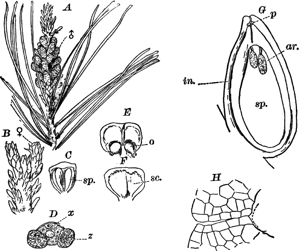
Fig. 77.—Scotch pine (except E and F). A, end of
a branch bearing a cluster of male flowers (♂), × ½. B, a similar
branch, with two young female flowers (♀), natural size. C, a scale
from a male flower, showing the two sporangia (sp.); × 5. D, a
single ripe pollen spore (microspore), showing the vegetative cell
(x), × 150. E, a similar scale, from a female flower of the
Austrian pine, seen from within, × 4. o, the sporangium (ovule).
F, the same, seen from the back, showing the scale (sc.) attached
to the back. G, longitudinal section through a full-grown ovule of
the Scotch pine. p, a pollen spore sending down its tube to the
archegonia (ar.). sp. the prothallium (endosperm), filling up the
embryo sac, × 10. H, the neck of the archegonium, × 150.
The ripe pollen spores (Fig. 77, D) are oval cells provided with a
double wall, the outer one giving rise to two peculiar bladder-like
appendages (z). Like the microspores of the smaller club mosses, a
small cell is cut off from the body of the spore (x). These pollen
spores are carried by the wind to the ovules, where they germinate.The wall of the ripe sporangium or pollen sac is composed of a single
layer of cells in most places, and these cells are provided with
thickened ridges which have to do with opening the pollen sac.We have already examined in some detail the structure of the
macrosporangium or ovule. In the full-grown ovule the macrospore,
which in the seed plants is generally known as the “embryo sac,” is
completely filled with the prothallium or “endosperm.” In the upper
part of the prothallium several large archegonia are formed in much
the same way as in the pteridophytes. The egg cell is very large, and
appears of a yellowish color, and filled with large drops that give it
a peculiar aspect. There is a large nucleus, but it is not always
readily distinguished from the other contents of the egg cell. The
neck of the archegonium is quite long, but does not project above the
surface of the prothallium (Fig. 77, H).
The pollen spores are produced in great numbers, and many of them fall
upon the female flowers, which when ready for pollination have the
scales somewhat separated. The pollen spores now sift down to the base
of the scales, and finally reach the opening of the ovule, where they
germinate. No spermatozoids are produced, the seed plants differing in
this respect from all pteridophytes. The pollen spore bursts its
outer coat, and sends out a tube which penetrates for some distance
into the tissue of the ovule, acting very much as a parasitic fungus
would do, and growing at the expense of the tissue through which it
grows. After a time growth ceases, and is not resumed until the
development of the female prothallium and archegonia is nearly
complete, which does not occur until more than a year from the time
the pollen spore first reaches the ovule. Finally the pollen tube
penetrates down to and through the open neck of the archegonium, until
it comes in contact with the egg cell. These stages can only be seen
by careful sections through a number of ripe ovules, but the track of
the pollen tube is usually easy to follow, as the cells along it are
often brown and apparently dead (Fig. 77, G).
Classification of the Gymnosperms.
There are three classes of the gymnosperms: I., cycads (Cycadeæ);
II., conifers (Coniferæ); III., joint firs (Gnetaceæ). All of the
gymnosperms of the northern United States belong to the second order,
but representatives of the others are found in the southern and
southwestern states.
The cycads are palm-like forms having a single trunk crowned by a
circle of compound leaves. Several species are grown for ornament in
conservatories, and a few species occur native in Florida, but
otherwise do not occur within our limits.
Fig. 78.—Illustrations of gymnosperms. A, fruiting
leaf of a cycad (Cycas), with macrosporangia (ovules) (ov.), × ¼.
B, leaf of Gingko, × ½. C, branch of hemlock (Tsuga), with a
ripe cone, × 1. D, red cedar (Juniperus), × 1. E, Arbor-vitæ
(Thuja), × 1.
The spore-bearing leaves usually form cones, recalling somewhat in
structure those of the horse-tails, but one of the commonest
cultivated species (Cycas revoluta) bears the ovules, which are very
large, upon leaves that are in shape much like the ordinary ones
(Fig. 78, A).
Of the conifers, there are numerous familiar forms, including all our
common evergreen trees. There are two sub-orders,—the true conifers
and the yews. In the latter there is no true cone, but the ovules are
borne singly at the end of a branch, and the seed in the yew (Taxus)
is surrounded by a bright red, fleshy integument. One species of yew,
a low, straggling shrub, occurs sparingly in the northern states, and
is the only representative of the group at the north. The European yew
and the curious Japanese Gingko (Fig. 78, B) are sometimes met
with in cultivation.
Of the true conifers, there are a number of families, based on
peculiarities in the leaves and cones. Some have needle-shaped leaves
and dry cones like the firs, spruces, hemlock (Fig. 78, C). Others
have flattened, scale-like leaves, and more or less fleshy cones, like
the red cedar (Fig. 78, D) and Arbor-vitæ (E).
A few of the conifers, such as the tamarack or larch (Larix) and
cypress (Taxodium), lose their leaves in the autumn, and are not,
therefore, properly “evergreen.”
The conifers include some of the most valuable as well as the largest
of trees. Their timber, especially that of some of the pines, is
particularly valuable, and the resin of some of them is also of much
commercial importance. Here belong the giant red-woods (Sequoia) of
California, the largest of all American trees.
The joint firs are comparatively small plants, rarely if ever reaching
the dimensions of trees. They are found in various parts of the world,
but are few in number, and not at all likely to be met with by the
ordinary student. Their flowers are rather more highly differentiated
than those of the other gymnosperms, and are said to show some
approach in structure to those of the angiosperms.
CHAPTER XV.
SPERMAPHYTES.
Class II.—Angiosperms.
The angiosperms include an enormous assemblage of plants, all those
ordinarily called “flowering plants” belonging here. There is almost
infinite variety shown in the form and structure of the tissues and
organs, this being particularly the case with the flowers. As already
stated, the ovules, instead of being borne on open carpels, are
enclosed in a cavity formed by a single closed carpel or several
united carpels. To the organ so formed the name “pistil” is usually
applied, and this is known as “simple” or “compound,” as it is
composed of one or of two or more carpels. The leaves bearing the
pollen spores are also much modified, and form the so-called
“stamens.” In addition to the spore-bearing leaves there are usually
other modified leaves surrounding them, these being often brilliantly
colored and rendering the flower very conspicuous. To these leaves
surrounding the sporophylls, the general name of “perianth” or
“perigone” is given. The perigone has a twofold purpose, serving both
to protect the sporophylls, and, at least in bright-colored flowers,
to attract insects which, as we shall see, are important agents in
transferring pollen from one flower to another.
When we compare the embryo sac (macrospore) of the angiosperms with
that of the gymnosperms a great difference is noticed, there being
much more difference than between the latter and the higher
pteridophytes. Unfortunately there are very few plants where the
structure of the embryo sac can be readily seen without very skilful
manipulation.
Fig. 79.—A, ripe ovule of Monotropa uniflora, in
optical section, × 100. m, micropyle. e, embryo sac. B, the
embryo sac, × 300. At the top is the egg apparatus, consisting of the
two synergidæ (s), and the egg cell (o). In the centre is the
“endosperm nucleus” (k). At the bottom, the “antipodal
cells” (g).
There are, however, a few plants in which the ovules are very small
and transparent, so that they may be mounted whole and examined alive.
The best plant for this purpose is probably the “Indian pipe” or
“ghost flower,” a curious plant growing in rich woods, blossoming in
late summer. It is a parasite or saprophyte, and entirely destitute of
chlorophyll, being pure white throughout. It bears a single nodding
flower at the summit of the stem. (Another species much like it, but
having several brownish flowers, is shown in Figure 115, L.)If this plant can be had, the structure of the ovule and embryo sac
may be easily studied, by simply stripping away the tissue bearing the
numerous minute ovules, and mounting a few of them in water, or water
to which a little sugar has been added.The ovules are attached to a stalk, and each consists of about two
layers of colorless cells enclosing a central, large, oblong cell
(Fig. 79, A, E), the embryo sac or macrospore. If the ovule is
from a flower that has been open for some time, we shall find in the
centre of the embryo sac a large nucleus (k) (or possibly two which
afterward unite into one), and at each end three cells. Those at the
base (g) probably represent the prothallium, and those at the upper
end a very rudimentary archegonium, here generally called the “egg
apparatus.”Of the three cells of the “egg apparatus” the lower (o) one is the
egg cell; the others are called “synergidæ.” The structure of the
embryo sac and ovules is quite constant among the angiosperms, the
differences being mainly in the shape of the ovules, and the degree to
which its coverings or integuments are developed.The pollen spores of many angiosperms will germinate very easily in a
solution of common sugar in water: about fifteen per cent of sugar is
the best. A very good plant for this purpose is the sweet pea, whose
pollen germinates very rapidly, especially in warm weather. The spores
may be sown in a little of the sugar solution in any convenient
vessel, or in a hanging drop suspended in a moist chamber, as
described for germinating the spores of the slime moulds. The tube
begins to develop within a few minutes after the spores are placed in
the solution, and within an hour or so will have reached a
considerable length. Each spore has two nuclei, but they are less
evident here than in some other forms (Fig. 79).
The upper part of the pistil is variously modified, having either
little papillæ which hold the pollen spores, or are viscid. In either
case the spores germinate when placed upon this receptive part
(stigma) of the pistil, and send their tubes down through the tissues
of the pistil until they reach the ovules, which are fertilized much
as in the gymnosperms.
The effect of fertilization extends beyond the ovule, the ovary and
often other parts of the flower being affected, enlarging and often
becoming bright-colored and juicy, forming the various fruits of the
angiosperms. These fruits when ripe may be either dry, as in the case
of grains of various kinds, beans, peas, etc.; or the ripe fruit may
be juicy, serving in this way to attract animals of many kinds which
feed on the juicy pulp, and leave the hard seeds uninjured, thus
helping to distribute them. Common examples of these fleshy fruits are
offered by the berries of many plants; apples, melons, cherries, etc.,
are also familiar examples.
The seeds differ a good deal both in regard to size and the degree to
which the embryo is developed at the time the seed ripens.
Classification of the Angiosperms.
The angiosperms are divided into two sub-classes: I. Monocotyledons
and II. Dicotyledons.
The monocotyledons comprise many familiar plants, both ornamental and
useful. They have for the most part elongated, smooth-edged leaves
with parallel veins, and the parts of the flower are in threes in the
majority of them. As their name indicates, there is but one cotyledon
or seed leaf, and the leaves from the first are alternate. As a rule
the embryo is very small and surrounded by abundant endosperm.
The most thoroughly typical members of the sub-class are the lilies
and their relatives. The one selected for special study here, the
yellow adder-tongue, is very common in the spring; but if not
accessible, almost any liliaceous plant will answer. Of garden
flowers, the tulip, hyacinth, narcissus, or one of the common lilies
may be used; of wild flowers, the various species of Trillium
(Fig. 83, A) are common and easily studied forms, but the leaves are
not of the type common to most monocotyledons.
The yellow adder-tongue (Erythronium americanum) (Fig. 81) is one of
the commonest and widespread of wild flowers, blossoming in the
northern states from about the middle of April till the middle of May.
Most of the plants found will not be in flower, and these send up but
a single, oblong, pointed leaf. The flowering plant has two similar
leaves, one of which is usually larger than the other. They seem to
come directly from the ground, but closer examination shows that they
are attached to a stem of considerable length entirely buried in the
ground. This arises from a small bulb (B) to whose base numerous
roots (r) are attached. Rising from between the leaves is a slender,
leafless stalk bearing a single, nodding flower at the top.
The leaves are perfectly smooth, dull purplish red on the lower side,
and pale green with purplish blotches above. The epidermis may be very
easily removed, and is perfectly colorless. Examined closely,
longitudinal rows of whitish spots may be detected: these are the
breathing pores.
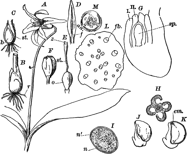
Fig. 81.—A, plant of the yellow adder-tongue
(Erythronium americanum), × ⅓. B, the bulb of the same, × ½. r,
roots. C, section of B. st. the base of the stem bearing the
bulb for next year (b) at its base. D, a single petal and stamen,
× ½. f, the filament. an. anther. E, the gynœcium (pistil), × 1.
o, ovary. st. style. z, stigma. F, a full-grown fruit, × ½.
G, section of a full-grown macrosporangium (ovule), × 25: i, ii, the
two integuments. sp. macrospore (embryo sac). H, cross-section of
the ripe anther, × 12. I, a single pollen spore, × 150, showing the
two nuclei (n, nʹ). J, a ripe seed, × 2. K, the same, in
longitudinal section. em. the embryo. L, cross-section of the
stem, × 12. fb. fibro-vascular bundle. M, diagram of the flower.
A cross-section of the stem shows numerous whitish areas scattered
through it. These are the fibro-vascular bundles which in the
monocotyledons are of a simple type. The bulb is composed of thick
scales, which are modified leaves, and on cutting it lengthwise, we
shall probably find the young bulb of next year (Fig. C, b)
already forming inside it, the young bulb arising as a bud at the
base of the stem of the present year.
The flower is made up of five circles of very much modified leaves,
three leaves in each set. The two outer circles are much alike, but
the three outermost leaves are slightly narrower and strongly tinged
with red on the back, completely concealing the three inner ones
before the flower expands. The latter are pure yellow, except for a
ridge along the back, and a few red specks near the base inside. These
six leaves constitute the perigone of the flower; the three outer are
called sepals, the inner ones petals.
The next two circles are composed of the sporophylls bearing the
pollen spores.[12] These are the stamens, and taken collectively are
known as the “Andrœcium.” Each leaf or stamen consists of two
distinct portions, a delicate stalk or “filament” (D, f), and the
upper spore-bearing part, the “anther” (an.). The anther in the
freshly opened flower has a smooth, red surface; but shortly after,
the flower opens, splits along each side, and discharges the pollen
spores. A section across the anther shows it to be composed of four
sporangia or pollen sacs attached to a common central axis
(“connective”) (Fig. H).
The central circle of leaves, the carpels (collectively the
“gynœcium”) are completely united to form a compound pistil (Fig. 81,
E). This shows three distinct portions, the ovule-bearing portion
below (o), the “ovary,” a stalk above (st.), the “style,” and the
receptive portion (z) at the top, the “stigma.” Both stigma and
ovary show plainly their compound nature, the former being divided
into three lobes, the latter completely divided into three chambers,
as well as being flattened at the sides with a more or less decided
seam at the three angles. The ovules, which are quite large, are
arranged in two rows in each chamber of the ovary, attached to the
central column (“placenta”).
The flowers open for several days in succession, but only when the sun
is shining. They are visited by numerous insects which carry the
pollen from one flower to another and deposit it upon the stigma,
where it germinates, and the tube, growing down through the long
style, finally reaches the ovules and fertilizes them. Usually only a
comparatively small number of the seeds mature, there being almost
always a number of imperfect ones in each pod. The pod or fruit (F)
is full-grown about a month after the flower opens, and finally
separates into three parts, and discharges the seeds. These are quite
large (Fig. 81, J) and covered with a yellowish brown outer coat,
and provided with a peculiar, whitish, spongy appendage attaching it
to the placenta. A longitudinal section of a ripe seed (K) shows the
very small, nearly triangular embryo (em.), while the rest of the
cavity of the seed is filled with a white, starch-bearing tissue, the
endosperm.
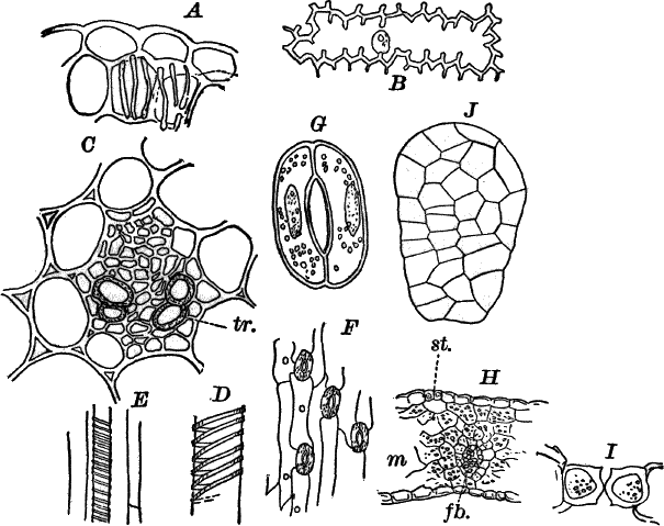
Fig. 82.—Erythronium. A, a portion of the wall of
the anther, × 150. B, a single epidermal cell from the petal, × 150.
C, cross-section of a fibro-vascular bundle of the stem, × 150.
tr. vessels. D, E, longitudinal section of the same, showing the
markings of the vessels, × 150. F, a bit of the epidermis from the
lower surface of a leaf, showing the breathing pores, × 50. G, a
single breathing pore, × 200. H, cross-section of a leaf, × 50.
st. a breathing pore. m, the mesophyll. fb. a vein. I,
cross-section of a breathing pore, × 200. J, young embryo, × 150.
A microscopical examination of the tissues of the plant shows them to
be comparatively simple, this being especially the case with the
fibro-vascular system.The epidermis of the leaf is readily removed, and examination shows it
to be made up of oblong cells with large breathing pores in rows. The
breathing pores are much larger than any we have yet seen, and are of
the type common to most angiosperms. The ordinary epidermal cells are
quite destitute of chlorophyll, but the two cells (guard cells)
enclosing the breathing pore contain numerous chloroplasts, and the
oblong nuclei of these cells are usually conspicuous (Fig. 82, G).
By placing a piece of the leaf between pieces of pith, and making a
number of thin cross-sections at right angles to the longer axis of
the leaf, some of the breathing pores will probably be cut across, and
their structure may be then better understood. Such a section is shown
in Figure 82, I.The body of the leaf is made up of chlorophyll-bearing cells of
irregular shape and with large air spaces between (H, m). The
veins traversing this tissue are fibro-vascular bundles of a type
structure similar to that of the stem, which will be described
presently.The stem is made up principally of large cells with thin walls, which
in cross-section show numerous small, triangular, intercellular spaces
(i) at the angles. These cells contain, usually, more or less
starch. The fibro-vascular bundles (C) are nearly triangular in
section, and resemble considerably those of the field horse-tail, but
they are not penetrated by the air channel, found in the latter. The
xylem, as in the pine, is toward the outside of the stem, but the
boundary between xylem and phloem is not well defined, there being no
cambium present. In the xylem are a number of vessels (C, tr.) at
once distinguishable from the other cells by their definite form, firm
walls, and empty cavity. The vessels in longitudinal sections show
spiral and ringed thickenings. The rest of the xylem cells, as well as
those of the phloem, are not noticeably different from the cells of
the ground tissue, except for their much smaller size, and absence of
intercellular spaces.The structure of the leaves of the perigone is much like that of the
green leaves, but the tissues are somewhat reduced. The epidermis of
the outer side of the sepals has breathing pores, but these are absent
from their inner surface, and from both sides of the petals. The walls
of the epidermal cells of the petals are peculiarly thickened by
apparent infoldings of the wall (B), and these cells, as well as
those below them, contain small, yellow bodies (chromoplasts) to which
the bright color of the flower is due. The red specks on the base of
the perigone leaves, as well as the red color of the back of the
sepals, the stalk, and leaves are due to a purplish red cell sap
filling the cells at these points.The filaments or stalks of the stamens are made up of very delicate
colorless cells, and the centre is traversed by a single
fibro-vascular bundle, which is continued up through the centre of the
anther. To study the latter, thin cross-sections should be made and
mounted in water. Each of the four sporangia, or pollen sacs, is
surrounded on the outside by a wall, consisting of two layers of
cells, becoming thicker in the middle of the section where the single
fibro-vascular bundle is seen (Fig. 81, H). On opening, the cavities
of the adjacent sporangia are thrown together. The inner cells of the
wall are marked by thickened bars, much as we saw in the pine
(Fig. 82, A), and which, like these, are formed shortly before the
pollen sacs open. The pollen spores (Fig. 81, I) are large, oval
cells, having a double wall, the outer one somewhat heavier than the
inner one, but sufficiently transparent to allow a clear view of the
interior, which is filled with very dense, granular protoplasm in
which may be dimly seen two nuclei (n, ni.), showing that here
also there is a division of the spore contents, although no wall is
present. The spores do not germinate very readily, and are less
favorable for this purpose than those of some other monocotyledons.
Among the best for this purpose are the spiderwort (Tradescantia)
and Scilla.Owing to the large size and consequent opacity of the ovules, as well
as to the difficulty of getting the early stages, the development and
finer structure of the ovule will not be discussed here. The
full-grown ovule may be readily sectioned, and a general idea of its
structure obtained. A little potash may be used to advantage in this
study, carefully washing it away when the section is sufficiently
cleared. We find now that the ovule is attached to a stalk (funiculus)
(Fig. 81, G, f), the body of the ovule being bent up so as to lie
against the stalk. Such an inverted ovule is called technically,
“anatropous.” The ovule is much enlarged where the stalk bends. The
upper part of the ovule is on the whole like that of the pine, but
there are two integuments (i, ii) instead of the single one found in
the pine.As the seed develops, the embryo sac (G, sp.) enlarges so as to
occupy pretty much the whole space of the seed. At first it is nearly
filled with a fluid, but a layer of cells is formed, lining the walls,
and this thickens until the whole space, except what is occupied by
the small embryo, is filled with them. These are called the “endosperm
cells,” but differ from the endosperm cells of the gymnosperms, in the
fact that they are not developed until after fertilization, and can
hardly, therefore, be regarded as representing the prothallium of the
gymnosperms and pteridophytes. These cells finally form a firm tissue,
whose cells are filled with starch that forms a reserve supply of food
for the embryo plant when the seed germinates. The embryo (Fig. 81,
K, em., Fig. 82, J), even when the seed is ripe, remains very
small, and shows scarcely any differentiation. It is a small,
pear-shaped mass of cells, the smaller end directed toward the upper
end of the embryo sac.
The integuments grow with the embryo sac, and become brown and hard,
forming the shell of the seed. The stalk of the ovule also enlarges,
and finally forms the peculiar, spongy appendage of the seeds already
noticed (Fig. 81, J, K).
CHAPTER XVI.
CLASSIFICATION OF THE MONOCOTYLEDONS.
In the following chapter no attempt will be made to give an exhaustive
account of the characteristics of each division of the monocotyledons,
but only such of the most important ones as may serve to supplement
our study of the special one already examined. The classification
here, and this is the case throughout the spermaphytes, is based
mainly upon the characters of the flowers and fruits.
The classification adopted here is that of the German botanist
Eichler, and seems to the author to accord better with our present
knowledge of the relationships of the groups than do the systems that
are more general in this country. According to Eichler’s
classification, the monocotyledons may be divided into seven groups;
viz., I. Liliifloræ; II. Enantioblastæ; III. Spadicifloræ;
IV. Glumaceæ; V. Scitamineæ; VI. Gynandræ; VII. Helobiæ.
Order I.—Liliifloræ.
The plants of this group agree in their general structure with the
adder’s-tongue, which is a thoroughly typical representative of the
group; but nevertheless, there is much variation among them in the
details of structure. While most of them are herbaceous forms (dying
down to the ground each year), a few, among which may be mentioned the
yuccas (“bear grass,” “Spanish bayonet”) of our southern states,
develop a creeping or upright woody stem, increasing in size from year
to year. The herbaceous forms send up their stems yearly from
underground bulbs, tubers, e.g. Trillium (Fig. 83, A), or
thickened, creeping stems, or root stocks (rhizomes). Good examples of
the last are the Solomon’s-seal (Fig. 83, B), Medeola (C, D),
and iris (Fig. 84 A). One family, the yams (Dioscoreæ), of which
we have one common native species, the wild yam (Dioscorea villosa),
have broad, netted-veined leaves and are twining plants, while another
somewhat similar family (Smilaceæ) climb by means of tendrils at the
bases of the leaves. Of the latter the “cat-brier” or “green-brier” is
a familiar representative.
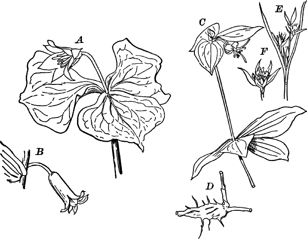
Fig. 83.—Types of Liliifloræ. A, Trillium, × ¼.
B, single flower of Solomon’s-seal (Polygonatum), × 1. C, upper
part of a plant. D, underground stem (rhizome) of Indian cucumber
root (Medeola), × ½. E, a rush (Juncus), × 1. F, a single
flower, × 2. A–D, Liliaceæ; E, Juncaceæ.
The flowers are for the most part conspicuous, and in plan like that
of the adder’s-tongue; but some, like the rushes (Fig. 83, E), have
small, inconspicuous flowers; and others, like the yams and smilaxes,
have flowers of two kinds, male and female.
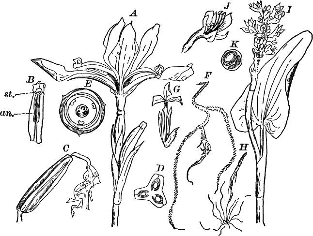
Fig. 84.—Types of Liliifloræ. A, flower of the
common blue-flag (Iris), × ½ (Iridaceæ). B, the petal-like upper
part of the pistil, seen from below, and showing a stamen (an.).
st. the stigma, × ½. C, the young fruit, × ½. D, section of the
same, × 1. E, diagram of the flower. F, part of a plant of the
so-called “gray moss” (Tillandsia), × ½ (Bromeliaceæ). G, a
single flower, × 2. H, a seed, showing the fine hairs attached to
it, × 1. I, plant of pickerel-weed (Pontederia), × ¼
(Pontederiaceæ). J, a single flower, × 1. K, section of the
ovary, × 4.
The principal family of the Liliifloræ is the Liliaceæ, including
some of the most beautiful of all flowers. All of the true lilies
(Lilium), as well as the day lilies (Funkia, Hemerocallis) of
the gardens, tulips, hyacinths, lily-of-the-valley, etc., belong here,
as well as a number of showy wild flowers including several species of
tiger-lilies (Lilium), various species of Trillium (Fig. 83, A),
Solomon’s-seal (Polygonatum) (Fig. 83, B), bellwort (Uvularia),
and others. In all of these, except Trillium, the perigone leaves
are colored alike, and the leaves parallel-veined; but in the latter
the sepals are green and the leaves broad and netted-veined. The fruit
of the Liliaceæ may be either a pod, like that of the
adder’s-tongue, or a berry, like that of asparagus or Solomon’s-seal.
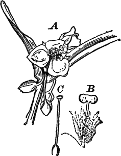
Fig. 85.—Enantioblastæ. A, inflorescence of the
common spiderwort (Tradescantia), × ½ (Commelyneæ). B, a single
stamen, showing the hairs attached to the filament, × 2. C, the
pistil, × 2.
Differing from the true lilies in having the bases of the perigone
leaves adherent to the surface of the ovary, so that the latter is
apparently below the flower (inferior), and lacking the inner circle
of stamens, is the iris family (Iridaceæ), represented by the wild
blue-flag (Iris versicolor) (Fig. 84, A, E), as well as by
numerous cultivated species. In iris the carpels are free above and
colored like the petals (B), with the stigma on the under side. Of
garden flowers the gladiolus and crocus are the most familiar
examples, besides the various species of iris; and of wild flowers the
little “blue-eyed grass” (Sisyrinchium).
The blue pickerel-weed (Pontederia) is the type of a family of which
there are few common representatives (Fig. 84, I, K).
The last family of the order is the Bromeliaceæ, all inhabitants of
the warmer parts of the globe, but represented in the southern states
by several forms, the commonest of which is the so-called “gray moss”
(Tillandsia) (Fig. 84, F, H). Of cultivated plants
the pineapple, whose fruit consists of a fleshy mass made up of the
crowded fruits and the fleshy flower stalks, is the best known.
Order II.—Enantioblastæ.
The second order of the monocotyledons, Enantioblastæ, includes very
few common plants. The most familiar examples are the various species
of Tradescantia (Fig. 88), some of which are native, others exotic.
Of the cultivated forms the commonest is one sometimes called
“wandering-jew,” a trailing plant with zigzag stems, and oval, pointed
leaves forming a sheath about each joint. Another common one is the
spiderwort already referred to. In this the leaves are long and
pointed, but also sheathing at the base. When the flowers are showy,
as in these, the sepals and petals are different, the former being
green. The flowers usually open but once, and the petals shrivel up as
the flower fades. There are four families of the order, the spiderwort
belonging to the highest one, Commelyneæ.
Order III.—Spadicifloræ.
The third order of the monocotyledons, Spadicifloræ, is a very large
one, and includes the largest and the smallest plants of the whole
sub-class. In all of them the flowers are small and often very
inconspicuous; usually, though not always, the male and female flowers
are separate, and often on different plants. The smallest members of
the group are little aquatics, scarcely visible to the naked eye, and
of extremely simple structure, but nevertheless these little plants
produce true flowers. In marked contrast to these are the palms, some
of which reach a height of thirty metres or more.
The flowers in most of the order are small and inconspicuous, but
aggregated on a spike (spadix) which may be of very large size. Good
types of the order are the various aroids (Aroideæ), of which the
calla (Richardia) is a very familiar cultivated example. Of wild
forms the sweet-flag (Acorus), Jack-in-the-pulpit (Arisæma)
(Fig. 86, A, D), skunk-cabbage (Symplocarpus), and wild calla
may be noted. In Arisæma (Fig. 86, A) the flowers are borne only
on the base of the spadix, and the plant is diœcious. The flowers are
of the simplest structure, the female consisting of a single carpel,
and the male of four stamens (C, D). While the individual flowers
are destitute of a perigone, the whole inflorescence (cluster of
flowers) is surrounded by a large leaf (spathe), which sometimes is
brilliantly colored, this serving to attract insects. The leaves of
the aroids are generally large and sometimes compound, the only
instance of true compound leaves among the monocotyledons (Fig. 86,
B).
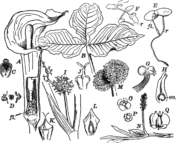
Fig. 86.—Types of Spadicifloræ. A, inflorescence
of Jack-in-the-pulpit (Arisæma, Aroideæ). The flowers (fl.) are
at the base of a spike (spadix), surrounded by a sheath (spathe),
which has been cut away on one side in order to show the flowers, × ½.
B, leaf of the same plant, × ¼. C, vertical section of a female
flower, × 2. D, three male flowers, each consisting of four stamens,
× 2. E, two plants of a duck-weed (Lemna), the one at the left is
in flower, × 4. F, another common species. L, Trisulea, × 1.
G, male flower of E, × 25. H, optical section of the female
flower, showing the single ovule (ov.), × 25. I, part of the
inflorescence of the bur-reed (Sparganium), with female flowers, × ½
(Typhaceæ). J, a single, female flower, × 2. K, a ripe fruit,
× 1. L, longitudinal section of the same. M, two male flowers,
× 1. N, a pond-weed (Potomogeton), × 1 (Naiadaceæ). O, a
single flower, × 2. P, the same, with the perianth removed, × 2.
Q, fruit of the same, × 2.
Probably to be regarded as reduced aroids are the duck-weeds
(Lemnaceæ) (Fig. 86, F, H), minute floating plants without any
differentiation of the plant body into stem and leaves. They are
globular or discoid masses of cells, most of them having roots; but
one genus (Wolffia) has no roots nor any trace of fibro-vascular
bundles. The flowers are reduced to a single stamen or carpel (Figs.
E, G, H).
The cat-tail (Typha) and bur-reed (Sparganium) (Fig. 86, I, L)
are common representatives of the family Typhaceæ, and the
pond-weeds (Naias and Potomogeton) are common examples of the
family Naiadeæ. These are aquatic plants, completely submerged
(Naias), or sometimes partially floating (Potomogeton). The latter
genus includes a number of species with leaves varying from linear
(very narrow and pointed) to broadly oval, and are everywhere common
in slow streams.
The largest members of the group are the screw-pines (Pandaneæ) and
the palms (Palmæ). These are represented in the United States by
only a few species of the latter family, confined to the southern and
southwestern portions. The palmettoes (Sabal and Chamærops) are
the best known.
Both the palms and screw-pines are often cultivated for ornament, and
as is well known, in the warmer parts of the world the palms are among
the most valuable of all plants. The date palm (Phœnix dactylifera)
and the cocoanut (Cocos nucifera) are the best known. The apparently
compound (“pinnate” or feather-shaped) leaves of many palms are not
strictly compound; that is, they do not arise from the branching of an
originally single leaf, but are really broad, undivided leaves, which
are closely folded like a fan in the bud, and tear apart along the
folds as the leaf opens.
Although these plants reach such a great size, an examination of the
stem shows that it is built on much the same plan as that of the other
monocotyledons; that is, the stem is composed of a mass of soft,
ground tissue through which run many small isolated, fibro-vascular
bundles. A good idea of this structure may be had by cutting across a
corn-stalk, which is built on precisely the same pattern.
Order IV.—Glumaceæ.
The plants of this order resemble each other closely in their habit,
all having long, narrow leaves with sheathing bases that surround the
slender, distinctly jointed stem which frequently has a hard, polished
surface. The flowers are inconspicuous, borne usually in close spikes,
and destitute of a perigone or having this reduced to small scales or
hairs. The flowers are usually surrounded by more or less dry leaves
(glumes, paleæ) which are closely set, so as to nearly conceal the
flowers. The flowers are either hermaphrodite or unisexual.
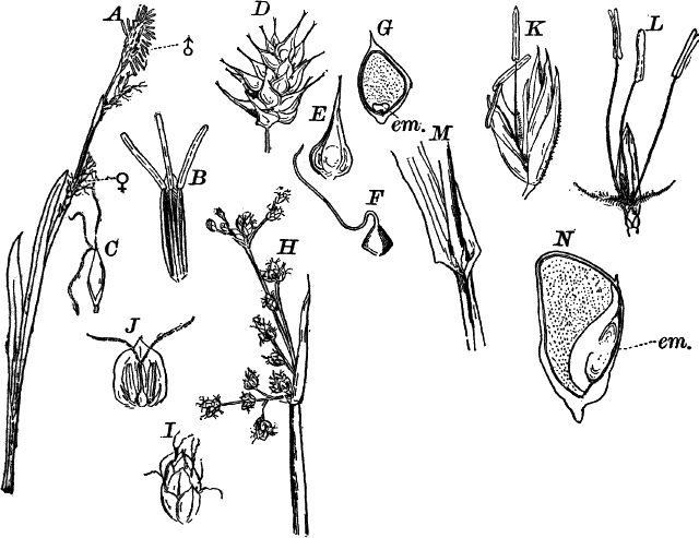
Fig. 87.—Types of Glumaceæ. A, a sedge, Carex
(Cyperaceæ). ♂, the male; ♀, the female flowers, × ½. B, a single
male flower, × 2. C, a female flower, × 2. D, fruiting spike of
another Carex, × ½. E, a single fruit, × 1. F, the same, with
the outer envelope removed, and slightly enlarged. G, section of
F, × 3. em. the embryo. H, a bulrush, Scirpus (Cyperaceæ),
× ½. I, a single spikelet, × 2. J, a single flower, × 3. K, a
spikelet of flowers of the common orchard grass, Dactylis
(Gramineæ), × 2. L, a single flower, × 2. M, the base of a leaf,
showing the split sheath encircling the stem, × 1. N, section of a
kernel of corn, showing the embryo (em.), × 2.
There are two well-marked families, the sedges (Cyperaceæ) and the
grasses (Gramineæ). The former have solid, often triangular stems,
and the sheath at the base of the leaves is not split. The commonest
genera are Carex (Fig. 87, A, G) and Cyperus, of which there
are many common species, differing very little and hard to
distinguish. There are several common species of Carex which blossom
early in the spring, the male flowers being quite conspicuous on
account of the large, yellow anthers. The female flowers are in
similar spikes lower down, where the pollen readily falls upon them,
and is caught by the long stigmas. In some other genera, e.g. the
bulrushes (Scirpus) (Fig. 87, H), the flowers are hermaphrodite,
i.e. contain both stamens and pistils. The fruit (Fig. 87, F) is
seed-like, but really includes the wall of the ovary as well, which is
grown closely to the enclosed seed. The embryo is small, surrounded by
abundant endosperm (Fig. 87, G). Very few of the sedges are of any
economic importance, though one, the papyrus of Egypt, was formerly
much valued for its pith, which was manufactured into paper.
The second family, the grasses, on the contrary, includes the most
important of all food plants, all of the grains belonging here. They
differ mainly from the sedges in having, generally, hollow,
cylindrical stems, and the sheath of the leaves split down one side;
the leaves are in two rows, while those of the sedges are in three.
The flowers (Fig. 87, L) are usually perfect; the stigmas, two in
number and like plumes, so that they readily catch the pollen which is
blown upon them. A few, like the Indian corn, have the flowers
unisexual; the male flowers are at the top of the stem forming the
“tassel,” and the female flowers lower down forming the ear. The
“silk” is composed of the enormously lengthened stigmas. The fruits
resemble those of the sedges, but the embryo is usually larger and
placed at one side of the endosperm (N, em.).
While most of the grasses are comparatively small plants, a few of
them are almost tree-like in their proportions, the species of bamboo
(Bambusa) sometimes reaching a height of twenty to thirty metres,
with stems thirty to forty centimetres in diameter.
Order V.—Scitamineæ.
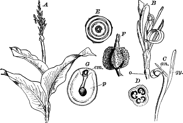
Fig. 88.—Scitamineæ. A, upper part of a flowering
plant of Indian shot (Canna), much reduced in size (Cannaceæ).
B, a single flower, × ½. C, the single stamen (an.), and
petal-like pistil (gy.), × 1. D, section of the ovary, × 2. E,
diagram of the flower. The place of the missing stamens is indicated
by small circles. F, fruit, × ½. G, section of an unripe seed.
em. embryo. p, perisperm, × 2.
The plants of this order are all inhabitants of the warmer parts of
the earth, and only a very few occur within the limits of the United
States, and these confined to the extreme south. They are extremely
showy plants, owing to their large leaves and brilliant flowers, and
for this reason are cultivated extensively. Various species of Canna
(Fig. 88) are common in gardens, where they are prized for their
large, richly-colored leaves, and clusters of scarlet, orange, or
yellow flowers. The leafy stems arise from thick tubers or root
stocks, and grow rapidly to a height of two metres or more in the
larger species. The leaves, as in all the order, are very large, and
have a thick midrib with lateral veins running to the margin. The
young leaves are folded up like a trumpet. The flowers are irregular
in form, and in Canna only a single stamen is found; or if more are
present, they are reduced to petal-like rudiments. The single, perfect
stamen (Fig. 88, C, an.) has the filament broad and colored like
the petals, and the anther attached to one side. The pistil (gy.) is
also petal-like. There are three circles of leaves forming the
perigone, the two outer being more or less membranaceous, and only the
three inner petal-like in texture. The ovary (o) is inferior, and
covered on the outside with little papillæ that afterward form short
spines on the outside of the fruit (F).
The seeds are large, but the embryo is very small. A section of a
nearly ripe seed shows the embryo (em.) occupying the upper part of
the embryo sac which does not nearly fill the seed and contains no
endosperm. The bulk of the seed is derived from the tissue of the body
of the ovule, which in most seeds becomes entirely obliterated by the
growth of the embryo sac. The cells of this tissue become filled with
starch, and serve the same purpose as the endosperm of other seeds.
This tissue is called “perisperm.”
Of food plants belonging to this order, the banana (Musa) is much
the most important. Others of more or less value are species of
arrowroot (Maranta) and ginger (Zingiber).
There are three families: I. Musaceæ (banana family);
II. Zingiberaceæ (ginger family); and III. Cannaceæ (Canna,
Maranta).
Order VI.—Gynandræ.
By far the greater number of the plants of this order belong to the
orchis family (Orchideæ), the second family of the order
(Apostasieæ), being a small one and unrepresented in the United
States. The orchids are in some respects the most highly specialized
of all flowers, and exhibit wonderful variety in the shape and color
of the flowers, which are often of extraordinary beauty, and show
special contrivances for cross-fertilization that are without parallel
among flowering plants.
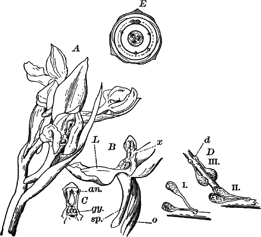
Fig. 89.—Gynandræ. A, inflorescence of the showy
orchis (Orchis spectabilis), × 1 (Orchideæ). B, a single flower,
with the upper leaves of the perianth turned back to show the column
(x). sp. the spur attached to the lower petal or lip. o, the
ovary, × 1. C, the column seen from in front. an. the stamen.
gy. the stigmatic surface, × 1. D, the two pollen masses attached
to a straw, which was inserted into the flower, by means of the viscid
disc (d): i, the masses immediately after their withdrawal; ii, iii,
the same a few minutes later, showing the change in position. E,
diagram of the flower; the position of the missing stamens indicated
by small circles.
The flowers are always more or less bilaterally symmetrical
(zygomorphic). The ovary is inferior, and usually twisted so as to
turn the flower completely around. There are two sets of perigone
leaves, three in each, and these are usually much alike except the
lower (through the twisting of the ovary) of the inner set. This
petal, known as the “lip” or “labellum,” is usually larger than the
others, and different in color, as well as being frequently of
peculiar shape. In many of them it is also prolonged backward in a
hollow spur (see Fig. 89, B). In all of the orchids except the
lady’s-slippers (Cypripedium) (Fig. 90, B), only one perfect
stamen is developed, and this is united with the three styles to form
a special structure known, as the “column” or “gynostemium” (Fig. 89,
B, C). The pollen spores are usually aggregated into two or four
waxy masses (“pollinia,” sing. pollinium), which usually can only be
removed by the agency of insects upon which all but a very few orchids
are absolutely dependent for the pollination of the flowers.
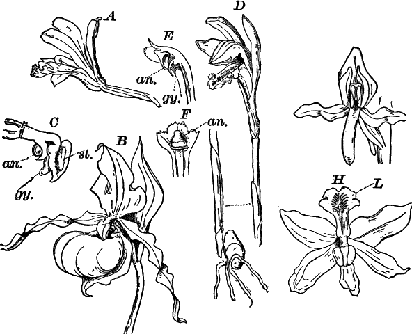
Fig. 90.—Forms of Orchideæ. A, putty-root
(Aplectrum), × 1. B, yellow lady’s-slipper (Cypripedium), × ½.
C, the column of the same, × 1. an. one of the two perfect
stamens. st. sterile, petal-like stamen. gy.. stigma. D,
Arethusa, × ½. E, section of the column, × 1: an. stamen. gy.
stigma. F, the same, seen from in front. G, Habenaria, × 1. H,
Calopogon, × 1. In the last the ovary is not twisted, so that the
lip (L) lies on the upper side of the flower.
In the lady-slippers there are two fertile stamens, and a third
sterile one has the form of a large triangular shield terminating the
column (Fig. 90, C, st.).
The ovules of the orchids are extremely small, and are only partly
developed at the time the flower opens, the pollen tube growing very
slowly and the ovules maturing as it grows down through the tissues of
the column. The ripe seeds are excessively numerous, but so fine as to
look like dust.
The orchids are mostly small or moderate-sized plants, few of them
being more than a metre or so in height. All of our native species,
with the exception of a few from the extreme south, grow from fibrous
roots or tubers, but many tropical orchids, as is well known, are
“epiphytes”; that is, they grow upon the trunks and branches of trees.
One genus, Vanilla, is a twining epiphyte; the fruit of this plant
furnishes the vanilla of commerce. Aside from this plant, the
economical value of the orchids is small, although a few of them are
used medicinally, but are not specially valuable.
Of the five thousand species known, the great majority are inhabitants
of the tropics, but nevertheless there are within the United States a
number of very beautiful forms. The largest and showiest are the
lady’s-slippers, of which we have six species at the north. The most
beautiful is the showy lady’s-slipper (Cypripedium spectabile),
whose large, pink and white flowers rival in beauty many of the
choicest tropical orchids. Many of the Habenarias, including the
yellow and purple fringed orchids, are strikingly beautiful as are the
Arethuseæ (Arethusa, Pogonia, Calopogon). The last of these
(Fig. 90, H) differs from all our other native orchids in having the
ovary untwisted so that the labellum lies on the upper side of the
flower.
A number of the orchids are saprophytic, growing in soil rich in
decaying vegetable matter, and these forms are often nearly or quite
destitute of chlorophyll, being brownish or yellowish in color, and
with rudimentary leaves. The coral roots (Corallorhiza), of which
there are several species, are examples of these, and another closely
related form, the putty-root (Aplectrum) (Fig. 90, A), has the
flowering stems like those of Corallorhiza, but there is a single,
large, plaited leaf sent up later.
Order VII.—Helobiæ.
The last order of the monocotyledons is composed of marsh or water
plants, some of which recall certain of the dicotyledons. Of the three
families, the first, Juncagineæ, includes a few inconspicuous plants
with grass-like or rush-like leaves, and small, greenish or yellowish
flowers (e.g. arrow-grass, Triglochin).
The second family (Alismaceæ) contains several large and showy
species, inhabitants of marshes. Of these the water-plantain
(Alisma), a plant with long-stalked, oval, ribbed leaves, and a
much-branched panicle of small, white flowers, is very common in
marshes and ditches, and the various species of arrowhead
(Sagittaria) are among the most characteristic of our marsh plants.
The flowers are unisexual; the female flowers are usually borne at the
base of the inflorescence, and the male flowers above. The gynœcium
(Fig. 91, B) consists of numerous, separate carpels attached to a
globular receptacle. The sepals are green and much smaller than the
white petals. The leaves (F) are broad, and, besides the thickened,
parallel veins, have numerous smaller ones connecting these.
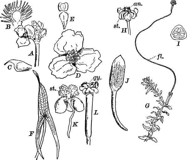
Fig. 91.—Types of Helobiæ. A, inflorescence of
arrowhead (Sagittaria), with a single female flower, × ½
(Alismaceæ). B, section through the gynœcium, showing the numerous
single carpels, × 3. C, a ripe fruit, × 3. D, a male flower, × 1.
E, a single stamen, × 3. F, a leaf of Sagittaria variabilis,
× ⅙. G, ditch-moss (Elodea), with a female flower (fl.), × ½.
(Hydrocharideæ). H, the flower, × 2. an. the rudimentary
stamens. st. the stigma. I, cross-section of the ovary, × 4. J,
male inflorescence of eel-grass (Vallisneria), × 1. K, a single
expanded male flower, × 12. st. the stamen. L, a female flower,
× 1. gy. the stigma.
The last family is the Hydrocharideæ. They are submersed aquatics,
or a few of them with long-stalked, floating leaves. Two forms, the
ditch-moss (Elodea) (Fig. 91, G, I) and eel-grass
(Vallisneria) are very common in stagnant or slow-running water. In
both of these the plants are completely submersed, but there is a
special arrangement for bringing the flowers to the surface of the
water. Like the arrowhead, the flowers are unisexual, but borne on
different plants. The female flowers (H, L) are comparatively
large, especially in Vallisneria, and are borne on long stalks, by
means of which they reach the surface of the water, where they expand
and are ready for pollination. The male flowers (Fig. 91, J, K)
are extremely small and borne, many together, surrounded by a
membranous envelope, the whole inflorescence attached by a short
stalk. When the flowers are ready to open, they break away from their
attachment, and the envelope opens, allowing them to escape, and they
immediately rise to the surface where they expand and collect in great
numbers about the open female flowers. Sometimes these are so abundant
during the flowering period (late in summer) that the surface of the
water looks as if flour had been scattered over it. After pollination
is effected, the stem of the female flower coils up like a spring,
drawing the flower beneath the water where the fruit ripens.
The cells of these plants show very beautifully the circulation of the
protoplasm, the movement being very marked and continuing for a long
time under the microscope. To see this the whole leaf of Elodea, or
a section of that of Vallisneria, may be used.
CHAPTER XVII.
DICOTYLEDONS.
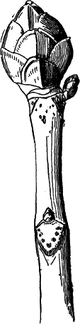
Fig. 92.—End of a branch of a horsechestnut in winter,
showing the buds covered by the thick, brown scale leaves, × 1.
The second sub-class of the angiosperms, the dicotyledons, receive
their name from the two opposite seed leaves or cotyledons with which
the young plant is furnished. These leaves are usually quite different
in shape from the other leaves, and not infrequently are very thick
and fleshy, filling nearly the whole seed, as may be seen in a bean or
pea. The number of the dicotyledons is very large, and very much the
greater number of living spermaphytes belong to this group. They
exhibit much greater variety in the structure of the flowers than the
monocotyledons, and the leaves, which in the latter are with few
exceptions quite uniform in structure, show here almost infinite
variety. Thus the leaves may be simple (undivided); e.g. oak, apple;
or compound, as in clover, locust, rose, columbine, etc. The leaves
may be stalked or sessile (attached directly to the stem), or even
grown around the stem, as in some honeysuckles. The edges of the
leaves may be perfectly smooth (“entire”), or they may be variously
lobed, notched, or wavy in many ways. As many of the dicotyledons are
trees or shrubs that lose their leaves annually, special leaves are
developed for the protection of the young leaves during the winter.
These have the form of thick scales, and often are provided with
glands secreting a gummy substance which helps render them
water-proof. These scales are best studied in trees with large, winter
buds, such as the horsechestnut (Fig. 92), hickory, lilac, etc. On
removing the hard, scale leaves, the delicate, young leaves, and often
the flowers, may be found within the bud. If we examine a young shoot
of lilac or buckeye, just as the leaves are expanding in the spring, a
complete series of forms may be seen from the simple, external scales,
through immediate forms, to the complete foliage leaf. The veins of
the leaves are almost always much-branched, the veins either being
given off from one main vein or midrib (feather-veined or
pinnate-veined), as in an apple leaf, or there may be a number of
large veins radiating from the base of the leaf, as in the scarlet
geranium or mallow. Such leaves are said to be palmately veined.
Some of them are small herbaceous plants, either upright or prostrate
upon the ground, over which they may creep extensively, becoming
rooted at intervals, as in the white clover, or sending out special
runners, as is seen in the strawberry. Others are woody stemmed
plants, persisting from year to year, and often becoming great trees
that live for hundreds of years. Still others are climbing plants,
either twining their stems about the support, like the morning-glory,
hop, honeysuckle, and many others, or having special organs (tendrils)
by which they fasten themselves to the support. These tendrils
originate in different ways. Sometimes, as in the grape and Virginia
creeper, they are reduced branches, either coiling about the support,
or producing little suckers at their tips by which they cling to walls
or the trunks of trees. Other tendrils, as in the poison ivy and the
true ivy, are short roots that fasten themselves firmly in the
crevices of bark or stones. Still other tendrils, as those of the
sweet-pea and clematis, are parts of the leaf.
The stems may be modified into thorns for protection, as we see in
many trees and shrubs, and parts of leaves may be similarly changed,
as in the thistle. The underground stems often become much changed,
forming bulbs, tubers, root stocks, etc. much as in the
monocotyledons. These structures are especially found in plants which
die down to the ground each year, and contain supplies of nourishment
for the rapid growth of the annual shoots.
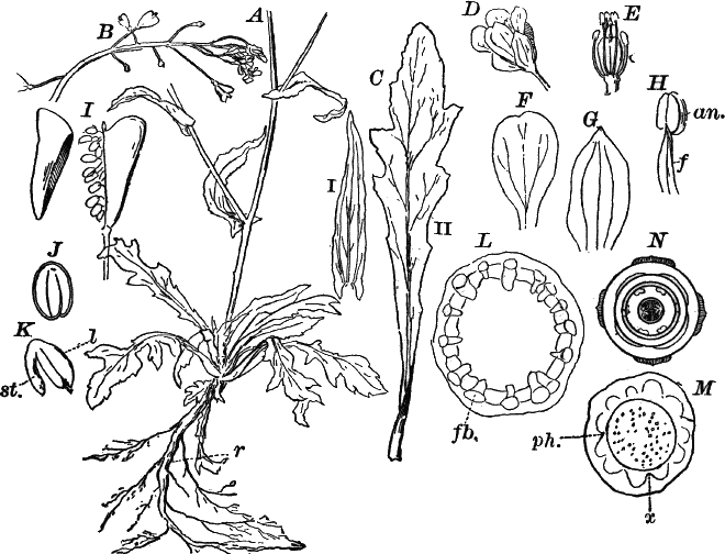
Fig. 93.—A, base of a plant of shepherd’s-purse
(Capsella bursa-pastoris), × ½. r, the main root. B, upper part
of the inflorescence, × 1. C, two leaves: i, from the upper part;
ii, from the base of the plant, × 1. D, a flower, × 3. E, the
same, with sepals and petals removed, × 3. F, petal. G, sepal.
H, stamen, × 10. f, filament. an. anther. I, a fruit with one
of the valves removed to show the seeds, × 4. J, longitudinal
section of a seed, × 8. K, the embryo removed from the seed, × 8.
l, the first leaves (cotyledons). st. the stem ending in the root.
L, cross-section of the stem, × 20. fb. fibro-vascular bundle.
M, a similar section of the main root, × 15. N, diagram of the
flower.
The structure of the tissues, and the peculiarities of the flower and
fruit, will be better understood by a somewhat careful examination of
a typical dicotyledon, and a comparison with this of examples of the
principal orders and families.
One of the commonest of weeds, and at the same time one of the most
convenient plants for studying the characteristics of the
dicotyledons, is the common shepherd’s-purse (Capsella
bursa-pastoris) (Figs. 93–95).
The plant grows abundantly in waste places, and is in flower nearly
the year round, sometimes being found in flower in midwinter, after a
week or two of warm weather. It is, however, in best condition for
study in the spring and early summer. The plant may at once be
recognized by the heart-shaped pods and small, white, four-petaled
flowers. The plant begins to flower when very small, but continues to
grow until it forms a much-branching plant, half a metre or more in
height. On pulling up the plant, a large tap-root (Fig. 93, A, r)
is seen, continuous with the main stem above ground. The first root of
the seedling plant continues here as the main root of the plant, as
was the case with the gymnosperms, but not with the monocotyledons.
From this tap-root other small ones branch off, and these divide
repeatedly, forming a complex root system. The main root is very tough
and hard, owing to the formation of woody tissue in it. A
cross-section slightly magnified (Fig. 93, M), shows a round,
opaque, white, central area (x), the wood, surrounded by a more
transparent, irregular ring (ph.), the phloem or bast; and outside
of this is the ground tissue and epidermis.
The lower leaves are crowded into a rosette, and are larger than those
higher up, from which they differ also in having a stalk (petiole),
while the upper leaves are sessile. The outline of the leaves varies
much in different plants and in different parts of the same plant,
being sometimes almost entire, sometimes divided into lobes almost to
the midrib, and between these extremes all gradations are found. The
larger leaves are traversed by a strong midrib projecting strongly on
the lower side of the leaf, and from this the smaller veins branch.
The upper leaves have frequently two smaller veins starting from the
base of the leaf, and nearly parallel with the midrib (C i). The
surface of the leaves is somewhat roughened with hairs, some of which,
if slightly magnified, look like little white stars.
Magnifying slightly a thin cross-section of the stem, it shows a
central, ground tissue (pith), whose cells are large enough to be seen
even when very slightly enlarged. Surrounding this is a ring of
fibro-vascular bundles (L, fb.), appearing white and opaque, and
connected by a more transparent tissue. Outside of the ring of
fibro-vascular bundles is the green ground tissue and epidermis.
Comparing this with the section of the seedling pine stem, a
resemblance is at once evident, and this arrangement was also noticed
in the stem of the horse-tail.
Branches are given off from the main stem, arising at the point where
the leaves join the stem (axils of the leaves), and these may in turn
branch. All the branches terminate finally in an elongated
inflorescence, and the separate flowers are attached to the main axis
of the inflorescence by short stalks. This form of inflorescence is
known technically as a “raceme.” Each flower is really a short branch
from which the floral leaves arise in precisely the same way as the
foliage leaves do from the ordinary branches. There are five sets of
floral leaves: I. four outer perigone leaves (sepals) (F), small,
green, pointed leaves traversed by three simple veins, and together
forming the calyx; II. four larger, white, inner perigone leaves
(petals) (G), broad and slightly notched at the end, and tapering to
the point of attachment. The petals collectively are known as the
“corolla.” The veins of the petals fork once; III. and IV. two sets of
stamens (E), the outer containing two short, and the inner, four
longer ones arranged in pairs. Each stamen has a slender filament
(H, f) and a two-lobed anther (an.). The innermost set consists
of two carpels united into a compound pistil. The ovary is oblong,
slightly flattened so as to be oval in section, and divided into two
chambers. The style is very short and tipped by a round, flattened
stigma.
The raceme continues to grow for a long time, forming new flowers at
the end, so that all stages of flowers and fruit may often be found in
the same inflorescence.
The flowers are probably quite independent of insect aid in
pollination, as the stamens are so placed as to almost infallibly shed
their pollen upon the stigma. This fact, probably, accounts for the
inconspicuous character of the flowers.
After fertilization is effected, and the outer floral leaves fall off,
the ovary rapidly enlarges, and becomes heart-shaped and much
flattened at right angles to the partition. When ripe, each half falls
away, leaving the seeds attached by delicate stalks (funiculi, sing.
funiculus) to the edges of the membranous partition. The seeds are
small, oval bodies with a shining, yellow-brown shell, and with a
little dent at the end where the stalk is attached. Carefully dividing
the seed lengthwise, or crushing it in water so as to remove the
embryo, we find it occupies the whole cavity of the seed, the young
stalk (st.) being bent down against the back of one of the
cotyledons (f).
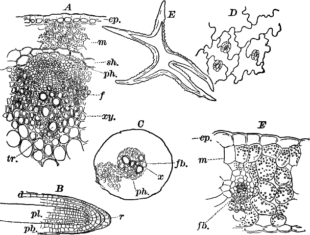
Fig. 94.—A, cross-section of the stem of the
shepherd’s-purse, including a fibro-vascular bundle, × 150. ep.
epidermis. m, ground tissue. sh. bundle sheath. ph. phloem.
xy. xylem. tr. a vessel. B, a young root seen in optical
section, × 150. r, root cap. d, young epidermis. pb. ground.
pl. young fibro-vascular bundle. C cross section of a small root,
× 150. fb. fibro-vascular bundle. D, epidermis from the lower side
of the leaf, × 150. E, a star-shaped hair from the surface of the
leaf, × 150. F, cross-section of a leaf, × 150. ep. epidermis.
m, ground tissue. fb. section of a vein.
A microscopic examination of a cross-section of the older root shows
that the central portion is made up of radiating lines of thick-walled
cells (fibres) interspersed with lines of larger, round openings
(vessels). There is a ring of small cambium cells around this merging
into the phloem, which is composed of irregular cells, with pretty
thick, but soft walls. The ground tissue is composed of large, loose
cells, which in the older roots are often ruptured and partly dried
up. The epidermis is usually indistinguishable in the older roots. To
understand the early structure of the roots, the smallest rootlets
obtainable should be selected. The smallest are so transparent that
the tips may be mounted whole in water, and will show very
satisfactorily the arrangement of the young tissues. The tissues do
not here arise from a single, apical cell, as we found in the
pteridophytes, but from a group of cells (the shaded cells in Fig. 94,
B). The end of the root, as in the fern, is covered with a root cap
(r) composed of successive layers of cells cut off from the growing
point. The rest of the root shows the same division of the tissues
into the primary epidermis (dermatogen) (d), young fibro-vascular
cylinder (plerome) (pl.), and young ground tissue (periblem)
(pb.). The structure of the older portions of such a root is
not very easy to study, owing to difficulty in making good
cross-sections of so small an object. By using a very sharp razor, and
holding perfectly straight between pieces of pith, however,
satisfactory sections can be made. The cells contain so much starch as
to make them almost opaque, and potash should be used to clear them.
The fibro-vascular bundle is of the radial type, there being two
masses of xylem (xy.) joined in the middle, and separating the two
phloem masses (ph.), some of whose cells are rather thicker walled
than the others. The bundle sheath is not so plain here as in the
fern. The ground tissue is composed of comparatively large cells with
thickish, soft walls, that contain much starch. The epidermis usually
dies while the root is still young. In the larger roots the early
formation of the cambium ring, and the irregular arrangement of the
tissues derived from its growth, soon obliterate all traces of the
primitive arrangement of the tissues. Making a thin cross-section of
the stem, and magnifying strongly, we find bounding the section a
single row of epidermal cells (Fig. 94, A, ep.) whose walls,
especially the outer ones, are strongly thickened. Within these are
several rows of thin-walled ground-tissue cells containing numerous
small, round chloroplasts. The innermost row of these cells (sh.)
are larger and have but little chlorophyll. This row of cells forms a
sheath around the ring of fibro-vascular bundles very much as is the
case in the horse-tail. The separate bundles are nearly triangular in
outline, the point turned inward, and are connected with each other by
masses of fibrous tissue (f), whose thickened walls have a peculiar,
silvery lustre. Just inside of the bundle sheath there is a row of
similar fibres marking the outer limit of the phloem (ph.). The rest
of the phloem is composed of very small cells. The xylem is composed
of fibrous cells with yellowish walls and numerous large vessels
(tr.). The central ground tissue (pith) has large, thin-walled cells
with numerous intercellular spaces, as in the stem of Erythronium.
Some of these cells contain a few scattered chloroplasts in the very
thin, protoplasmic layer lining their walls, but the cells are almost
completely filled with colorless cell sap.A longitudinal section shows that the epidermal cells are much
elongated, the cells of the ground tissue less so, and in both the
partition walls are straight. In the fibrous cells, both of the
fibro-vascular bundle and those lying between, the end walls are
strongly oblique. The tracheary tissue of the xylem is made up of
small, spirally-marked vessels, and larger ones with thickened rings
or with pits in the walls. The small, spirally-marked vessels are
nearest the centre, and are the first to be formed in the young
bundle.The epidermis of the leaves is composed of irregular cells with wavy
outlines like those of the ferns. Breathing pores, of the same type as
those in the ferns and monocotyledons, are found on both surfaces, but
more abundant and more perfectly developed on the lower surface of the
leaf. Owing to their small size they are not specially favorable for
study. The epidermis is sparingly covered with unicellular hairs, some
of which are curiously branched, being irregularly star-shaped. The
walls of these cells are very thick, and have little protuberances
upon the outer surface (Fig. 93, E).Cross-sections of the leaf may be made between pith as already
directed; or, by folding the leaf carefully several times, the whole
can be easily sectioned. The structure is essentially as in the
adder-tongue, but the epidermal cells appear more irregular, and the
fibro-vascular bundles are better developed. They are like those of
the stem, but somewhat simpler. The xylem lies on the upper side.The ground tissue is composed, as in the leaves we have studied, of
chlorophyll-bearing, loose cells, rather more compact upon the upper
side. (In the majority of dicotyledons the upper surface of the leaves
is nearly or quite destitute of breathing pores, and the cells of the
ground tissue below the upper epidermis are closely packed, forming
what is called the “palisade-parenchyma” of the leaf.)
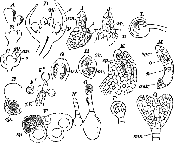
Fig. 95.—A–D, successive stages in the development
of the flower of Capsella, × 50. A, surface view. B–D, optical
sections. s, sepals, p, petals. an. stamens. gy. pistil. E,
cross-section of the young anther, × 180. sp. spore mother cells.
F, cross-section of full-grown anther. sp. pollen spores, × 50.
Fʹ, four young pollen spores, × 300. Fʺ, pollen spores germinating
upon the stigma, × 300. pt. pollen tube. G, young pistil in
optical section, × 25. H, cross-section of a somewhat older one. ov.
ovules. I–L, development of the ovule. sp. embryo sac
(macrospore). I–K, × 150. L, × 50. M, embryo sac of a full-grown
ovule, × 150. Sy. Synergidæ. o, egg cell. n, endosperm
nucleus. ant. antipodal cells. N–Q, development of the embryo,
× 150. sus. suspensor.
The shepherd’s-purse is an admirable plant for the study of the
development of the flower which is much the same in other angiosperms.
To study this, it is only necessary to teaze out, in a drop of water,
the tip of a raceme, and putting on a cover glass, examine with a
power of from fifty to a hundred diameters. In the older stages it is
best to treat with potash, which will render the young flowers quite
transparent. The young flower (Fig. 95, A) is at first a little
protuberance composed of perfectly similar small cells filled with
dense protoplasm. The first of the floral leaves to appear are the
sepals which very early arise as four little buds surrounding the
young flower axis (Fig. 95, A, B). The stamens (C, an.) next
appear, being at first entirely similar to the young sepals. The
petals do not appear until the other parts of the flower have reached
some size, and the first tracheary tissue appears in the
fibro-vascular bundle of the flower stalk (D). The carpels are more
or less united from the first, and form at first a sort of shallow cup
with the edges turned in (D, gy.). This cup rapidly elongates, and
the cavity enlarges, becoming completely closed at the top where the
short style and stigma develop. The ovules arise in two lines on the
inner face of each carpel, and the tissue which bears them (placenta)
grows out into the cavity of the ovary until the two placentæ meet in
the middle and form a partition completely across the ovary (Fig. 95,
H).The stamens soon show the differentiation into filament and anther,
but the former remains very short until immediately before the flowers
are ready to open. The anther develops four sporangia (pollen sacs),
the process being very similar to that in such pteridophytes as the
club mosses. Each sporangium (Fig. E, F) contains a central mass
of spore mother cells, and a wall of three layers of cells. The spore
mother cells finally separate, and the inner layer of the wall cells
becomes absorbed much as we saw in the fern, and the mass of mother
cells thus floats free in the cavity of the sporangium. Each one now
divides in precisely the same way as in the ferns and gymnosperms,
into four pollen spores. The anther opens as described for
Erythronium.By carefully picking to pieces the young ovaries, ovules in all stages
of development may be found, and on account of their small size and
transparency, show beautifully their structure. Being perfectly
transparent, it is only necessary to mount them in water and cover.The young ovule (I, J) consists of a central, elongated body
(nucellus), having a single layer of cells enclosing a large central
cell (the macrospore or embryo sac) (sp.). The base of the nucellus
is surrounded by two circular ridges (i, ii) of which the inner is at
first higher than the outer one, but later (K, L), the latter
grows up above it and completely conceals it as well as the nucellus.
One side of the ovule grows much faster than the other, so that it is
completely bent upon itself, and the opening between the integuments
is brought close to the base of the ovule (Fig. 95, L). This opening
is called the “micropyle,” and allows the pollen tube to enter.The full-grown embryo sac shows the same structure as that already
described in Monotropa (page 276), but as the walls of the
full-grown ovule are thicker here, its structure is rather difficult
to make out. The ripe stigma is covered with little papillæ (Fig. 95,
F) that hold the pollen spores which may be found here sending out
the pollen tube. By carefully opening the ovary and slightly crushing
it in a drop of water, the pollen tube may sometimes be seen growing
along the stalk of the ovule until it reaches and enters the
micropyle.To study the embryo a series of young fruits should be selected, and
the ovules carefully dissected out and mounted in water, to which a
little caustic potash has been added. The ovule will be thus rendered
transparent, and by pressing gently on the cover glass with a needle
so as to flatten the ovule slightly, there is usually no trouble in
seeing the embryo lying in the upper part of the embryo sac, and by
pressing more firmly it can often be forced out upon the slide. The
potash should now be removed as completely as possible with blotting
paper, and pure water run under the cover glass.The fertilized egg cell first secretes a membrane, and then divides
into a row of cells (N) of which the one nearest the micropyle is
often much enlarged. The cell at the other end next enlarges and
becomes divided by walls at right angles to each other into eight
cells. This globular mass of cells, together with the cell next to it,
is the embryo plant, the row of cells to which it is attached taking
no further part in the process, and being known as the “suspensor.”
Later the embryo becomes indented above and forms two lobes (Q),
which are the beginnings of the cotyledons. The first root and the
stem arise from the cells next the suspensor.
CHAPTER XVIII.
CLASSIFICATION OF DICOTYLEDONS.
Division I.—Choripetalæ.
Nearly all of the dicotyledons may be placed in one of two great
divisions distinguished by the character of the petals. In the first
group, called Choripetalæ, the petals are separate, or in some
degenerate forms entirely absent. As familiar examples of this group,
we may select the buttercup, rose, pink, and many others.
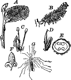
Fig. 96.—Iulifloræ. A, male; B, female
inflorescence of a willow, Salix (Amentaceæ), × ½. C, a single
male flower, × 2. D, a female flower, × 2. E, cross-section of the
ovary, × 8. F, an opening fruit. G, single seed with its hairy
appendage, × 2.
The second group (Sympetalæ or Gamopetalæ) comprises those
dicotyledons whose flowers have the petals more or less completely
united into a tube. The honeysuckles, mints, huckleberry, lilac, etc.,
are familiar representatives of the Sympetalæ, which includes the
highest of all plants.
The Choripetalæ may be divided into six groups, including twenty-two
orders. The first group is called Iulifloræ, and contains numerous,
familiar plants, mostly trees. In these plants, the flowers are small
and inconspicuous, and usually crowded into dense catkins, as in
willows (Fig. 96) and poplars, or in spikes or heads, as in the
lizard-tail (Fig. 97, G), or hop (Fig. 97, I). The individual
flowers are very small and simple in structure, being often reduced to
the gynœcium or andræcium, carpels and stamens being almost always in
separate flowers. The outer leaves of the flower (sepals and petals)
are either entirely wanting or much reduced, and never differentiated
into calyx and corolla.
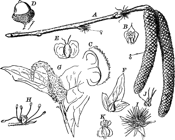
Fig. 97.—Types of Iulifloræ. A, branch of hazel,
Corylus (Cupuliferæ), × 1. ♂, male; ♀, female inflorescence. B,
a single male flower, × 3. C, section of the ovary of a female
flower, × 25. D, acorn of red oak, Quercus (Cupuliferæ), × ½.
E, seed of white birch, Betula (Betulaceæ), × 3. F, fruit of
horn-bean, Carpinus (Cupuliferæ), × 1. G, lizard-tail, Saururus
(Saurureæ), × ¼. H, a single flower, × 2. I, female
inflorescence of the hop, Humulus (Cannabineæ), × 1. J, a single
scale with two flowers, × 1. K, a male flower of a nettle, Urtica
(Urticaceæ), × 5.
In the willows (Fig. 96) the stamens are bright-colored, so that the
flowers are quite showy, and attract numerous insects which visit them
for pollen and nectar, and serve to carry the pollen to the pistillate
flowers, thus insuring their fertilization. In the majority of the
group, however, the flowers are wind-fertilized. An excellent example
of this is seen in the common hazel (Fig. 97, A). The male flowers
are produced in great numbers in drooping catkins at the ends of the
branches, shedding the pollen in early spring before the leaves
unfold. The female flowers are produced on the same branches, but
lower down, and in much smaller numbers. The stigmas are long, and
covered with minute hairs that catch the pollen which is shaken out
in clouds every time the plant is shaken by the wind, and falls in a
shower over the stigmas. A similar arrangement is seen in the oaks,
hickories, and walnuts.
There are three orders of the Iulifloræ: Amentaceæ, Piperineæ,
and Urticinæ. The first contains the birches (Betulaceæ); oaks,
beeches, hazels, etc. (Cupuliferæ); walnuts and hickories
(Juglandeæ); willows and poplars (Salicaceæ). They are all trees
or shrubs; the fruit is often a nut, and the embryo is very large,
completely filling it.
The Piperineæ are mostly tropical plants, and include the pepper
plant (Piper), as well as other plants with similar properties. Of
our native forms, the only common one is the lizard-tail (Saururus),
not uncommon in swampy ground. In these plants, the calyx and corolla
are entirely absent, but the flowers have both carpels and stamens
(Fig. 97, H).
The Urticinæ include, among our common plants, the nettle family
(Urticaceæ); plane family (Plataneæ), represented by the sycamore
or buttonwood (Platanus); the hemp family (Cannabineæ); and the
elm family (Ulmaceæ). The flowers usually have a calyx, and may
have only stamens or carpels, or both. Sometimes the part of the stem
bearing the flowers may become enlarged and juicy, forming a
fruit-like structure. Well-known examples of this are the fig and
mulberry.
The second group of the Choripetalæ is called Centrospermæ, and
includes but a single order comprising seven families, all of which,
except one (Nyctagineæ), are represented by numerous native species.
The latter comprises mostly tropical plants, and is represented in our
gardens by the showy “four-o’clock” (Mirabilis). In this plant, as
in most of the order, the corolla is absent, but here the calyx is
large and brightly colored, resembling closely the corolla of a
morning-glory or petunia. The stamens are usually more numerous than
the sepals, and the pistil, though composed of several carpels, has,
as a rule, but a single cavity with the ovules arising from the base,
though sometimes the ovary is several celled.
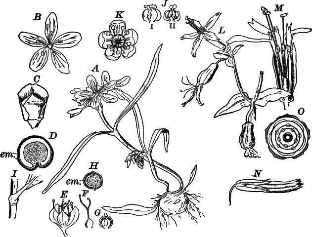
Fig. 98.—Types of Centrospermæ. A, plant of
spring-beauty, Claytonia (Portulacaceæ), × ½. B, a single
flower, × 1. C, fruit, with the sepals removed, × 2. D, section of
the seed, showing the curved embryo (em.), × 5. E, single flower
of smart-weed, Polygonum (Polygonaceæ), × 2. F, the pistil, × 2.
G, section of the ovary, showing the single ovule, × 4. H, section
of the seed, × 2. I, base of the leaf, showing the sheath, × 1. J,
flower of pig-weed, Chenopodium (Chenopodiaceæ), × 3: i, from
without; ii, in section. K, flower of the poke-weed, Phytolacca
(Phytolaccaceæ), × 2. L, fire-pink, Silene (Caryophyllaceæ),
× ½. M, a flower with half of the calyx and corolla removed, × 1.
N, ripe fruit of mouse-ear chick-weed, Cerastium
(Caryophyllaceæ), opening by ten teeth at the summit, × 2. O,
diagram of the flower of Silene.
The first family (Polygoneæ) is represented by the various species
of Polygonum (knotgrass, smart-weed, etc.), and among cultivated
plants by the buckwheat (Fagopyrum). The goose-foot or pig-weed
(Chenopodium) among native plants, and the beet and spinach of the
gardens are examples of the family Chenopodiaceæ. Nearly resembling
the last is the amaranth family (Amarantaceæ), of which the showy
amaranths and coxcombs of the gardens, and the coarse, green amaranth
or pig-weed are representatives.
The poke-weed (Phytolacca) (Fig. 98, K), so conspicuous in autumn
on account of its dark-purple clusters of berries and crimson stalks,
is our only representative of the family Phytolaccaceæ. The two
highest families are the purslane family (Portulacaceæ) and pink
family (Caryophylleæ). These are mostly plants with showy flowers in
which the petals are large and conspicuous, though some of the pink
family, e.g. some chick-weeds, have no petals. Of the purslane
family the portulacas of the gardens, and the common purslane or
“pusley,” and the spring-beauty (Claytonia) (Fig. 98, A) are the
commonest examples. The pink family is represented by many common and
often showy plants. The carnation, Japanese pinks, and sweet-william,
all belonging to the genus Dianthus, of which there are also two or
three native species, are among the showiest of the family. The genera
Lychnis and Silene (Fig. 98, L) also contain very showy species.
Of the less conspicuous genera, the chick-weeds (Cerastium and
Stellaria) are the most familiar.
The third group of the Choripetalæ (the Aphanocyclæ) is a very
large one and includes many common plants distributed among five
orders. The lower ones have all the parts of the flower entirely
separate, and often indefinite in number; the higher have the gynœcium
composed of two or more carpels united to form a compound pistil.
The first order (Polycarpæ) includes ten families, of which the
buttercup family (Ranunculaceæ) is the most
familiar. The plants of this family show much variation in the details
of the flowers, which are usually showy, but the general plan is much
the same. In some of them, like the anemones (Fig. 99, A), clematis,
and others, the corolla is absent, but the sepals are large and
brightly colored so as to appear like petals. In the columbine
(Aquilegia) (Fig. 99, F) the petals are tubular, forming
nectaries, and in the larkspur (Fig. 99, T) one of the sepals is
similarly changed.
Representing the custard-apple family (Anonaceæ) is the curious
papaw (Asimina), common in many parts of the United States
(Fig. 100, A). The family is mainly a tropical one, but this species
extends as far north as southern Michigan.
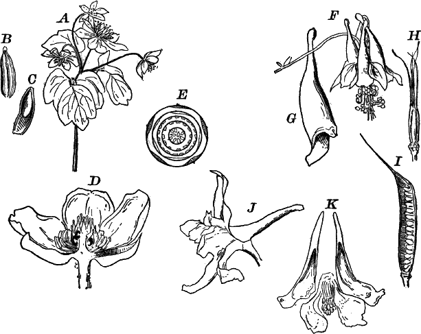
Fig. 99.—Types of Aphanocyclæ (Polycarpæ), family
Ranunculaceæ. A, Rue anemone (Anemonilla), × ½. B, a fruit,
× 2. C, section of the same. D, section of a buttercup flower
(Ranunculus), × 1½. E, diagram of buttercup flower. F, wild
columbine (Aquilegia), × ½. G, one of the spur-shaped petals, × 1.
H, the five pistils, × 1. I, longitudinal section of the fruit,
× 1. J, flower of larkspur (Delphinium), × 1. K, the four petals
and stamens, after the removal of the five colored and petal-like
sepals, × 1.
The magnolia family (Magnoliaceæ) has several common members, the
most widely distributed being, perhaps, the tulip-tree
(Liriodendron) (Fig. 100, C), much valued for its timber. Besides
this there are several species of magnolia, the most northerly species
being the sweet-bay (Magnolia glauca) of the Atlantic States, and
the cucumber-tree (M. acuminata); the great magnolia
(M. grandiflora) is not hardy in the northern states.
The sweet-scented shrub (Calycanthus) (Fig. 100, G) is the only
member of the family Calycanthaceæ found within our limits. It grows
wild in the southern states, and is cultivated for its sweet-scented,
dull, reddish flowers.
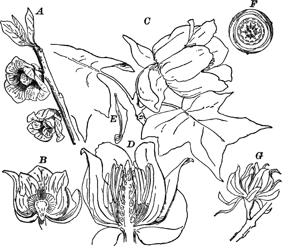
Fig. 100.—Types of Aphanocyclæ (Polycarpæ). A,
branch of papaw, Asimina (Anonaceæ), × ½. B, section of the
flower, × 1. C, flower and leaf of tulip-tree, Liriodendron
(Magnoliaceæ), × ⅓. D, section of a flower, × ½. E, a ripe
fruit, × 1. F, diagram of the flower. G, flower of the
sweet-scented shrub, Calycanthus (Calycanthaceæ), × ½
The barberry (Berberis) (Fig. 101, A) is the type of the family
Berberideæ, which also includes the curious mandrake or may-apple
(Podophyllum) (Fig. 101, D), and the twin-leaf or rheumatism-root
(Jeffersonia), whose curious seed vessel is shown in Figure 101,
G. The fruit of the barberry and may-apple are edible, but the root
of the latter is poisonous.
The curious woody twiner, moon-seed (Menispermum) (Fig. 101, I),
is the sole example in the northern states of the family Menispermeæ
to which it belongs. The flowers are diœcious, and the pistillate
flowers are succeeded by black fruits looking like grapes. The
flattened, bony seed is curiously sculptured, and has the embryo
curled up within it.
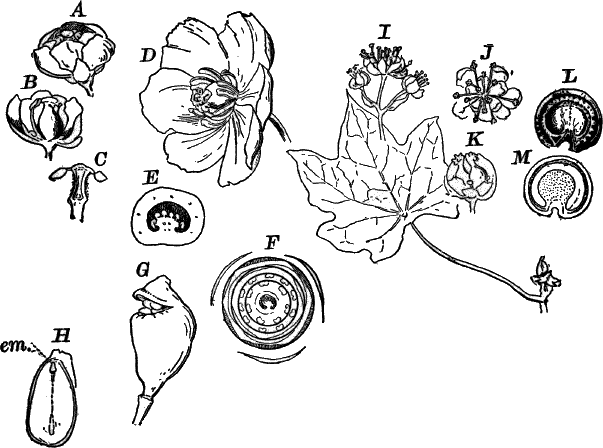
Fig. 101.—Types of Aphanocyclæ (Polycarpæ). A–H,
Berberidaceæ. A, flower of barberry (Berberis), × 2. B, the
same in section. C, a stamen, showing the method of opening, × 3.
D, flower of may-apple (Podophyllum), × ½. E, section of the
ovary of D, × 1. F, diagram of the flower. G, ripe fruit of
twin-leaf (Jeffersonia), opening by a lid, × ½. H, section of
seed, showing the embryo (em.), × 2. I, young leaf and cluster of
male flowers of moon-seed, Menispermum (Menispermeæ), × 1. J, a
single male flower, × 2. K, section of a female flower, × 2. L,
ripe seed, × 1. M, section of L, showing the curved embryo.
The last two families of the order, the laurel family (Laurineæ) and
the nutmeg family (Myristicineæ) are mostly tropical plants,
characterized by the fragrance of the bark, leaves, and fruit. The
former is represented by the sassafras and spice-bush, common
throughout the eastern United States. The latter has no members within
our borders, but is familiar to all through the common nutmeg, which
is the seed of Myristica fragrans of the East Indies. “Mace” is the
“aril” or covering of the seed of the same plant.
The second order of the Aphanocyclæ comprises a number of aquatic
plants, mostly of large size, and is known as the Hydropeltidinæ.
The flowers and leaves are usually very large, the latter usually
nearly round in outline, and frequently with the stalk inserted near
the middle. The leaves of the perigone are numerous, and sometimes
merge gradually into the stamens, as we find in the common white
water-lily (Castalia).
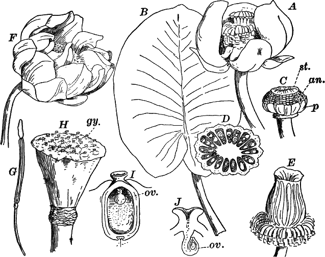
Fig. 102.—Types of Aphanocyclæ (Hydropeltidinæ).
A, yellow water-lily, Nymphæa (Nymphæaceæ), × ½. B, a leaf of
the same, × ⅙. C, freshly opened flower, with the large petal-like
sepals removed, × ½. p, petals. an. stamens. st. stigma. D,
section of the ovary, × 2. E, young fruit, × ½. F, lotus,
Nelumbo (Nelumbieæ). × ⅙. G, a stamen, × 1. H, the large
receptacle, with the separate pistils sunk in its surface, × ½. I,
section of a single pistil, × 2. ov. the ovule. J, upper part of a
section through the stigma and ovule (ov.), × 4.
There are three families, all represented within the United States.
The first (Nelumbieæ) has but a single species, the yellow lotus or
nelumbo (Nelumbo lutea), common in the waters of the west and
southwest, but rare eastward (Fig. 101, F). In this flower, the end
of the flower axis is much enlarged, looking like the rose of a
watering-pot, and has the large, separate carpels embedded in its
upper surface. When ripe, each forms a nut-like fruit which is edible.
There are but two species of Nelumbo known, the second one
(N. speciosa) being a native of southeastern Asia, and probably
found in ancient times in Egypt, as it is represented frequently in
the pictures and carvings of the ancient Egyptians. It differs mainly
from our species in the color of its flowers which are red instead of
yellow. It has recently been introduced into New Jersey where it has
become well established in several localities.
The second family (Cabombeæ) is also represented at the north by but
one species, the water shield (Brasenia), not uncommon in marshes.
Its flowers are quite small, of a dull-purple color, and the leaves
oval in outline and centrally peltate, i.e. the leaf stalk inserted
in the centre. The whole plant is covered with a transparent
gelatinous coat.
The third family (Nymphæaceæ) includes the common white water-lilies
(Castalia) and the yellow water-lilies (Nymphæa) (Fig. 102, A).
In the latter the petals are small and inconspicuous (Fig. 102, C,
p), but the sepals are large and showy. In this family the carpels,
instead of being separate, are united into a large compound pistil.
The water-lilies reach their greatest perfection in the tropics, where
they attain an enormous size, the white, blue, or red flowers of some
species being thirty centimetres or more in diameter, and the leaves
of the great Victoria regia of the Amazon reaching two metres or
more in width.
The third order of the Aphanocyclæ (Rhœadinæ or Crucifloræ)
comprises a number of common plants, principally characterized by
having the parts of the flowers in twos or fours, so that they are
more or less distinctly cross-shaped, whence the name Crucifloræ.
There are four families, of which the first is the poppy family
(Papaveraceæ), including the poppies, eschscholtzias, Mexican or
prickly poppy (Argemone), etc., of the gardens, and the blood-root
(Sanguinaria), celandine poppy (Stylophorum), and a few other wild
plants (see Fig. 103, A–I). Most of the family have a colored juice
(latex), which is white in the poppy, yellow in celandine and
Argemone, and orange-red in the blood-root. From the latex of the
opium poppy the opium of commerce is extracted.
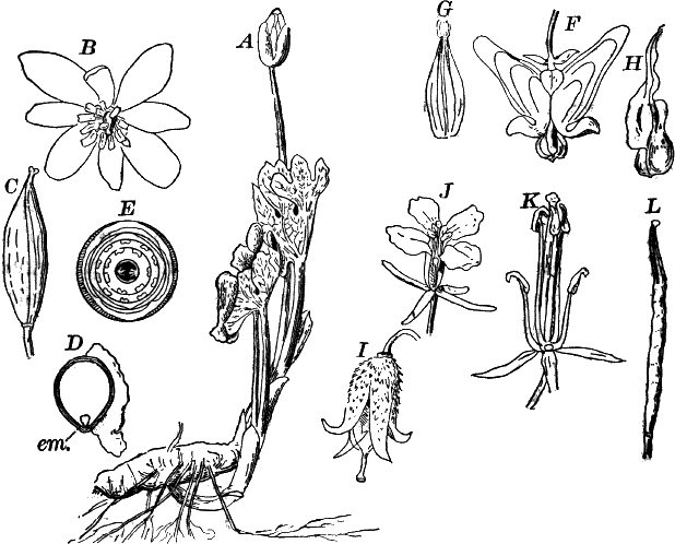
Fig. 103.—Types of Aphanocyclæ (Rhœdinæ). A,
plant of blood-root, Sanguinaria (Papaveraceæ), × ⅓. B, a single
flower, × 1. C, fruit, × ½. D, section of the seed. em. embryo,
× 2. E, diagram of the flower. F, flower of Dutchman’s breeches,
Dicentra (Fumariaceæ), × 1. G, group of three stamens of the
same, × 2. H, one of the inner petals, × 2. I, fruit of celandine
poppy, Stylophorum (Papaveraceæ), × ½. J, flower of mustard,
Brassica (Cruciferæ), × 1. K, the same, with the petals removed,
× 2. L, fruit of the same, × 1.
The second family, the fumitories (Fumariaceæ) are delicate, smooth
plants, with curious flowers and compound leaves. The garden
bleeding-heart (Dicentra spectabilis) and the pretty, wild
Dicentras (Fig. 103, F) are familiar to nearly every one.
Other examples are the mountain fringe (Adlumia), a climbing
species, and several species of Corydalis, differing mainly from
Dicentra in having the corolla one-sided.
The mustard family (Cruciferæ) comprises by far the greater part of
the order. The shepherd’s-purse, already studied, belongs here, and
may be taken as a type of the family. There is great uniformity in all
as regards the flowers, so that the classification is based mainly on
differences in the fruit and seeds. Many of the most valuable garden
vegetables, as well as a few more or less valuable wild plants, are
members of the family, which, however, includes some troublesome
weeds. Cabbages, turnips, radishes, with all their varieties, belong
here, as well as numerous species of wild cresses. A few like the
wall-flower (Cheiranthus) and stock (Matthiola) are cultivated for
ornament.
The last family is the caper family (Capparideæ), represented by
only a few not common plants. The type of the order is Capparis,
whose pickled flower-buds constitute capers.
The fourth order (Cistifloræ) of the Aphanocyclæ is a very large
one, but the majority of the sixteen families included in it are not
represented within our limits. The flowers have the sepals and petals
in fives, the stamens either the same or more numerous.
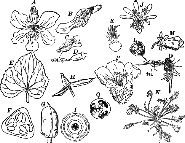
Fig. 104.—Types of Aphanocyclæ (Cistifloræ). A,
flower of wild blue violet, Viola (Violaceæ), × 1. B, the lower
petal prolonged behind into a sac or spur, × 1. C, the stamens, × 2.
D, pistil, × 2. E, a leaf, × ½. F, section of the ovary, × 2.
G, the fruit, × 1. H, the same after it has opened, × 1. I,
diagram of the flower. J, flower of mignonette, Reseda
(Resedaceæ), × 2. K, a petal, × 3. L, cross-section of the
ovary, × 3. M, fruit, × 1. N, plant of sundew, Drosera
(Droseraceæ), × ½. O, a leaf that has captured a mosquito, × 2.
P, flower of another species (D. filiformis), × 2. Q,
cross-section of the ovary, × 4.
Among the commoner members of the order are the mignonettes
(Resedaceæ) and the violets (Violaceæ), of which the various wild
and cultivated species are familiar plants (Fig. 104, A, M). The
sundews (Droseraceæ) are most extraordinary plants, growing in boggy
land over pretty much the whole world. They are represented in
the United States by several species of sundew (Drosera), and the
still more curious Venus’s-flytrap (Dionæa) of North Carolina. The
leaves of the latter are sensitive, and composed of two parts which
snap together like a steel trap. If an insect lights upon the leaf,
and touches certain hairs upon its upper surface, the two parts snap
together, holding the insect tightly. A digestive fluid is secreted by
glands upon the inner surface of the leaf, and in a short time the
captured insect is actually digested and absorbed by the leaves. The
same process takes place in the sundew (Fig. 104, N) where, however,
the mechanism is somewhat different. Here the tentacles, with which
the leaf is studded, secrete a sticky fluid which holds any small
insect that may light upon it. The tentacles now slowly bend inward
and finally the edges of the leaf as well, until the captured insect
is firmly held, when a digestive process, similar to that in Dionœa,
takes place. This curious habit is probably to be explained from the
position where the plant grows, the roots being in water where there
does not seem to be a sufficient supply of nitrogenous matter for the
wants of the plant, which supplements the supply from the bodies of
the captured insects.
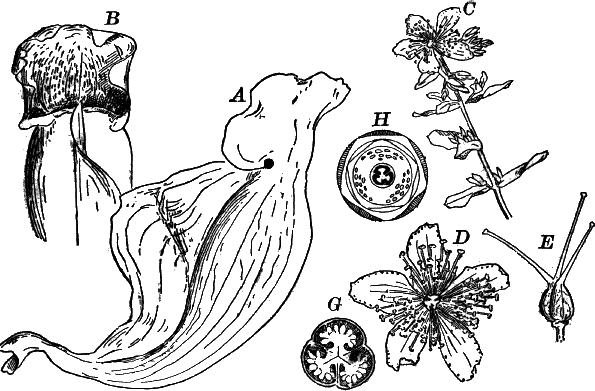
Fig. 105.—Types of Aphanocyclæ (Cistifloræ). A,
B, leaves of the pitcher-plant, Sarracenia (Sarraceniaceæ). A,
from the side; B, from in front, × ½. C, St. John’s-wort
(Hypericum), × ½. D, a flower, × 1. E, the pistil, × 2. G,
cross-section of the ovary, × 4. H, diagram of the flower.
Similar in their habits, but differing much in appearance from the
sundews, are the pitcher-plants (Sarraceniaceæ), of which one
species (Sarracenia purpurea) is very common in peat bogs throughout
the northern United States. In this species (Fig. 105, A, B), the
leaves form a rosette, from the centre of which arises in early summer
a tall stalk bearing a single, large, nodding, dark-reddish flower
with a curious umbrella-shaped pistil. The leaf stalk is hollow and
swollen, with a broad wing on one side, and the blade of the leaf
forms a sort of hood at the top. The interior of the pitcher is
covered above with stiff, downward-pointing hairs, while below it is
very smooth. Insects readily enter the pitcher, but on attempting to
get out, the smooth, slippery wall at the bottom, and the stiff,
downward-directed hairs above, prevent their escape, and they fall
into the fluid which fills the bottom of the cup and are drowned, the
leaf absorbing the nitrogenous compounds given off during the process
of decomposition. There are other species common in the southern
states, and a California pitcher-plant (Darlingtonia) has a colored
appendage at the mouth of the pitcher which serves to lure insects
into the trap.
Another family of pitcher-plants (Nepentheæ) is found in the warmer
parts of the old world, and some of them are occasionally cultivated
in greenhouses. In these the pitchers are borne at the tips of the
leaves attached to a long tendril.
Two other families of the order contain familiar native plants, the
rock-rose family (Cistaceæ), and the St. John’s-worts
(Hypericaceæ). The latter particularly are common plants, with
numerous showy yellow flowers, the petals usually marked with black
specks, and the leaves having clear dots scattered through them. The
stamens are numerous, and often in several distinct groups (Fig. 105,
C, D).
The last order of the Aphanocyclæ (the Columniferæ) has three
families, of which two, the mallows (Malvaceæ), and the lindens
(Tiliaceæ), include well-known species. Of the former, the various
species of mallows (Fig. 106, A) belonging to the genus Malva are
common, as well as some species of Hibiscus, including the showy
swamp Hibiscus or rose-mallow (H. moscheutos), common in salt
marshes and in the fresh-water marshes of the great lake region. The
hollyhock and shrubby Althæa are familiar cultivated plants of this
order, and the cotton-plant (Gossypium) also belongs here. In all of
these the stamens are much branched, and united into a tube enclosing
the style. Most of them are characterized also by the development of
great quantities of a mucilaginous matter within their tissues.
The common basswood (Tilia) is the commonest representative of the
family Tiliaceæ (Fig. 106, G). The nearly related European linden,
or lime-tree, is sometimes planted. Its leaves are ordinarily somewhat
smaller than our native species, which it, however, closely resembles.
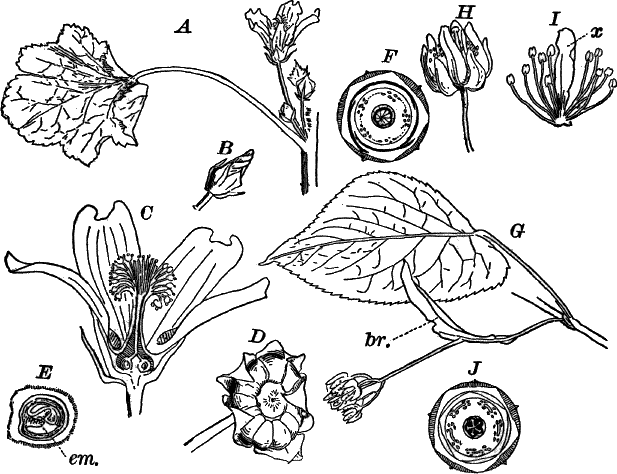
Fig. 106.—Types of Aphanocyclæ (Columniferæ). A,
flower and leaf of the common mallow, Malva (Malvaceæ), × ½. B,
a flower bud, × 1. C, section of a flower, × 2. D, the fruit, × 2.
E, section of one division of the fruit, with the enclosed seed,
× 3. em. the embryo. F, diagram of the flower. G, leaf and
inflorescence of the basswood, Tilia (Tiliaceæ), × ⅓. br. a
bract. H, a single flower, × 1. I, group of stamens, with
petal-like appendage (x), × 2. J, diagram of the flower.
The fourth group of the Choripetalæ is the Eucyclæ. The flowers
most commonly have the parts in fives, and the stamens are never more
than twice as many as the sepals. The carpels are usually more or less
completely united into a compound pistil. There are four orders,
comprising twenty-five families.
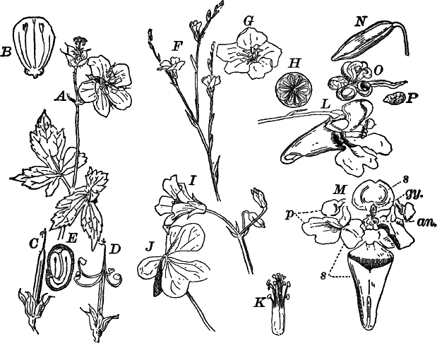
Fig. 107.—Types of Eucyclæ (Gruinales). A, wild
crane’s-bill Geranium (Geraniaceæ), × ½. B, a petal, × 1. C,
the young fruit, the styles united in a column, × ½. D, the ripe
fruit, the styles separating to discharge the seeds, × ½. E, section
of a seed, × 2. F, wild flax. Linum (Linaceæ), × ½. G, a
single flower, × 2. H, cross-section of the young fruit, × 3. I,
flower. J, leaf of wood-sorrel, Oxalis (Oxalideæ), × 1. K, the
stamens and pistil, × 2. L, flower of jewel-weed, Impatiens
(Balsamineæ), × 1. M, the same, with the parts separated. p,
petals. s, sepals. an. stamens. gy. pistil. N, fruit, × 1.
O, the same, opening. P, a seed, × 2.
The first order (Gruinales) includes six families, consisting for
the most part of plants with conspicuous flowers. Here belong the
geraniums (Fig. 107, A), represented by the wild geraniums and
crane’s-bill, and the very showy geraniums (Pelargonium) of the
gardens. The nasturtiums (Tropæolum) represent another family,
mostly tropical, and the wood-sorrels (Oxalis) (Fig. 107, I) are
common, both wild and cultivated. The most useful member of the order
is unquestionably the common flax (Linum), of which there are also
several native species (Fig. 107, F). These are types of the flax
family (Linaceæ). Linen is the product of the tough, fibrous inner
bark of L. usitatissimum, which has been cultivated for its fibre
from time immemorial. The last family is the balsam family
(Balsamineæ). The jewel-weed or touch-me-not
(Impatiens), so called from the sensitive pods which
spring open on being touched, is very common in moist ground
everywhere (Fig. 107, L–P). The garden balsam, or lady’s slipper, is
a related species (I. balsamina).
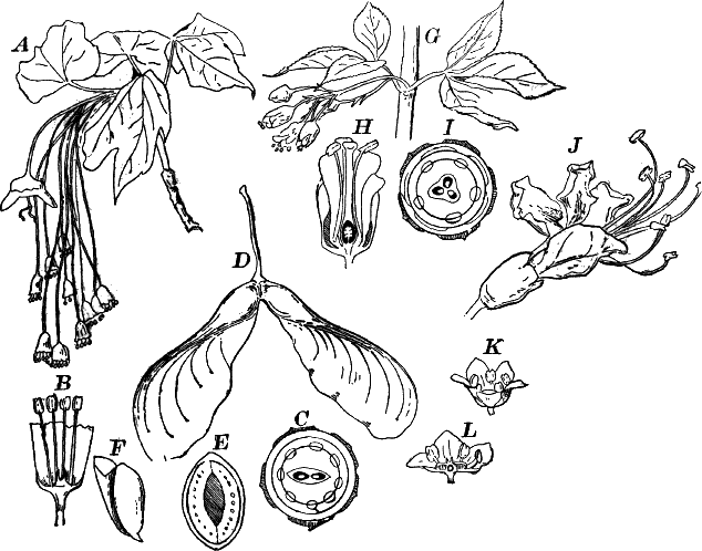
Fig. 108.—Eucyclæ (Terebinthinæ, Æsculinæ). A,
leaves and flowers of sugar-maple, Acer (Aceraceæ), × ½. B, a
male flower, × 2. C, diagram of a perfect flower. D, fruit of the
silver-maple, × ½. E, section across the seed, × 2. F, embryo
removed from the seed, × 1. G, leaves and flowers of bladder-nut,
Staphylea, (Sapindaceæ), × ½. H, section of a flower, × 2. I,
diagram of the flower. J, flower of buckeye (Æsculus), × 1½. K,
flower of smoke-tree, Rhus (Anacardiaceæ), × 3. L, the same, in
section.
The second order (Terebinthinæ) contains but few common plants.
There are six families, mostly inhabitants of the warmer parts of the
world. The best-known members of the order are the orange, lemon,
citron, and their allies. Of our native plants the prickly ash
(Zanthoxylum), and the various species of sumach (Rhus), are the
best known. In the latter genus belong the poison ivy
(R. toxicodendron) and the poison dogwood (R. venenata). The
Venetian sumach or smoke-tree (R. Cotinus) is commonly planted for
ornament.
The third order of the Eucyclæ, the Æsculinæ, embraces six
families, of which three, the horsechestnuts, etc. (Sapindaceæ), the
maples (Aceraceæ), and the milkworts (Polygalaceæ), have several
representatives in the northern United States. Of the first the
buckeye (Æsculus) (Fig. 108, J) and the bladder-nut (Staphylea)
(Fig. 108, G) are the commonest native genera, while the
horsechestnut (Æsculus hippocastanum) is everywhere planted.
The various species of maple (Acer) are familiar examples of the
Aceraceæ (see Fig. 106, A, F).
The fourth and last order of the Eucyclæ, the Frangulinæ, is
composed mainly of plants with inconspicuous flowers, the stamens as
many as the petals. Not infrequently they are diœcious, or in some,
like the grape, some of the flowers may be unisexual while others are
hermaphrodite (i.e. have both stamens and pistil). Among the
commoner plants of the order may be mentioned the spindle-tree, or
burning-bush, as it is sometimes called (Euonymus) (Fig. 109, A),
and the climbing bitter-sweet (Celastrus) (Fig. 109, D), belonging
to the family Celastraceæ; the holly and black alder, species of
Ilex, are examples of the family Aquifoliaceæ; the various species
of grape (Vitis), the Virginia creeper (Ampelopsis quinquefolia),
and one or two other cultivated species of the latter, represent the
vine family (Vitaceæ or Ampelidæ), and the buckthorn (Rhamnus)
is the type of the Rhamnaceæ.
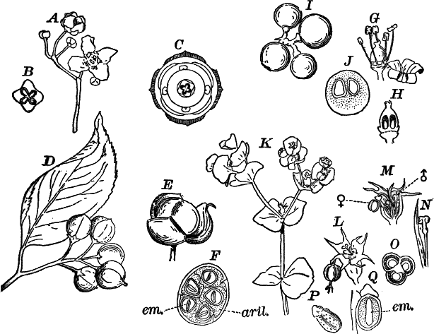
Fig. 109.—Eucylæ (Frangulinæ), Tricoccæ. A,
flowers of spindle-tree, Euonymus, (Celastraceæ), × 1. B,
cross-section of the ovary, × 2. C, diagram of the flower. D, leaf
and fruit of bitter-sweet (Celastrus), × ½. E, fruit opening and
disclosing the seeds. F, section of a nearly ripe fruit, showing the
seeds surrounded by the scarlet integument (aril). em. the embryo,
× 1. G, flower of grape-vine, Vitis (Vitaceæ), × 2. The corolla
has fallen off. H, vertical section of the pistil, × 2. I, nearly
ripe fruits of the frost-grape, × 1. J, cross-section of young
fruit, × 2. K, a spurge, Euphorbia (Euphorbiaceæ), × ½. L,
single group of flowers, surrounded by the corolla-like involucre,
× 3. M, section of the same, ♂, male flowers; ♀, female flowers.
N, a single male flower, × 5. O, cross-section of ovary, × 6. P,
a seed, × 2. Q, longitudinal section of the seed, × 3. em.
embryo.
The fifth group of the Choripetalæ is a small one, comprising but a
single order (Tricoccæ). The flowers are small and inconspicuous,
though sometimes, as in some Euphorbias and the showy Poinsettia
of the greenhouses, the leaves or bracts surrounding the inflorescence
are conspicuously colored, giving the whole the appearance of a large,
showy, single flower. In northern countries the plants are mostly
small weeds, of which the various spurges or Euphorbias are the most
familiar. These plants (Fig. 109, K) have the small flowers
surrounded by a cup-shaped involucre (L, M) so that the whole
inflorescence looks like a single flower. In the spurges, as in the
other members of the order, the flowers are very simple, being often
reduced to a single stamen or pistil (Fig. 109, M, N). The plants
generally abound in a milky juice which is often poisonous. This juice
in a number of tropical genera is the source of India-rubber. Some
genera like the castor-bean (Ricinus) and Croton are cultivated
for their large, showy leaves.
The water starworts (Callitriche), not uncommon in stagnant water,
represent the family Callitrichaceæ, and the box (Buxus) is the
type of the Buxaceæ.
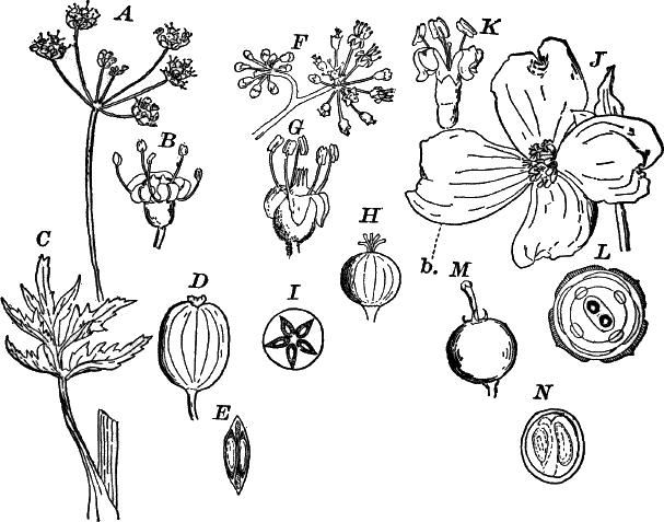
Fig. 110.—Types of Calycifloræ (Umbellifloræ).
A, inflorescence of wild parsnip, Pastinaca (Umbelliferæ), × ½.
B, single flower of the same, × 3. C, a leaf, showing the
sheathing base, × ¼. D, a fruit, × 2. E, cross-section of D.
F, part of the inflorescence of spikenard, Aralia (Araliaceæ),
× 1. G, a single flower of the same, × 3. H, the fruit, × 2. I,
cross-section of the H. J, inflorescence of dogwood, Cornus
(Corneæ). The cluster of flowers is surrounded by four white bracts
(b), × ⅓. K, a single flower of the same, × 2. L, diagram of the
flower. M, young fruit of another species (Cornus stolonifera)
(red osier), × 2. N, cross-section of M.
The last and highest group of the Choripetalæ, the Calycifloræ,
embraces a very large assemblage of familiar plants, divided into
eight orders and thirty-two families. With few exceptions, the floral
axis grows up around the ovary, carrying the outer floral leaves above
it, and the ovary appears at the bottom of a cup around whose edge the
other parts of the flower are arranged. Sometimes, as in the fuchsia,
the ovary is grown to the base of the cup or tube, and thus looks as
if it were outside the flower. Such an ovary is said to be “inferior”
in distinction from one that is entirely free from the tube, and thus
is evidently within the flower. The latter is the so-called “superior”
ovary. The carpels are usually united into a compound pistil, but may
be separate, as in the stonecrop (Fig. 111, E), or strawberry
(Fig. 114, C).
The first order of the Calycifloræ (Umbellifloræ) has the flowers
small, and usually arranged in umbels, i.e. several stalked flowers
growing from a common point. The ovary is inferior, and there is a
nectar-secreting disc between the styles and the stamens. Of the three
families, the umbel-worts or Umbelliferæ is the commonest. The
flowers are much alike in all (Fig. 110, A, B), and nearly all
have large, compound leaves with broad, sheathing bases. The stems are
generally hollow. So great is the uniformity of the flowers and plant,
that the fruit (Fig. 110, D) is generally necessary before the plant
can be certainly recognized. This is two-seeded in all, but differs
very much in shape and in the development of oil channels, which
secrete the peculiar oil that gives the characteristic taste to the
fruits of such forms as caraway, coriander, etc. Some of them, like
the wild parsnip, poison hemlock, etc., are violent poisons, while
others like the carrot are perfectly wholesome.
The wild spikenard (Aralia) (Fig. 110, F), ginseng, and the true
ivy (Hedera) are examples of the Araliaceæ, and the various
species of dogwood (Cornus) (Fig. 110, J–N) represent the dogwood
family (Corneæ).
The second order (Saxifraginæ) contains eight families, including a
number of common wild and cultivated plants. The true saxifrages are
represented by several wild and cultivated species of Saxifraga, the
little bishop’s cap or mitre-wort (Mitella) (Fig. 111, D), and
others. The wild hydrangea (Fig. 111, F) and the showy garden
species represent the family Hydrangeæ. In these some of the flowers
are large and showy, but with neither stamens nor pistils (neutral),
while the small, inconspicuous flowers of the central part of the
inflorescence are perfect. In the garden varieties, all of the flowers
are changed, by selection, into the showy, neutral ones. The syringa
or mock orange (Philadelphus) (Fig. 111, I), the gooseberry, and
currants (Ribes) (Fig. 111, A), and the stonecrop (Sedum)
(Fig. 111, E) are types of the families Philadelpheæ, Ribesieæ,
and Crassulaceæ.
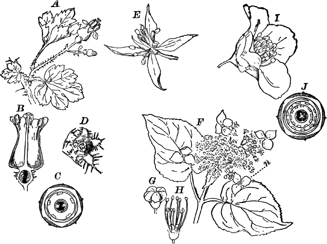
Fig. 111.—Calycifloræ (Saxifraginæ): A, flowers
and leaves of wild gooseberry, Ribes (Ribesieæ), × 1. B,
vertical section of the flower, × 2. C, diagram of the flower. D,
flower of bishop’s-cap, Mitella (Saxifragaceæ), × 3. E, flower
of stonecrop, Sedum (Crassulaceæ), × 2. F, flowers and leaves of
hydrangea (Hydrangeæ), × ½. n, neutral flower. G, unopened
flower, × 2. H, the same, after the petals have fallen away. I,
flower of syringa, Philadelphus (Philadelpheæ), × 1. J, diagram
of the flower.
The third order (Opuntieæ) has but a single family, the cacti
(Cactaceæ). These are strictly American in their distribution, and
inhabit especially the dry plains of the southwest, where they reach
an extraordinary development. They are nearly or quite leafless, and
the fleshy, cylindrical, or flattened stems are usually beset with
stout spines. The flowers (Fig. 112, A) are often very showy, so
that many species are cultivated for ornament and are familiar to
every one. The beautiful night-blooming cereus, of which there are
several species, is one of these. A few species of prickly-pear
(Opuntia) occur as far north as New York, but most are confined to
the hot, dry plains of the south and southwest.
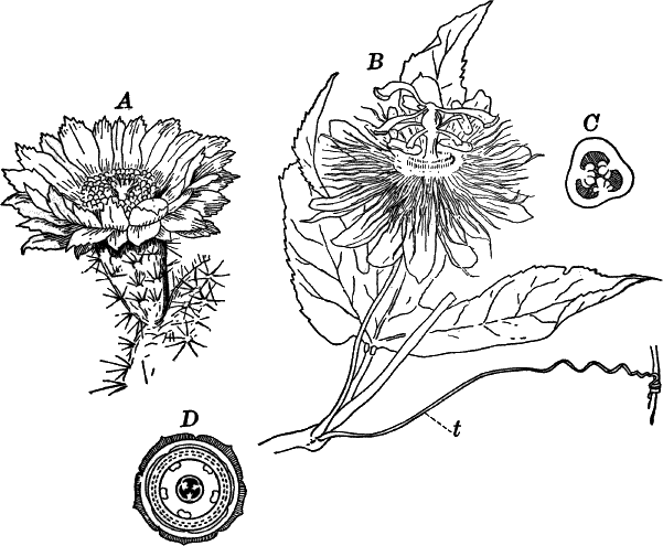
Fig. 112.—Calycifloræ, Opuntieæ (Passiflorinæ).
A, flower of a cactus, Mamillaria (Cactaceæ) (from “Gray’s
Structural Botany”). B, leaf and flower of a passion-flower,
Passiflora (Passifloraceæ), × ½. t, a tendril. C,
cross-section of the ovary, × 2. D, diagram of the flower.
The fourth order (Passiflorinæ) are almost without exception
tropical plants, only a very few extending into the southern United
States. The type of the order is the passion-flower (Passiflora)
(Fig. 112, B), whose numerous species are mostly inhabitants of
tropical America, but a few reach into the United States. The only
other members of the order likely to be met with by the student are
the begonias, of which a great many are commonly cultivated as house
plants on account of their fine foliage and flowers. The leaves are
always one-sided, and the flowers monœcious.[13] Whether the begonias
properly belong with the Passiflorinæ has been questioned.
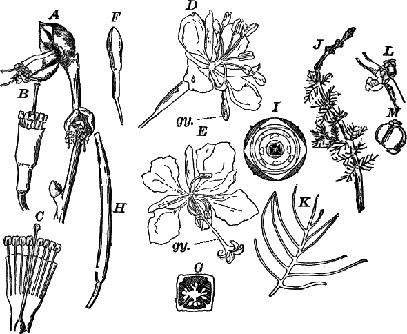
Fig. 113.—Calycifloræ (Myrtifloræ, Thymelinæ).
A, flowering branch of moosewood, Dirca
(Thymelæaceæ), × 1. B, a single flower, × 2. C,
the same, laid open. D, a young flower of willow herb, Epilobium
(Onagraceæ), × 1. The pistil (gy.) is not yet ready for
pollination. E, an older flower, with receptive pistil. F, an
unopened bud, × 1. G , cross-section of the ovary, × 4. H, a young
fruit, × 1. I, diagram of the flower. J, flowering branch of water
milfoil, Myriophyllum (Haloragidaceæ), × ½. K, a single leaf,
× 1. L, female flowers of the same, × 2. M, the fruit, × 2.
The fifth order (Myrtifloræ) have regular four-parted flowers with
usually eight stamens, but sometimes, through branching of the
stamens, these appear very numerous. The myrtle family, the members of
which are all tropical or sub-tropical, gives name to the order. The
true myrtle (Myrtus) is sometimes cultivated for its pretty glossy
green leaves and white flowers, as is also the pomegranate whose
brilliant, scarlet flowers are extremely ornamental. Cloves are the
dried flower-buds of an East-Indian myrtaceous tree (Caryophyllus).
In Australia the order includes the giant gum-trees
(Eucalyptus), the largest of all known trees, exceeding
in size even the giant trees of California.
Among the commoner Myrtifloræ, the majority belong to the two
families Onagraceæ and Lythraceæ. The former includes the evening
primroses (Œnothera), willow-herb (Epilobium) (Fig. 113, D),
and fuchsia; the latter, the purple loosestrife (Lythrum) and swamp
loosestrife (Nesæa). The water-milfoil (Myriophyllum) (Fig. 113,
J) is an example of the family Haloragidaceæ, and the Rhexias of
the eastern United States represent with us the family Melastomaceæ.
The sixth order of the Calycifloræ is a small one (Thymelinæ),
represented in the United States by very few species. The flowers are
four-parted, the calyx resembling a corolla, which is usually absent.
The commonest member of the order is the moosewood (Dirca)
(Fig. 113, A), belonging to the first of the three families
(Thymelæaceæ). Of the second family (Elæagnaceæ), the commonest
example is Shepherdia, a low shrub having the leaves covered with
curious, scurfy hairs that give them a silvery appearance. The third
family (Proteaceæ) has no familiar representatives.
The seventh order (Rosifloræ) includes many well-known plants, all
of which may be united in one family (Rosaceæ), with several
sub-families. The flowers are usually five-parted with from five to
thirty stamens, and usually numerous, distinct carpels. In the apple
and pear (Fig. 114, I), however, the carpels are more or less grown
together; and in the cherry, peach, etc., there is but a single carpel
giving rise to a single-seeded stone-fruit (drupe) (Fig. 114, E,
H). In the strawberry (Fig. 114, A), rose (G), cinquefoil
(Potentilla), etc., there are numerous distinct, one-seeded carpels,
and in Spiræa (Fig. 114, F) there are five several-seeded carpels,
forming as many dry pods when ripe. The so-called “berry” of the
strawberry is really the much enlarged flower axis, or “receptacle,”
in which the little one-seeded fruits are embedded, the latter being
what are ordinarily called the seeds.
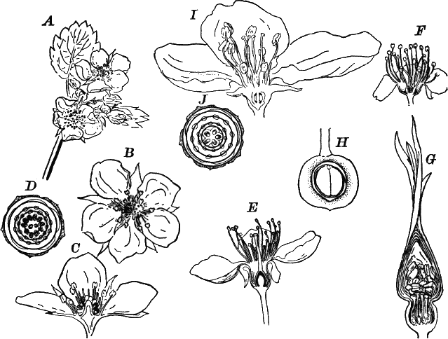
Fig. 114.—Calycifloræ (Rosifloræ). A,
inflorescence of strawberry (Fragaria), × ½. B, a single flower,
× 1. C, section of B. D, floral diagram. E, vertical section
of a cherry-flower (Prunus), × 1. F, vertical section of the
flower of Spiræa, × 2. G, vertical section of the bud of a wild
rose (Rosa), × 1. H, vertical section of the young fruit, × 1.
I, section of the flower of an apple (Pyrus), × 1. J, floral
diagram of apple.
From the examples given, it will be seen that the order includes not
only some of the most ornamental, cultivated plants, but the majority
of our best fruits. In addition to those already given, may be
mentioned the raspberry, blackberry, quince, plum, and apricot.
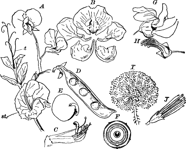
Fig. 115.—Calycifloræ (Leguminosæ). A, flowers
and leaf of the common pea, Pisum (Papilionaceæ), × ½. t,
tendril. st. stipules. B, the petals, separated and displayed,
× 1. C, flower, with the calyx and corolla removed, × 1. D, a
fruit divided lengthwise, × ½. E, the embryo, with one of the
cotyledons removed, × 2. F, diagram of the flower. G, flower of
red-bud, Cercis (Cæsalpinaceæ), × 2. H, the same, with calyx and
corolla removed. I, inflorescence of the sensitive-brier,
Schrankia (Mimosaceæ), × 1. J, a single flower, × 2.
The last order of the Calycifloræ and the highest of the
Choripetalæ is the order Leguminosæ, of which the bean, pea,
clover, and many other common plants are examples. In most of our
common forms the flowers are peculiar in shape, one of the petals
being larger than the others, and covering them in the bud. This
petal is known as the standard. The two lateral petals are known as
the wings, and the two lower and inner are generally grown together
forming what is called the “keel” (Fig. 115, A, B). The stamens,
ten in number, are sometimes all grown together into a tube, but
generally the upper one is free from the others (Fig. 115, C). There
is but one carpel which forms a pod with two valves when ripe
(Fig. 115, D). The seeds are large, and the embryo fills the seed
completely. From the peculiar form of the flower, they are known as
Papilionaceæ (papilio, a butterfly). Many of the Papilionaceæ
are climbers, either having twining stems, as in the common beans, or
else with part of the leaf changed into a tendril as in the pea
(Fig. 115, A), vetch, etc. The leaves are usually compound.
Of the second family (Cæsalpineæ), mainly tropical, the honey locust
(Gleditschia) and red-bud (Cercis) (Fig. 115, G) are the
commonest examples. The flowers differ mainly from the Papilionaceæ
in being less perfectly papilionaceous, and the stamens are almost
entirely distinct (Fig. 115, H). The last family (Mimosaceæ) is
also mainly tropical. The acacias, sensitive-plant (Mimosa), and the
sensitive-brier of the southern United States (Schrankia) (Fig. 115,
I) represent this family. The flowers are quite different from the
others of the order, being tubular and the petals united, thus
resembling the flowers of the Sympetalæ. The leaves of Mimosa and
Schrankia are extraordinarily sensitive, folding up if irritated.
CHAPTER XIX.
CLASSIFICATION OF DICOTYLEDONS (Continued).
Division II.—Sympetalæ.
The Sympetalæ or Gamopetalæ are at once distinguished from the
Choripetalæ by having the petals more or less united, so that the
corolla is to some extent tubular. In the last order of the
Choripetalæ we found a few examples (Mimosaceæ) where the same
thing is true, and these form a transition from the Choripetalæ to
the Sympetalæ.
There are two great divisions, Isocarpæ and Anisocarpæ. In the
first the carpels are of the same number as the petals and sepals; in
the second fewer. In both cases the carpels are completely united,
forming a single, compound pistil. In the Isocarpæ there are usually
twice as many stamens as petals, occasionally the same number.
There are three orders of the Isocarpæ, viz., Bicornes,
Primulinæ, and Diospyrinæ. The first is a large order with six
families, including many very beautiful plants, and a few of some
economic value. Of the six families, all but one (Epacrideæ) are
represented in the United States. Of these the Pyrolaceæ includes
the pretty little pyrolas and prince’s-pine (Chimaphila) (Fig. 116,
J); the Monotropeæ has as its commonest examples, the curious
Indian-pipe (Monotropa uniflora), and pine-sap (M. hypopitys)
(Fig. 116, L). These grow on decaying vegetable matter, and are
quite devoid of chlorophyll, the former species being pure white
throughout (hence a popular name, “ghost flower”); the latter is
yellowish. The magnificent rhododendrons and azaleas (Fig. 116, F),
and the mountain laurel (Kalmia) (Fig. 116, I), belong to the
Rhodoraceæ. The heath family (Ericaceæ), besides the true heaths
(Erica, Calluna), includes the pretty trailing-arbutus or
may-flower (Epigæa), Andromeda, Oxydendrum (Fig. 116, E),
wintergreen (Gaultheria), etc. The last family is represented by the
cranberry (Vaccinium) and huckleberry (Gaylussacia).
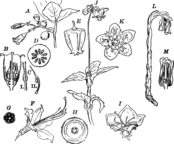
Fig. 116.—Types of Isocarpous sympetalæ
(Bicornes). A, flowers, fruit, and leaves of huckleberry,
Gaylussacia (Vaccinieæ), × 1. B, vertical section of the flower,
× 3. C, a stamen: i, from in front; ii, from the side, × 4. D,
cross-section of the young fruit, × 2. E, flower of sorrel-tree,
Oxydendrum (Ericaceæ), × 2. F, flower of azalea
(Rhododendron), × ½. G, cross-section of the ovary, × 3. H,
diagram of the flower. I, flower of mountain laurel (Kalmia), × 1.
J, prince’s-pine, Chimaphila (Pyrolaceæ), × ½. K, a single
flower, × 1. L, plant of pine-sap, Monotropa, (Monotropeæ), × ½.
M, section of a flower, × 1.
The second order, the primroses (Primulinæ), is principally
represented in the cooler parts of the world by the true primrose
family (Primulaceæ), of which several familiar plants may be
mentioned. The genus Primula includes the European primrose and
cowslip, as well as two or three small American species, and the
commonly cultivated Chinese primrose. Other genera are Dodecatheon,
of which the beautiful shooting-star (D. Meadia) (Fig. 117, A) is
the best known. Something like this is Cyclamen, sometimes
cultivated as a house plant. The moneywort (Lysimachia nummularia)
(Fig. 117, D), as well as other species, also belongs here.
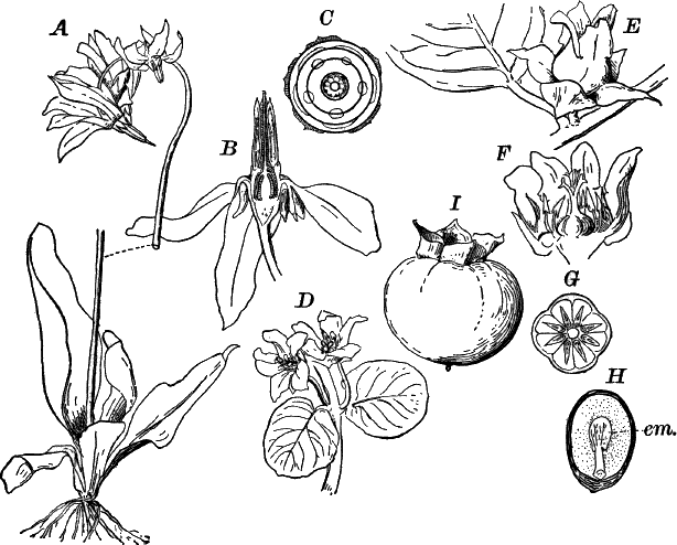
Fig. 117.—Isocarpous sympetalæ (Primulinæ,
Diospyrinæ). A, shooting-star, Dodecatheon (Primulaceæ), × ½.
B, section of a flower, × 1. C, diagram of the flower. D,
Moneywort, Lysimachia (Primulaceæ), × ½. E, a perfect flower of
the persimmon, Diospyros (Ebenaceæ), × 1. F, the same, laid open:
section of the young fruit, × 2. H, longitudinal section of a ripe
seed, × 1. em. the embryo. I, fruit, × ½.
The sea-rosemary (Statice) and one or two cultivated species of
plumbago are the only members of the plumbago family (Plumbagineæ)
likely to be met with. The remaining families of the Primulinæ are
not represented by any common plants.
The third and last order of the Isocarpous sympetalæ has but a
single common representative in the United States; viz., the persimmon
(Diospyros) (Fig. 117, E). This belongs to the family Ebenaceæ,
to which also belongs the ebony a member of the same genus as the
persimmon, and found in Africa and Asia.
The second division of the Sympetalæ (the Anisocarpæ) has usually
but two or three carpels, never as many as the petals. The stamens are
also never more than five, and very often one or more are abortive.
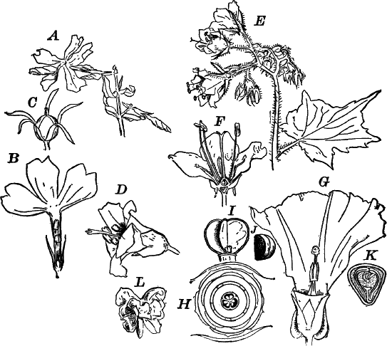
Fig. 118.—Types of Anisocarpous sympetalæ
(Tubifloræ). A, flower and leaves of wild phlox (Polemoniaceæ),
× ½. B, section of a flower, × 1. C, fruit, × 1. D, flower of
blue valerian (Polemonium), × 1. E, flowers and leaf of
water-leaf, Hydrophyllum (Hydrophyllaceæ), × ½. F, section of a
flower, × 1. G, flower of wild morning-glory, Convolvulus
(Convolvulaceæ), × ½. One of the bracts surrounding the calyx and
part of the corolla are cut away. H, diagram of the flower. I, the
fruit of a garden morning-glory, from which the outer wall has fallen,
leaving only the inner membranous partitions, × 1. J, a seed, × 1.
K, cross-section of a nearly ripe seed, showing the crumpled embryo,
× 2. L, an embryo removed from a nearly ripe seed, and spread out;
one of the cotyledons has been partially removed, × 1.
The first order (Tubifloræ) has, as the name indicates, tubular
flowers which show usually perfect, radial symmetry
(Actinomorphism). There are five families, all represented by
familiar plants. The first (Convolvulaceæ) has as its type the
morning-glory (Convolvulus) (Fig. 118, G), and the nearly related
Ipomœas of the gardens. The curious dodder (Cuscuta), whose
leafless, yellow stems are sometimes very conspicuous, twining over
various plants, is a member of this family which has lost its
chlorophyll through parasitic habits. The sweet potato (Batatas) is
also a member of the morning-glory family. The numerous species, wild
and cultivated, of phlox (Fig. 118, A), and the blue valerian
(Polemonium) (Fig. 118, D), are examples of the family
Polemoniaceæ.
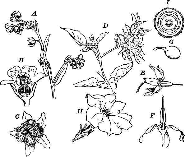
Fig. 119.—Anisocarpous sympetalæ (Tubifloræ). A,
inflorescence of hound’s-tongue, Cynoglossum (Borragineæ), × ½.
B, section of a flower, × 2. C, nearly ripe fruit, × 1. D,
flowering branch of nightshade, Solanum (Solaneæ), × ½. E, a
single flower, × 1. F, section of the flower, × 2. G, young fruit,
× 1. H, flower of Petunia (Solaneæ), × ½. I, diagram of the
flower.
The third family (Hydrophyllaceæ) includes several species of
water-leaf (Hydrophyllum) (Fig. 118, E) and Phacelia, among our
wild flowers, and species of Nemophila, Whitlavia and others from
the western states, but now common in gardens.
The Borage family (Borragineæ) includes the forget-me-not
(Myosotis) and a few pretty wild flowers, e.g. the orange-flowered
puccoons (Lithospermum); but it also embraces a
number of the most troublesome weeds, among which are the
hound’s-tongue (Cynoglossum) (Fig. 119, A), and the
“beggar’s-ticks” (Echinospermum), whose prickly
fruits (Fig. 119, C) become detached on the slightest provocation,
and adhere to whatever they touch with great tenacity. The flowers in
this family are arranged in one-sided inflorescences which are coiled
up at first and straighten as the flowers expand.
The last family (Solaneæ) includes the nightshades (Solanum)
(Fig. 119, D), to which genus the potato (S. tuberosum) and the
egg-plant (S. Melongena) also belong. Many of the family contain a
poisonous principle, e.g. the deadly nightshade (Atropa), tobacco
(Nicotiana), stramonium (Datura), and others. Of the cultivated
plants, besides those already mentioned, the tomato (Lycopersicum),
and various species of Petunia (Fig. 119, H), Solanum, and
Datura are the commonest.
The second order of the Anisocarpæ consists of plants whose flowers
usually exhibit very marked, bilateral symmetry (Zygomorphism). From
the flower often being two-lipped (see Fig. 120), the name of the
order (Labiatifloræ) is derived.
Of the nine families constituting the order, all but one are
represented within our limits, but the great majority belong to two
families, the mints (Labiatæ) and the figworts (Scrophularineæ).
The mints are very common and easily recognizable on account of their
square stems, opposite leaves, strongly bilabiate flowers, and the
ovary splitting into four seed-like fruits (Fig. 120, D, F).
The great majority of them, too, have the surface covered with
glandular hairs secreting a strong-scented volatile oil, giving the
peculiar odor to these plants. The dead nettle (Lamium) (Fig. 120,
A) is a thoroughly typical example. The sage, mints, catnip, thyme,
lavender, etc., will recall the peculiarities of the family.
The stamens are usually four in number through the abortion of one of
them, but sometimes only two perfect stamens are present.
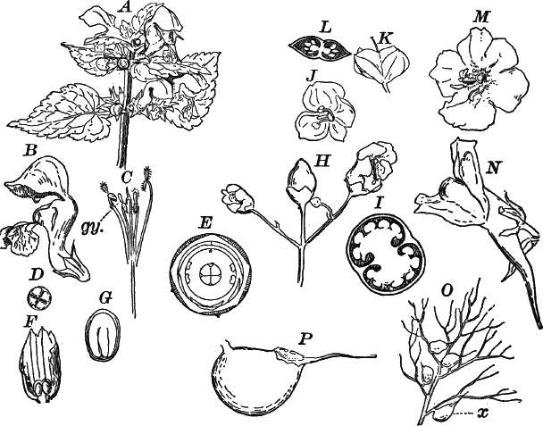
Fig. 120.—Anisocarpous sympetalæ (Labiatifloræ).
A, dead nettle, Lamium, (Labiatæ), × ½. B, a single flower,
× 1. C, the stamens and pistil, × 1. D, cross-section of the
ovary, × 2. E, diagram of the flower; the position of the absent
stamen is indicated by the small circle. F, fruit of the common
sage, Salvia (Labiatæ), × 1. Part of the persistent calyx has been
removed to show the four seed-like fruits, or nutlets. G, section of
a nutlet, × 3. The embryo fills the seed completely. H, part of an
inflorescence of figwort, Scrophularia (Scrophularineæ), × 1. I,
cross-section of the young fruit, × 2. J, flower of speedwell,
Veronica (Scrophularineæ), × 2. K, fruit of Veronica, × 2.
L, cross-section of K. M, flower of moth-mullein, Verbascum
(Scrophularineæ), × ½. N, flower of toad-flax, Linaria
(Scrophularineæ), × 1. O, leaf of bladder-weed, Utricularia
(Lentibulariaceæ), × 1. x, one of the “traps.” P, a single trap,
× 5.
The Scrophularineæ differ mainly from the Labiatæ in having round
stems, and the ovary not splitting into separate one-seeded fruits.
The leaves are also sometimes alternate. There are generally four
stamens, two long and two short, as in the labiates, but in the
mullein (Verbascum) (Fig. 120, M), where the flower is only
slightly zygomorphic, there is a fifth rudimentary stamen, while in
others (e.g. Veronica) (Fig. 120, J) there are but two stamens.
Many have large, showy flowers, as in the cultivated foxglove
(Digitalis), and the native species of Gerardia, mullein,
Mimulus, etc., while a few like the figwort, Scrophularia
(Fig. 120, H), and speedwells (Veronica) have duller-colored or
smaller flowers.
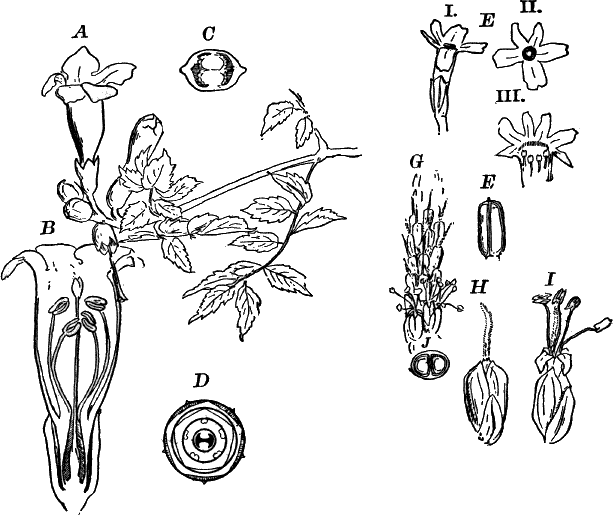
Fig. 121.—Anisocarpous sympetalæ (Labiatifloræ).
A, flowering branch of trumpet-creeper, Tecoma (Bignoniaceæ),
× ¼. B, a single flower, divided lengthwise, × ½. C, cross-section
of the ovary, × 2. D, diagram of the flower. E, flower of vervain,
Verbena (Verbenæ), × 2: i, from the side; ii, from in front; iii,
the corolla laid open. F, nearly ripe fruit of the same, × 2. G,
part of a spike of flowers of the common plantain, Plantago
(Plantagineæ), × 1; The upper flowers have the pistils mature, but
the stamens are not yet ripe. H, a flower from the upper (younger)
part of the spike. I, an older expanded flower, with ripe stamens,
× 3.
The curious bladder-weed (Utricularia) is the type of the family
Lentibulariaceæ, aquatic or semi-aquatic plants which possess
special contrivances for capturing insects or small water animals.
These in the bladder-weed are little sacs (Fig. 120, P) which act as
traps from which the animals cannot escape after being captured. There
does not appear to be here any actual digestion, but simply an
absorption of the products of decomposition, as in the pitcher-plant.
In the nearly related land form, Pinguicula, however, there is much
the same arrangement as in the sundew.
The family Gesneraceæ is mainly a tropical one, represented in the
greenhouses by the magnificent Gloxinia and Achimenes, but of
native plants there are only a few parasitic forms destitute of
chlorophyll and with small, inconspicuous flowers. The commonest of
these is Epiphegus, a much-branched, brownish plant, common in
autumn about the roots of beech-trees upon which it is parasitic, and
whence it derives its common name, “beech-drops.”
The bignonia family (Bignoniaceæ) is mainly tropical, but in our
southern states is represented by the showy trumpet-creeper (Tecoma)
(Fig. 121, A), the catalpa, and Martynia.
The other plants likely to be met with by the student belong either to
the Verbenaceæ, represented by the showy verbenas of the gardens,
and our much less showy wild vervains, also belonging to the genus
Verbena (Fig. 121, E); or to the plantain family (Plantagineæ),
of which the various species of plantain (Plantago) are familiar to
every one (Fig. 121, G, I). The latter seem to be forms in which
the flowers have become inconspicuous, and are wind fertilized, while
probably all of its showy-flowered relatives are dependent on insects
for fertilization.
The third order (Contortæ) of the Anisocarpæ includes five
families, all represented by familiar forms. The first, the olive
family (Oleaceæ), besides the olive, contains the lilac and jasmine
among cultivated plants, and the various species of ash (Fraxinus),
and the pretty fringe-tree (Chionanthus) (Fig. 122, A), often
cultivated for its abundant white flowers. The other families are the
Gentianaceæ including the true gentians (Gentiana) (Fig. 122,
F), the buck-bean (Menyanthes), the centauries
(Erythræa and Sabbatia), and several other less
familiar genera; Loganiaceæ, with the pink-root (Spigelia)
(Fig. 122, D), as the best-known example; Apocynaceæ including the
dog-bane (Apocynum) (Fig. 122, H), and in the gardens the oleander
and periwinkle (Vinca).

Fig. 122.—Anisocarpous sympetalæ (Contortæ). A,
flower of fringe-tree, Chionanthus (Oleaceæ), × 1. B, base of
the flower, with part of the calyx and corolla removed, × 2. C,
fruit of white ash, Fraxinus (Oleaceæ), × 1. D, flower of
pink-root, Spigelia (Loganiaceæ), × ½. E, cross-section of the
ovary, × 3. F, flower of fringed gentian, Gentiana
(Gentianaceæ), × ½. G, diagram of the flower. H, flowering
branch of dog-bane, Apocynum (Apocynaceæ), × ½. I, vertical
section of a flower, × 2. J, bud. K, flower of milk-weed,
Asclepias (Asclepiadaceæ), × 1. L, vertical section through the
upper part of the flower, × 2. gy. pistil. p, pollen masses. an.
stamen. M, a pair of pollen masses, × 6. N, a nearly ripe seed,
× 1.
The last family is the milk-weeds (Asclepiadaceæ), which have
extremely complicated flowers. Our numerous milk-weeds (Fig. 122, K)
are familiar representatives, and exhibit perfectly the peculiarities
of the family. Like the dog-banes, the plants contain a milky juice
which is often poisonous. Besides the true milk-weeds (Asclepias),
there are several other genera within the United States, but mostly
southern in their distribution. Many of them are twining plants and
occasionally cultivated for their showy flowers. Of the cultivated
forms, the wax-plant (Hoya), and Physianthus are the commonest.
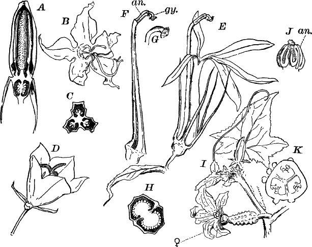
Fig. 123.—Anisocarpous sympetalæ (Campanulinæ).
A, vertical section of the bud of American bell-flower, Campanula
(Campanulaceæ), × 2. B, an expanded flower, × 1. The stamens have
discharged their pollen, and the stigma has opened. C, cross-section
of the ovary, × 3. D, flower of the Carpathian bell-flower
(Campanula Carpatica), × 1. E, flower of cardinal-flower,
Lobelia (Lobeliaceæ), × 1. F, the same, with the corolla and
sepals removed. an. the united anthers. gy. the tip of the pistil.
G, the tip of the pistil, × 2, showing the circle of hairs
surrounding the stigma. H, cross-section of the ovary, × 3. I, tip
of a branch of cucumber, Cucurbita (Cucurbitaceæ), with an
expanded female flower (♀). J, andrœcium of a male flower, showing
the peculiar convoluted anthers (an.), × 2. K, cross-section of
the ovary, × 2.
The fourth order (Campanulinæ) also embraces five families, but of
these only three are represented among our wild plants. The
bell-flowers (Campanula) (Fig. 123, A, D) are examples of the
family Campanulaceæ, and numerous species are common, both wild and
cultivated.
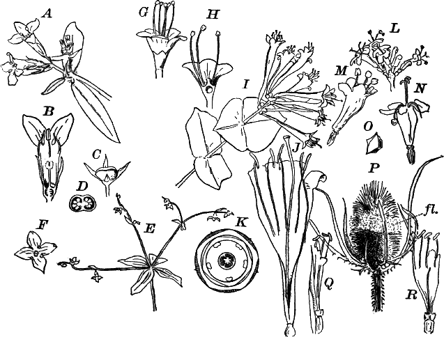
Fig. 124.—Anisocarpous sympetalæ (Aggregatæ). A,
flowering branch of Houstonia purpurea, × 1 (Rubiaceæ). B,
vertical section of a flower, × 2. C, fruit of bluets (Houstonia
cœrulea), × 1. D, cross-section of the same. E, bedstraw,
Galium (Rubiaceæ), × ½. F, a single flower, × 2. G, flower of
arrow-wood, Viburnum (Caprifoliaceæ), × 2. H, the same, divided
vertically. I, flowering branch of trumpet honeysuckle,
Lonicera (Caprifoliaceæ), × ½. J, a single flower,
the upper part laid open, × 1. K, diagram of the flower. L, part
of the inflorescence of valerian, Valeriana, (Valerianeæ), × 1.
M, young; N, older flower, × 2. O, cross-section of the young
fruit; one division of the three contains a perfect seed, the others
are crowded to one side by its growth. P, inflorescence of teasel,
Dipsacus (Dipsaceæ), × ¼. fl. flowers. Q, a single flower,
× 1. R, the same, with the corolla laid open.
The various species of Lobelia, of which the splendid
cardinal-flower (L. Cardinalis) (Fig. 123, E) is one of the most
beautiful, represent the very characteristic family Lobeliaceæ.
Their milky juice contains more or less marked poisonous properties.
The last family of the order is the gourd family (Cucurbitaceæ),
represented by a few wild species, but best known by the many
cultivated varieties of melons, cucumbers, squashes, etc. They are
climbing or running plants, and provided with tendrils. The flowers
are usually unisexual, sometimes diœcious, but oftener monœcious
(Fig. 123, I).
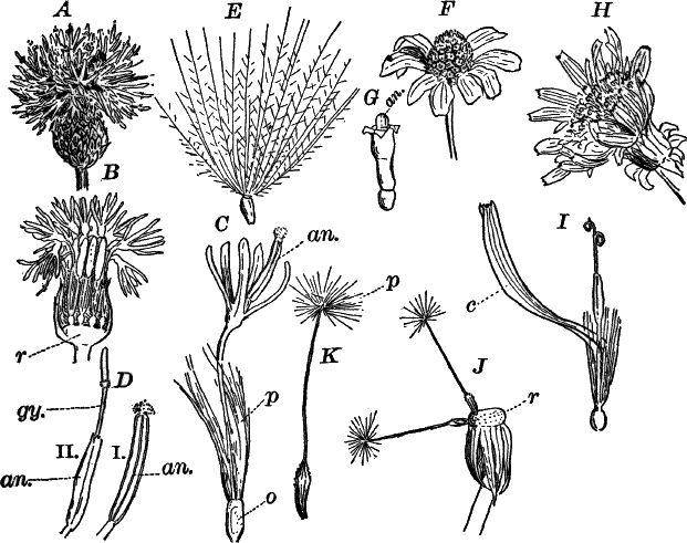
Fig. 125.—Anisocarpous sympetalæ (Aggregatæ).
Types of Compositæ. A, inflorescence of Canada thistle
(Cirsium), × 1. B, vertical section of A. r, the receptacle or
enlarged end of the stem, to which the separate flowers are attached.
C, a single flower, × 2. o, the ovary. p, the “pappus” (calyx
lobes). an. the united anthers. D, the upper part of the stamens
and pistil, × 3: i, from a young flower; ii, from an older one. an.
anthers. gy. pistil. E, ripe fruit, × 1. F, inflorescence of
may-weed (Maruta). The central part (disc) is occupied by perfect
tubular flowers (G), the flowers about the edge (rays) are sterile,
with the corolla much enlarged and white, × 2. G, a single flower
from the disc, × 3. H, inflorescence of dandelion (Taraxacum), the
flowers all alike, with strap-shaped corollas, × 1. I, a single
flower, × 2. c, the split, strap-shaped corolla. J, two ripe
fruits, still attached to the receptacle (r). The pappus is raised
on a long stalk, × 1. K, a single fruit, × 2.
The last and highest order of the Sympetalæ, and hence of the
dicotyledons, is known as Aggregatæ, from the tendency to have the
flowers densely crowded into a head, which not infrequently is closely
surrounded by bracts so that the whole inflorescence resembles a
single flower. There are six families, five of which have common
representatives, but the last family (Calycereæ) has no members
within our limits.
The lower members of the order, e.g. various Rubiaceæ (Fig. 124,
A, E), have the flowers in loose inflorescences, but as we examine
the higher families, the tendency for the flowers to become crowded
becomes more and more evident, and in the highest of our native forms
Dipsaceæ (Fig. 124, P) and Compositæ (Fig. 125) this is very
marked indeed. In the latter family, which is by far the largest of
all the angiosperms, including about ten thousand species, the
differentiation is carried still further. Among our native Compositæ
there are three well-marked types. The first of these may be
represented by the thistles (Fig. 125, A). The so-called flower of
the thistle is in reality a close head of small, tubular flowers
(Fig. 125, C), each perfect in all respects, having an inferior
one-celled ovary, five stamens with the anthers united, and a
five-parted corolla. The sepals (here called the “pappus”) (p) have
the form of fine hairs. These little flowers are attached to the
enlarged upper end of the flower stalk (receptacle, r), and are
surrounded by closely overlapping bracts or scale leaves which look
like a calyx; the flowers, on superficial examination, appear as
single petals. In other forms like the daisy and may-weed (Fig. 125,
F), only the central flowers are perfect, and the edge of the
inflorescence is composed of flowers whose corollas are split and
flattened out, but the stamens and sometimes the pistils are wanting
in these so-called “ray-flowers.” In the third group, of which the
dandelion (Fig. 125, H), chicory, lettuce, etc., are examples, all
of the flowers have strap-shaped, split corollas, and contain both
stamens and pistils.
The families of the Aggregatæ are the following: I. Rubiaceæ of
which Houstonia (Fig. 124, A), Galium (E), Cephalanthus
(button-bush), and Mitchella (partridge-berry) are examples;
II. Caprifoliaceæ, containing the honeysuckles (Lonicera)
(Fig. 124, I), Viburnum (G), snowberry (Symphoricarpus), and
elder (Sambucus); III. Valerianeæ, represented by the common
valerian (Valeriana) (Fig. 124, L); IV. Dipsaceæ, of which the
teasel (Dipsacus) (Fig. 124, P), is the type, and also species of
scabious (Scabiosa); V. Compositæ to which the innumerable,
so-called compound flowers, asters, golden-rods, daisies, sunflowers,
etc. belong; VI. Calycereæ.
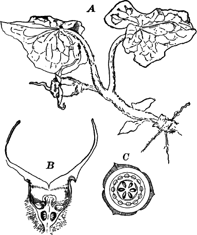
Fig. 126.—Aristolochiaceæ. A, plant of wild ginger
(Asarum), × ⅓. B, vertical section of the flower, × 1. C,
diagram of the flower.
Besides the groups already mentioned, there are several families of
dicotyledons whose affinities are very doubtful. They are largely
parasitic, e.g. mistletoe; or water plants, as the horned pond-weed
(Ceratophyllum). One family, the Aristolochiaceæ, represented by
the curious “Dutchman’s pipe” (Aristolochia sipho), a woody twiner
with very large leaves, and the common wild ginger (Asarum)
(Fig. 126), do not appear to be in any wise parasitic, but the
structure of their curious flowers differs widely from any other group
of plants.
CHAPTER XX.
FERTILIZATION OF FLOWERS.
If we compare the flowers of different plants, we shall find almost
infinite variety in structure, and this variation at first appears to
follow no fixed laws; but as we study the matter more thoroughly, we
find that these variations have a deep significance, and almost
without exception have to do with the fertilization of the flower.
In the simpler flowers, such as those of a grass, sedge, or rush among
the monocotyledons, or an oak, hazel, or plantain, among dicotyledons,
the flowers are extremely inconspicuous and often reduced to the
simplest form. In such plants, the pollen is conveyed from the male
flowers to the female by the wind, and to this end the former are
usually placed above the latter so that these are dusted with the
pollen whenever the plant is shaken by the wind. In these plants, the
male flowers often outnumber the female enormously, and the pollen is
produced in great quantities, and the stigmas are long and often
feathery, so as to catch the pollen readily. This is very beautifully
shown in many grasses.
If, however, we examine the higher groups of flowering plants, we see
that the outer leaves of the flower become more conspicuous, and that
this is often correlated with the development of a sweet fluid
(nectar) in certain parts of the flower, while the wind-fertilized
flowers are destitute of this as well as of odor.
If we watch any bright-colored or sweet-scented flower for any length
of time, we shall hardly fail to observe the visits of insects to it,
in search of pollen or honey, and attracted to the flower by its
bright color or sweet perfume. In its visits from flower to flower,
the insect is almost certain to transfer part of the pollen carried
off from one flower to the stigma of another of the same kind, thus
effecting pollination.
That the fertilization of a flower by pollen from another is
beneficial has been shown by many careful experiments which show that
nearly always—at least in flowers where there are special
contrivances for cross-fertilization—the number of seeds is greater
and the quality better where cross-fertilization has taken place, than
where the flower is fertilized by its own pollen. From these
experiments, as well as from very numerous studies on the structure of
the flower with reference to insect aid in fertilization, we are
justified in the conclusion that all bright-colored flowers are, to a
great extent, dependent upon insect aid for transferring the pollen
from one flower to another, and that many, especially those with
tubular or zygomorphic (bilateral) flowers are perfectly incapable of
self-fertilization. In a few cases snails have been known to be the
conveyers of pollen, and the humming-birds are known in some cases, as
for instance the trumpet-creeper (Fig. 121, A), to take the place of
insects.[14]
At first sight it would appear that most flowers are especially
adapted for self-fertilization; but in fact, although stamens and
pistils are in the same flower, there are usually effective
preventives for avoiding self-fertilization. In a few cases
investigated, it has been found that the pollen from the flower will
not germinate upon its own stigma, and in others it seems to act
injuriously. One of the commonest means of avoiding self-fertilization
is the maturing of stamens and pistils at different times. Usually the
stamens ripen first, discharging the pollen and withering before the
stigma is ready to receive it, e.g. willow-herb (Fig. 113, D),
campanula (Fig. 123, A, D), and pea; in the two latter, the pollen
is often shed before the flower opens. Not so frequently the stigmas
mature first, as in the plantain (Fig. 121, G).
In many flowers, the stamens, as they ripen, move so as to place
themselves directly before the entrance to the nectary, where they are
necessarily struck by any insect searching for honey; after the pollen
is shed, they move aside or bend downward, and their place is taken by
the pistil, so that an insect which has come from a younger flower
will strike the part of the body previously dusted with pollen against
the stigma, and deposit the pollen upon it. This arrangement is very
beautifully seen in the nasturtium and larkspur (Fig. 99, J).
The tubular flowers of the Sympetalæ are especially adapted for
pollination by insects with long tongues, like the bees and
butterflies, and in most of these flowers the relative position of the
stamens and pistil is such as to ensure cross-fertilization, which in
the majority of them appears to be absolutely dependent upon insect
aid.
The great orchid family is well known on account of the singular form
and brilliant colors of the flowers which have no equals in these
respects in the whole vegetable kingdom. As might be expected, there
are numerous contrivances for cross-fertilization among them, some of
which are so extraordinary as to be scarcely credible. With few
exceptions the pollen is so placed as to render its removal by insects
necessary. One of the simpler contrivances is readily studied in the
little spring-orchis (Fig. 89) or one of the Habenarias (Fig. 90,
G). In the first, the two pollen masses taper below where each is
attached to a viscid disc which is covered by a delicate membrane.
These discs are so placed that when an insect enters the flower and
thrusts its tongue into the spur of the flower, its head is brought
against the membrane covering the discs, rupturing it so as to expose
the disc which adheres firmly to the head or tongue of the insect,
the substance composing the disc hardening like cement on exposure to
the air. As the insect withdraws its tongue, one or both of the pollen
masses are dragged out and carried away. The action of the insect may
be imitated by thrusting a small grass-stalk or some similar body into
the spur of the flower, when on withdrawing it, the two pollen masses
will be removed from the flower. If we now examine these carefully, we
shall see that they change position, being nearly upright at first,
but quickly bending downward and forward (Fig. 89, D, ii, iii), so
that on thrusting the stem into another flower the pollen masses
strike against the sticky stigmatic surfaces, and a part of the pollen
is left adhering to them.
The last arrangement that will be mentioned here is one discovered by
Darwin in a number of very widely separated plants, and to which he
gave the name “heterostylism.” Examples of this are the primroses
(Primula), loosestrife (Lythrum), partridge-berry (Mitchella),
pickerel-weed (Pontederia), (Fig. 84, I), and others. In these
there are two, sometimes three, sets of flowers differing very much in
the relative lengths of stamens and pistil, those with long pistils
having short stamens and vice versa. When an insect visits a flower
with short stamens, that part is covered with pollen which in the
short-styled (but long-stamened) flower will strike the stigma, as the
pistil in one flower is almost exactly of the length of the stamens in
the other form. In such flowers as have three forms, e.g.
Pontederia, each flower has two different lengths of stamens, both
differing from the style of the same flower. Microscopic examination
has shown that there is great variation in the size of the pollen
spores in these plants, the large pollen from the long stamens being
adapted to the long style of the proper flower.
It will be found that the character of the color of the flower is
related to the insects visiting it. Brilliantly colored flowers are
usually visited by butterflies, bees, and similar day-flying insects.
Flowers opening at night are usually white or pale yellow, colors best
seen at night, and in addition usually are very strongly scented so
as to attract the night-flying moths which usually fertilize them.
Sometimes dull-colored flowers, which frequently have a very offensive
odor, are visited by flies and other carrion-loving insects, which
serve to convey pollen to them.
Occasionally, flowers in themselves inconspicuous are surrounded by
showy leaves or bracts which take the place of the petals of the
showier flowers in attracting insect visitors. The large dogwood
(Fig. 110, J), the calla, and Jack-in-the-pulpit (Fig. 86, A) are
illustrations of this.
CHAPTER XXI.
HISTOLOGICAL METHODS.
In the more exact investigations of the tissues, it is often necessary
to have recourse to other reagents than those we have used hitherto,
in order to bring out plainly the more obscure points of structure.
This is especially the case in studies in cell division in the higher
plants, where the changes in the dividing nucleus are very
complicated.
For studying these the most favorable examples for ready demonstration
are found in the final division of the pollen spores, especially of
some monocotyledons. An extremely good subject is offered by the
common wild onion (Allium Canadense), which flowers about the last
of May. The buds, which are generally partially replaced by small
bulbs, are enclosed in a spathe or sheath which entirely conceals
them. Buds two to three millimetres in length should be selected, and
these opened so as to expose the anthers. The latter should now be
removed to a slide, and carefully crushed in a drop of dilute acetic
acid (one-half acid to one-half distilled water). This at once fixes
the nuclei, and by examining with a low power, we can determine at
once whether or not we have the right stages. The spore mother cells
are recognizable by their thick transparent walls, and if the desired
dividing stages are present, a drop of staining fluid should be added
and allowed to act for about a minute, the preparation being covered
with a cover glass. After the stain is sufficiently deep, it should be
carefully withdrawn with blotting paper, and pure water run under the
cover glass.The best stain for acetic acid preparations is, perhaps, gentian
violet. This is an aniline dye readily soluble in water. For our
purpose, however, it is best to make a concentrated, alcoholic
solution from the dry powder, and dilute this as it is wanted. A drop
of the alcoholic solution is diluted with several times its volume of
weak acetic acid (about two parts of distilled water to one of the
acid), and a drop of this mixture added to the preparation. In this
way the nucleus alone is stained and is rendered very distinct,
appearing of a beautiful violet-blue color.If the preparation is to be kept permanently, the acid must all be
washed out, and dilute glycerine run under the cover glass. The
preparation should then be sealed with Canada balsam or some other
cement, but previously all trace of glycerine must be removed from the
slide and upper surface of the cover glass. It is generally best to
gently wipe the edge of the cover glass with a small brush moistened
with alcohol before applying the cement.
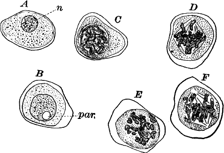
Fig. 127.—A, pollen mother cell of the wild onion.
n, nucleus. B–F, early stages in the division of the nucleus.
par. nucleolus; acetic acid, gentian violet, × 350.
If the spore mother cells are still quite young, we shall find the
nucleus (Fig. 127, A, n) comparatively small, and presenting a
granular appearance when strongly magnified. These granules, which
appear isolated, are really parts of filaments or segments, which are
closely twisted together, but scarcely visible in the resting nucleus.
On one side of the nucleus may usually be seen a large nucleolus
(called here, from its lateral position, paranucleus), and the whole
nucleus is sharply separated from the surrounding protoplasm by a thin
but evident membrane.The first indication of the approaching division of the nucleus is an
evident increase in size (B), and at the same time the colored
granules become larger, and show more clearly that they are in lines
indicating the form of the segments. These granules next become more
or less confluent, and the segments become very evident, appearing as
deeply stained, much-twisted threads filling the nuclear cavity
(Fig. 127, C), and about this time the nucleolus disappears.The next step is the disappearance of the nuclear membrane so that the
segments lie apparently free in the protoplasm of the cell. They
arrange themselves in a flat plate in the middle of the cell, this
plate appearing, when seen from the side, as a band running across the
middle of the cell. (Fig. 127, D, shows this plate as seen from the
side, E seen from above.)About the time the nuclear plate is complete, delicate lines may be
detected in the protoplasm converging at two points on opposite sides
of the cell, and forming a spindle-shaped figure with the nuclear
plate occupying its equator. This stage (D), is known as the
“nuclear spindle.” The segments of the nuclear plate next divide
lengthwise into two similar daughter segments (F), and these then
separate, one going to each of the new nuclei. This stage is not
always to be met with, as it seems to be rapidly passed over, but
patient search will generally reveal some nuclei in this condition.
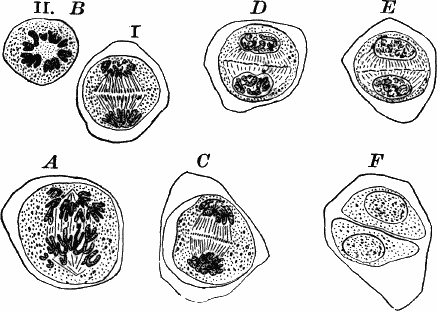
Fig. 128.—Later stages of nuclear divisions in the
pollen mother cell of wild onion, × 350. All the figures are seen from
the side, except B ii, which is viewed from the pole.
Although this is almost impossible to demonstrate, there are probably
as many filaments in the nuclear spindle as there are segments (in
this case about sixteen), and along these the nuclear segments travel
slowly toward the two poles of the spindle (Fig. 128, A, B). As
the two sets of segments separate, they are seen to be connected by
very numerous, delicate threads, and about the time the young nuclei
reach the poles of the nuclear spindle, the first trace of the
division wall appears in the form of isolated particles (microsomes),
which arise first as thickenings of these threads in the middle of
the cell, and appear in profile as a line of small granules not at
first extending across the cell, but later, reaching completely across
it (Fig. 128, C, E). These granules constitute the young cell wall
or “cell plate,” and finally coalesce to form a continuous membrane
(Fig. 128, F).The two daughter nuclei pass through the same changes, but in reverse
order that we saw in the mother nucleus previous to the formation of
the nuclear plate, and by the time the partition wall is complete the
nuclei have practically the same structure as the first stages we
examined (Fig. 128, F).[15]This complicated process of nuclear division is known technically as
“karyokinesis,” and is found throughout the higher animals as well as
plants.
The simple method of fixing and staining, just described, while giving
excellent results in many cases, is not always applicable, nor as a
rule are the permanent preparations so made satisfactory. For
permanent preparations, strong alcohol (for very delicate tissues,
absolute alcohol, when procurable, is best) is the most convenient
fixing agent, and generally very satisfactory. Specimens may be put
directly into the alcohol, and allowed to stay two or three days, or
indefinitely if not wanted immediately. When alcohol does not give
good results, specimens fixed with chromic or picric acid may
generally be used, and there are other fixing agents which will not be
described here, as they will hardly be used by any except the
professional botanist. Chromic acid is best used in a watery solution
(five per cent chromic acid, ninety-five per cent distilled water).
For most purposes a one per cent solution is best; in this the objects
remain from three or four to twenty-four hours, depending on size, but
are not injured by remaining longer. Picric acid is used as a
saturated solution in distilled water, and the specimen may remain for
about the same length of time as in the chromic acid. After the
specimen is properly fixed it must be thoroughly washed in several
waters, allowing it to remain in the last for twenty-four hours or
more until all trace of the acid has been removed, otherwise there is
usually difficulty in staining.
As staining agents many colors are used. The most useful are
hæmatoxylin, carmine, and various aniline colors, among which may be
mentioned, besides gentian violet, safranine, Bismarck brown, methyl
violet. Hæmatoxylin and carmine are prepared in various ways, but are
best purchased ready for use, all dealers in microscopic supplies
having them in stock. The aniline colors may be used either dissolved
in alcohol or water, and with all, the best stain, especially of the
nucleus, is obtained by using a very dilute, watery solution, and
allowing the sections to remain for twenty-four hours or so in the
staining mixture.
Hæmatoxylin and carmine preparations may be mounted either in
glycerine or balsam. (Canada balsam dissolved in chloroform is the
ordinary mounting medium.) In using glycerine it is sometimes
necessary to add the glycerine gradually, allowing the water to slowly
evaporate, as otherwise the specimens will sometimes collapse owing to
the too rapid extraction of the water from the cells. Aniline colors,
as a rule, will not keep in glycerine, the color spreading and finally
fading entirely, so that with most of them the specimens must be
mounted in balsam.
Glycerine mounts must be closed, which may be done with Canada balsam
as already described. The balsam is best kept in a wide-mouthed
bottle, specially made for the purpose, which has a glass cap covering
the neck, and contains a glass rod for applying the balsam.
Before mounting in balsam, the specimen must be completely freed from
water by means of absolute alcohol. (Sometimes care must be taken to
bring it gradually into the alcohol to avoid collapsing.[16]) If an
aniline stain has been used, it will not do to let it stay more than a
minute or so in the alcohol, as the latter quickly extracts the stain.
After dehydrating, the specimen should be placed on a clean slide in a
drop of clove oil (bergamot or origanum oil is equally good), which
renders it perfectly transparent, when a drop of balsam should be
dropped upon it, and a perfectly clean cover glass placed over the
preparation. The chloroform in which the balsam is dissolved will soon
evaporate, leaving the object embedded in a transparent film of balsam
between the slide and cover glass. No further treatment is necessary.
For the finer details of nuclear division or similar studies, balsam
mounts are usually preferable.
It is sometimes found necessary in sectioning very small and delicate
organs to embed them in some firm substance which will permit
sectioning, but these processes are too difficult and complicated to
be described here.
The following books of reference may be recommended. This list is, of
course, not exhaustive, but includes those works which will probably
be of most value to the general student.
- 1. Goebel. Outlines of Morphology and Classification.
- 2. Sachs. Physiology of Plants.
- 3. De Bary. Comparative Anatomy of Ferns and Phanerogams.
- 4. De Bary. Morphology and Biology of Fungi, Mycetozoa,
and Bacteria.
These four works are translations from the German,
and take the place of Sachs’s Text-book of Botany, a very
admirable work published first about twenty years ago,
and now somewhat antiquated. Together they constitute
a fairly exhaustive treatise on general botany.—New
York, McMillan & Co.
- 5. Gray. Structural Botany.—New York, Ivison & Co.
- 6. Goodale. Physiological Botany.—New York, Ivison & Co.
These two books cover somewhat the same ground as
1 and 2, but are much less exhaustive.
- 5. Strasburger. Das Botanische Practicum.—Jena.
Where the student reads German, the original is to be
preferred, as it is much more complete than the translations,
which are made from an abridgment of the original
work. This book and the next (7 and 8) are laboratory
manuals, and are largely devoted to methods of work.
- 7. Arthur, Barnes, and Coulter. Plant Dissection.—Holt
& Co., New York. - 8. Whitman. Methods in Microscopic Anatomy and Embryology.—Casino
& Co., Boston.
For identifying plants the following books may be mentioned:—
- Green algæ (exclusive of desmids, but including Cyanophyceæ
and Volvocineæ). - Wolle. Fresh-water Algæ of the United States.—Bethlehem,
Penn. - Desmids. Wolle. Desmids of the United States.—Bethlehem,
Penn. - The red and brown algæ are partially described in Farlow’s
New England Algæ. Report of United States Fish Commission,
1879.—Washington. - The Characeæ are being described by Dr. F. F. Allen of New
York. The first part has appeared. - The literature of the fungi is much scattered. Farlow and
Trelease have prepared a careful index of the American
literature on the subject. - Mosses. Lesquereux and James. Mosses of North America.—Boston,
Casino & Co. - Barnes. Key to the Genera of Mosses.—Bull. Purdue
School of Science, 1886. - Pteridophytes. Underwood. Our Native Ferns and their
Allies.—Holt & Co., New York. - Spermaphytes. Gray. Manual of the Botany of the Northern
United States. 6th edition, 1890. This also includes
the ferns, and the liverworts.—New York, Ivison & Co. - Coulter. Botany of the Rocky Mountains.—New York,
Ivison & Co. - Chapman. Flora of the Southern United States.—New
York, 1883. - Watson. Botany of California.
FOOTNOTES.
[1] For the mounting of permanent preparations, see Chapter XIX.
[2] The term “colony” is, perhaps, inappropriate, as the whole mass of
cells arises from a single one, and may properly be looked upon as an
individual plant.
[3] Algæ (sing. alga).
[4] “Host,” the plant or animal upon which a parasite lives.
[5] The antheridia, when present, arise as branches just below the oögonium,
and become closely applied to it, sometimes sending tubes through
its wall, but there has been no satisfactory demonstration of an actual
transfer of the contents of the antheridium to the egg cell.
[6] The filaments are attached to the surface of the leaf by suckers,
which are not so readily seen in this species as in some others. A mildew
growing abundantly in autumn on the garden chrysanthemum, however,
shows them very satisfactorily if a bit of the epidermis of a leaf on which
the fungus is just beginning to grow is sliced off with a sharp razor and
mounted in dilute glycerine, or water, removing the air with alcohol.
These suckers are then seen to be globular bodies, penetrating the outer
wall of the cell (Fig. 40).
[7] Sing. soredium.
[8] Sing. basidium.
[9] A vessel differs from a tracheid in being composed of several cells
placed end to end, the partitions being wholly or partially absorbed, so as
to throw the cells into close communication.
[10] In most conifers the symmetrical form of the young tree is maintained
as long as the tree lives.
[11] See the last chapter for details.
[12] The three outer stamens are shorter than the inner set.
[13] Monœcious: having stamens and carpels in different flowers, but on
the same plant.
[14] In a number of plants with showy flowers, e.g. violets, jewel-weed,
small, inconspicuous flowers are also formed, which are self-fertilizing.
These inconspicuous flowers are called “cleistogamous.”
[15] The division is repeated in the same way in each cell so that ultimately
four pollen spores are formed from each of the original mother cells.
[16] For gradual dehydrating, the specimens may be placed successively in
30 per cent, 50 per cent, 70 per cent, 90 per cent, and absolute alcohol.
INDEX.
| A | B | C | D | E | F | G | H | I | J | K | L | M |
| N | O | P | Q | R | S | T | U | V | W | X | Y | Z |
- Acacia, 209.
- Acer, -aceæ. See “Maple.”
- Acetic acid, 3, 59, 98, 138, 230.
- Achimenes, 218.
- Acorus. See “Sweet-flag.”
- Actinomorphic, 213.
- Adder-tongue, 116; Fig. 70. See also “Erythronium.”
- Adiantum. See “Maiden-hair.”
- Adlumia. See “Mountain-fringe.”
- Æsculinæ, 199.
- Æsculus. See “Buckeye,” “Horse-chestnut.”
- Aggregatæ, 222.
- Alcohol, 5, 31, 55, 83, 230, 233.
- Algæ, 4, 21.
- Alga-fungi. See “Phycomycetes.”
- Alisma, -ceæ. See “Water-plantain.”
- Allium. See “Wild onion.”
- Amaranth, 185.
- Amarantus, -aceæ. See “Amaranth.”
- Amœba, 7; Fig. 2.
- Ampelidæ. See “Vine.”
- Ampelopsis. See “Virginia creeper.”
- Anatomy, 3.
- Anatropous, 151.
- Andreæaceæ, 99, 100.
- Andrœcium, 148.
- Andromeda, 211.
- Anemone, 185.
- Angiocarpæ, 84.
- Angiosperm, 129, 143, 145.
- Aniline colors, 233.
- Anisocarpæ, 210, 213.
- Anonaceæ. See “Custard-apple.”
- Anther, 148, 175, 179.
- Antheridium, 27, 36, 39, 45, 51, 59, 68, 89, 96, 106, 122.
- Anthoceros, Anthoceroteæ, 91; Fig. 57.
- Aphanocyclæ, 185, 196.
- Aplectrum, 167; Fig. 90.
- Apocynum, -aceæ. See “Dog-bane.”
- Apostasieæ, 164.
- Apple, 145, 171, 206; Fig. 114.
- Apricot, 207.
- Aquilegia. See “Columbine.”
- Aralia, -aceæ. See “Spikenard.”
- Archegonium, 89, 97, 105, 122, 133, 140, 144.
- Archicarp, 138, 145.
- Arcyria, 13; Fig. 5.
- Arethusa, Arethuseæ, 166; Fig. 90.
- Argemone, 191.
- Aril, 189.
- Arisæma, 78, 157; Fig. 86.
- Aristolochia, -aceæ, 224.
- Aroid, Aroideæ, 157.
- Arrow-grass, 167.
- Arrowhead, 167; Fig. 91.
- Arrowroot, 163.
- Asarum. See “Wild ginger.”
- Asclepias, -daceæ. See “Milk-weed.”
- Ascobolus, 71–73; Fig. 43.
- Ascomycetes, 65, 66.
- Ascospore, 66.
- Ascus, 66, 69.
- Ash, 218; Fig. 122.
- Asimina. See “Papaw.”
- Aspidium, Fig. 70.
- Asplenium, 104; Fig. 70.
- Aster, 224.
- Atropa. See “Deadly nightshade.”
- Axil, 174.
- Azalea, 210; Fig. 116.
- Azolla, 117; Fig. 71.
- Bacteria, 15, 17, 19; Fig. 8.
- Balsam, Balsamineæ, 198.
- Bamboo, 162.
- Bambusa. See “Bamboo.”
- Banana, 163.
- Barberry, 17, 187; Fig. 101.
- Bark. See “Cortex.”
- Basidiomycetes, 77.
- Basidium, 77, 80, 83.
- Basswood, 195; Fig. 106.
- Bast. See “Phloem.”
- Batatas. See “Sweet-potato.”
- Batrachospermum, 53; Fig. 31.
- Bean, 207, 208.
- Bear-grass. See “Yucca.”
- Bee, 227, 228.
- Beech, 183.
- Beech-drops, 218.
- Beet, 184.
- Beggar’s-ticks, 215.
- Begonia, 3, 205.
- Bell-flower, 220, 226; Fig. 123.
- Bellwort, 156.
- Berberis, -ideæ. See “Barberry.”
- Bergamot oil, 234.
- Berry, 145, 156.
- Betulaceæ, 183.
- Bicornes, 210.
- Bignonia, -aceæ, 218.
- Biology, 2.
- Birch, 183.
- Bird’s-nest fungus. See “Cyathus.”
- Bishop’s cap, 202; Fig. 111.
- Bismarck brown, 233.
- Bitter-sweet, 199; Fig. 109.
- Black alder, 199.
- Blackberry, 207.
- Black fungi. See “Pyrenomycetes.”
- Bladder-nut, 199; Fig. 108.
- Bladder-weed, 33, 217; Fig. 120.
- Bleeding-heart. See “Dicentra.”
- Blood-root, 191; Fig. 103.
- Blue-eyed grass, 156.
- Blue-flag. See “Iris.”
- Blue-green slime, 15.
- Blue valerian. See “Polemonium.”
- Borage, 215.
- Borragineæ. See “Borage.”
- Bordered pits, 138.
- Botany defined, 2.
- systematic, 3.
- Botrychium. See “Grape fern.”
- Box, 201.
- Bract, 199, 222, 229.
- Brasenia. See “Water-shield.”
- Breathing pore, 91, 99, 113, 130, 147, 150, 177.
- Bromeliaceæ, 156.
- Bryophyte, 86.
- Buck-bean, 218.
- Buckeye, 171, 199.
- Buckthorn, 199.
- Buckwheat, 184.
- Budding, 64.
- Bulbochæte, 28; Fig. 16.
- Bulb, 146, 153, 172.
- Bulrush, 161; Fig. 87.
- Bundle-sheath, 110, 176.
- Burning-bush. See “Spindle-tree.”
- Bur-reed, 159; Fig. 86.
- Buttercup, 181, 185; Fig. 99.
- Butterfly, 227, 228.
- Button-bush, 223.
- Buttonwood. See “Sycamore.”
- Buxus, Buxaceæ. See “Box.”
- Cabbage, 192.
- Cabombeæ, 190.
- Cactus, Cactaceæ, 203; Fig. 112.
- Cæsalpineæ, 210.
- Calcium, 2.
- Calla, 157, 229.
- Callithamnion, 50–52; Fig. 29.
- Callitriche, -chaceæ. See “Water starwort.”
- Calluna. See “Heath.”
- Calopogon, 166; Fig. 91.
- Calycanthus, -aceæ, 187; Fig. 100.
- Calycereæ, 223.
- Calycifloræ, 200.
- Calyx, 174, 182.
- Cambium, 137–138, 175.
- Campanula. See “Bell-flower.”
- Campanulaceæ, 220.
- Campanulinæ, 220.
- Canada balsam, 230–234.
- Canada thistle, 224; Fig. 125.
- Canna, -aceæ, 162, 163; Fig. 88.
- Caper family, 194.
- Capparis, -ideæ. See “Caper.”
- Caprifoliaceæ, 223.
- Capsella. See “Shepherd’s-purse.”
- Caraway, 202.
- Carbon, 2, 95.
- Carbon-dioxides, 95.
- Cardinal-flower. See “Lobelia.”
- Carex, 161; Fig. 87.
- Carmine, 25, 233.
- Carnation, 185.
- Carpel, 148, 154, 175, 179.
- Carpophyll. See “Carpel.”
- Carpospore, 51–53.
- Carrot, 202.
- Caryophylleæ. See “Pink.”
- Caryophyllus. See “Clove.”
- Castalia, 189.
- Castor-bean, 200.
- Catalpa, 218.
- Cat-brier, 154.
- Catkin, 181.
- Catnip, 215.
- Cat-tail, 159.
- Cedar apple, Cedar rust. See “Gymnosporangium.”
- Celastraceæ, 199.
- Celastrus. See “Bitter-sweet.”
- Celery, 3.
- Cell, 6.
- Cellulose, 3.
- Centaury, 219.
- Centrospermæ, 183.
- Cephalanthus. See “Button-bush.”
- Cerastium. See “Chick-weed.”
- Ceratophyllum. See “Horned pond-weed.”
- Cercis. See “Red-bud.”
- Chamærops. See “Palmetto.”
- Chara, 38–40; Fig. 23.
- Characeæ, 21, 37, 40.
- Chareæ, 40.
- Cheiranthus. See “Wall-flower.”
- Chenopodium, -aceæ. See “Goose-foot.”
- Cherry, 15, 206; Fig. 114.
- Chicory, 223.
- Chick-weed, 185; Fig. 98.
- Chimaphila. See “Prince’s pine.”
- Chionanthus. See “Fringe-tree.”
- Chlorine, 2.
- Chlorococcum, 23; Fig. 12.
- Chloroform, 234.
- Chloroplast, 22, 45.
- Chlorophyll, 15.
- Chlorophyll body. See “Chloroplast.”
- Chlorophyceæ, 21.
- Chondrus. See “Irish moss.”
- Choripetalæ, 181, 208.
- Chromic acid, 25–35, 233.
- Chromoplast, 150.
- Cicinnobulus, 69; Fig. 39.
- Cilium, 8.
- Cinquefoil, 206.
- Cistaceæ. See “Rock-rose.”
- Cistifloræ, 192.
- Citron, 196.
- Citrus. See “Orange,” “Lemon.”
- Cladophora, 24, 25.
- Classification, 3–9.
- Clavaria, 85; Fig. 51.
- Claytonia. See “Spring-beauty.”
- Clematis, 185.
- Climbing plants, 171.
- Closterium, 33; Fig. 20.
- Clove, 205.
- Clove oil, 234.
- Clover, 207.
- Club moss, 116.
- Cluster-cup, 78.
- Cocos. See “Palm-coco,” 159.
- Coleochæte, 28; Fig. 17.
- Collateral fibro-vascular bundle, 135.
- Collema, 76; Fig. 44.
- Columella, 55.
- Columbine, 186; Fig. 99.
- Column, 165.
- Columniferæ, 195.
- Commelyneæ, 157.
- Compositæ, 223, 224.
- Compound flower, 224.
- Conceptacle, 45.
- Cone, 131.
- Conferva, 26.
- Confervaceæ, 21, 24.
- Conidium, 68.
- Conifer, 129, 140, 141.
- Coniferæ. See “Conifer.”
- Conjugatæ, 22–29.
- Connective, 148.
- Conocephalus. See “Liverwort, giant.”
- Contortæ, 218.
- Convolvulaceæ, 213.
- Convolvulus. See “Morning-glory.”
- Coprinus, 82–84; Fig. 48.
- Coral root, 167.
- Corallorhiza. See “Coral root.”
- Coriander, 202.
- Corn, 160, 161.
- Cornus, -aceæ. See “Dogwood.”
- Corolla, 174, 182.
- Cortex, 39, 130.
- Corydalis, 192.
- Cotton, 195.
- Cotyledon, 134, 146, 180.
- Cowslip, 211.
- Coxcomb, 185.
- Crab-apple, 77, 80.
- Cranberry, 211.
- Crassulaceæ, 203.
- Crane’s-bill, 3, 196; Fig. 107.
- Cress, 192.
- Croton, 200.
- Cruciferæ. See “Mustard family.”
- Crucifloræ. See “Rhœadinæ.”
- Cucumber, 221.
- Cucumber-tree. See “Magnolia.”
- Cucurbitaceæ. See “Gourd.”
- Cup fungi (“Discomycetes”), 71.
- Cupuliferæ, 183.
- Curl, 66.
- Currant, 203.
- Cuscuta. See “Dodder.”
- Custard-apple, 186.
- Cyanophyceæ. See “Blue-green slime.”
- Cyathus, 84; Fig. 50.
- Cycad, -eæ, 140.
- Cycas revoluta, 141; Fig. 71.
- Cyclamen, 212.
- Cynoglossum. See “Hound’s-tongue.”
- Cyperaceæ. See “Sedge.”
- Cyperus, 161.
- Cypress, 142.
- Cypripedium. See “Lady’s-slipper.”
- Cystopus. See also “White rust.”
- Daisy, 223.
- Dandelion, 66, 223; Fig. 125.
- Darlingtonia, 195.
- Datura. See “Stramonium.”
- Day lily, 155.
- Deadly nightshade, 215.
- Dead nettle, 215; Fig. 120.
- Delphinium. See “Larkspur.”
- Dermatogen, 176.
- Desmid, 33, 34; Fig. 20.
- Devil’s apron. See “Laminaria.”
- Dianthus. See “Pink.”
- Diatomaceæ, 41, 42; Figs. 24, 25.
- Dicentra, 192; Fig. 103.
- Dicotyledon, 145, 170, 181, 225.
- Digitalis. See “Foxglove.”
- Diœcious, 88.
- Dionæa. See “Venus’s fly-trap.”
- Dioscoreæ. See “Yam.”
- Dioscorea villosa, 154.
- Diospyros. See “Persimmon.”
- Diospyrinæ, 210.
- Dipsacus, -aceæ. See “Teasel.”
- Dirca. See “Moosewood.”
- Ditch-moss, 167; Fig. 91.
- Dodder, 214.
- Dodecatheon. See “Shooting-star.”
- Dog-bane, 219; Fig. 122.
- Dogwood, 202, 229; Fig. 110.
- Draparnaldia, 26; Fig. 14.
- Drosera -aceæ. See “Sun-dew.”
- Drupe. See “Stone-fruit.”
- Duck-weed, 159; Fig. 86.
- Dutchman’s pipe. See “Aristolochia.”
- Earth star. See “Geaster.”
- Ebenaceæ (ebony), 212.
- Echinospermum. See “Beggar’s-ticks.”
- Ectocarpus, 45, 47; Fig. 28.
- Eel-grass, 168, 169; Fig. 91.
- Egg apparatus, 144.
- Egg cell, 27, 36, 39, 45, 90, 106, 133, 144.
- Egg-plant, 215.
- Eichler, 153.
- Elater, 91, 122.
- Elder, 224.
- Elæagnaceæ, 206.
- Elm, 183.
- Elodea. See “Ditch-moss.”
- Embryo, 90, 97, 107, 133, 149, 180.
- Embryology, 3.
- Embryo sac, 143, 144, 151.
- Enantioblastæ, 153, 156; Fig. 85.
- Endosperm, 133, 146, 152.
- Entire leaves, 170.
- Entomophthoreæ, 57.
- Epacrideæ, 210.
- Epidermis, 91, 111, 112, 113, 122, 135, 137, 150, 177.
- Epigæa. See “Trailing arbutus.”
- Epilobium. See “Willow-herb.”
- Epiphegus. See “Beech-drops.”
- Epiphyte, 166.
- Equisetum, -tinæ. See “Horse-tail.”
- Ergot, 76.
- Erica, -aceæ. See “Heath.”
- Erysiphe, 70.
- Erythræa. See “Centaury.”
- Erythronium, 146–152; Fig. 81.
- Eschscholtzia, 191.
- Eucalyptus, 206.
- Eucyclæ, 196, 200.
- Eudorina, 20.
- Euglena, 11, 19; Fig. 9.
- Euonymus. See “Spindle-tree.”
- Euphorbia, 199; Fig. 109.
- Eurotium, 70; Fig. 42.
- Evening primrose, 206.
- Exoascus, 66.
- Fagopyrum. See “Buckwheat.”
- Feather-veined. See “Pinnate-veined.”
- Fern, 5, 102, 104, 116.
- flowering, 118; Fig. 70.
- lady, 104; Fig. 70.
- maiden-hair. See “Maiden-hair fern.”
- ostrich. See “Ostrich-fern.”
- sensitive, 104.
- true, 117.
- water. See “Water-fern.”
- Fertilization, 225.
- Fibre, 124, 175, 177.
- Fibro-vascular bundle, 107, 110, 121, 123, 135, 136, 147, 150, 159, 174.
- Fig, 183.
- Figwort, 215, 216; Fig. 120.
- Filament (of stamen), 148, 17.
- Filices. See “True ferns.”
- Filicineæ. See “Fern.”
- Fir, 142.
- Fission, 23.
- Flagellata, 19.
- Flagellum, 19.
- Flax, 197; Fig. 107.
- Flies, 229.
- Flower, 128, 131.
- Flowering-plant. See “Spermaphyte.”
- Forget-me-not, 215.
- Four-o’clock, 183.
- Foxglove, 217.
- Frangulinæ, 199.
- Fraxinus. See “Ash.”
- Fringe-tree, 218; Fig. 122.
- Fruit, 145.
- Fucaceæ, 43.
- Fuchsia, 201.
- Fucus, 42–46.
- Fumariaceæ. See “Fumitory.”
- Fumitory, 192.
- Funaria, 93–99; Figs. 58–62.
- gross anatomy, 93, 94.
- protonema, 93.
- “flower,” 94.
- structure of leaf, 94.
- chloroplasts, division of, 95.
- formation of starch in chloroplasts, 95.
- structure of stem, 96.
- root hairs, 96.
- buds, 96.
- antheridium spermatozoids, 96, 97.
- archegonium, 97.
- embryo, 98.
- capsule and spores, 98, 99.
- germination of spores, 99.
- Fungi, culture of, 5, 54.
- true. See “Mycomycetes.”
- alga. See “Phycomycetes.”
- Funiculus, 151, 175.
- Funkia. See “Day lily.”
- Galium, 223; Fig. 124.
- Gamopetalæ. See “Sympetalæ.”
- Gaultheria. See “Wintergreen.”
- Gaylussacia. See “Huckleberry.”
- Geaster, 84; Fig. 49.
- Gentian, 218; Fig. 122.
- Gentian violet, 4, 138, 231.
- Gentiana, -aceæ. See “Gentian.”
- Geranium, -aceæ, 3, 171, 196; Fig. 107.
- Gerardia, 217.
- Germ cell. See “Egg cell.”
- Gesneraceæ, 218.
- Ghost flower. See “Indian-pipe.”
- Gill, 83.
- Ginger, 163.
- Gingko, 142; Fig. 78.
- Gleditschia. See “Honey locust.”
- Gloxinia, 218.
- Glumaceæ, 153, 160; Fig. 87.
- Glume, 162.
- Glycerine, 4, 51, 55, 59, 67, 83, 98, 224, 231, 233.
- Gnetaceæ. See “Joint fir.”
- Golden-rod, 224.
- Gonium, 20.
- Gooseberry, 203; Fig. 111.
- Goose-foot, 184; Fig. 98.
- Gossypium. See “Cotton.”
- Gourd, 221.
- Gramineæ. See “Grass.”
- Grape, 171, 199; Fig. 109.
- Grape fern, 116; Fig. 70.
- Graphis, 75; Fig. 45.
- Grass, 161, 225; Fig. 87.
- Gray moss. See “Tillandsia.”
- Green-brier, 154.
- Green-felt. See “Vaucheria.”
- Green monad, 12, 19.
- Green slime, 21, 22; Fig. 11.
- Ground pine, 123; Fig. 73.
- Ground tissue, 110, 111, 113, 124, 137, 177, 178.
- Gruinales, 196.
- Guard cell, 113, 135, 150.
- Gulf weed. See “Sargassum.”
- Gum. See “Eucalyptus.”
- Gymnocarpæ, 84.
- Gymnosperm, 129, 141.
- Gymnosporangium, 79–81; Fig. 47.
- Gynandræ, 153, 164.
- Gynœcium, 148, 167.
- Gynostemium. See “Column.”
- Habenaria, 166, 227; Fig. 90.
- Hæmatoxylin, 233.
- Hair, 8, 177.
- Haloragidaceæ, 206.
- Hazel, 182, 183, 225; Fig. 97.
- Head, 181.
- Heath, 211.
- Helobiæ, 153, 167.
- Hemerocallis. See “Day lily.”
- Hemi-angiocarpæ, 84.
- Hemlock, 142; Fig. 78.
- Hemp, 183.
- Hepaticæ. See “Liverwort.”
- Hermaphrodite, 199.
- Heterocyst, 17.
- Heterostylism, 228.
- Hibiscus, 195.
- Hickory, 170, 183.
- Holly, 199.
- Hollyhock, 195.
- Honey locust, 209.
- Honeysuckle, 170, 172, 181, 223; Fig. 124.
- Hop, 171, 181; Fig. 97.
- Horned pond-weed, 224.
- Horse-chestnut, 170, 199.
- Horse-tail, 116–120.
- Hound’s-tongue, 215; Fig. 119.
- Houstonia, 223; Fig. 124.
- Hoya. See “Wax-plant.”
- Huckleberry, 181, 211; Fig. 116.
- Humming-bird, 226.
- Hyacinth, 146.
- Hydnum, 84; Fig. 51.
- Hydrangea, -geæ, 202; Fig. 111.
- Hydrocharideæ, 167.
- Hydrogen, 2, 95.
- Hydropeltidinæ, 189.
- Hydrophyllum, -aceæ. See “Water-leaf.”
- Hypericum, -aceæ. See “St. John’s-wort.”
- Ilex. See “Holly.”
- Impatiens. See “Jewel-weed,” “Balsam.”
- India-rubber, 200.
- Indian-pipe, 144, 210; Fig. 79.
- Indian turnip. See “Arisæma.”
- Indusium, 118.
- Inflorescence, 157.
- Integument, 133, 144, 151, 180.
- Intercellular space, 124, 135, 150.
- Internode, 39.
- Iodine, 4, 22, 31.
- Ipomœa, 213.
- Iridaceæ, 156.
- Iris, 154, 156; Fig. 84.
- Irish moss, 49.
- Isocarpæ, 210, 212.
- Isoetes. See “Quill-wort.”
- Iulifloræ, 181.
- Ivy, 202.
- Jack-in-the-pulpit. See “Arisæma.”
- Jasmine, 218.
- Jeffersonia. See “Twin-leaf.”
- Jewel-weed, 197; Fig. 107.
- Joint fir, 140, 142.
- Juncagineæ, 167.
- Juncus. See “Rush.”
- Jungermanniaceæ, 92; Fig. 57.
- Kalmia. See “Mountain laurel.”
- Karyokinesis, 233.
- Keel, 208.
- Kelp. See “Laminaria.”
- giant. See “Macrocystis.”
- Knotgrass. See “Polygonum.”
- Labellum. See “Lip.”
- Labiatæ. See “Mint.”
- Labiatifloræ, 215.
- Lady’s-slipper, 164, 166, 198; Fig. 90.
- Lamella, 83.
- Laminaria, 45, 47; Fig. 28.
- Lamium. See “Dead nettle.”
- Larch. See “Tamarack.”
- Larix. See “Tamarack.”
- Larkspur, 186, 227; Fig. 99.
- Latex, 191.
- Laurel, 188.
- Laurineæ. See “Laurel.”
- Lavender, 215.
- Leaf-green. See “Chlorophyll.”
- Leaf tendril, 171.
- Leaf thorn, 172.
- Leguminosæ, 207.
- Lemanea, 53; Fig. 31.
- Lemna. See “Duck-weed.”
- Lemon, 198.
- Lentibulariaceæ, 217.
- Lettuce, 223.
- Lichenes, 73; Figs. 44, 45.
- Ligula, 127.
- Ligulatæ, 125.
- Lilac, 170, 181, 218.
- Liliaceæ, 155.
- Liliifloræ, 153, 155; Fig. 83.
- Lilium. See “Lily.”
- Lily, 146, 155.
- Lily-of-the-valley, 155.
- Lime. See “Linden.”
- Linden, 195; Fig. 106.
- Linear, 159.
- Linum, -aceæ. See “Flax.”
- Lip, 165.
- Liriodendron. See “Tulip-tree.”
- Lithospermum. See “Puccoon.”
- Liverwort, 86.
- classification of, 91.
- horned. See “Anthoceroteæ.”
- giant, 91; Fig. 57.
- Lizard-tail, 181, 183; Fig. 97.
- Lobelia, -aceæ. 221; Fig. 123.
- Loganieæ, 219.
- Lonicera. See “Honeysuckle.”
- Loosestrife. See “Lythrum.”
- swamp. See “Nesæa.”
- Lotus. See “Nelumbo.”
- Lychnis, 185.
- Lycoperdon, 84; Fig. 49.
- Lycopersicum. See “Tomato.”
- Lycopodiaceæ. See “Ground pine.”
- Lycopodinæ. See “Club moss.”
- Lycopodium, 123.
- Lysimachia. See “Moneywort.”
- Lythrum, -aceæ, 206, 228.
- Mace, 189.
- Macrocystis, 48.
- Macrospore, 126, 127, 128, 143.
- Madotheca, 86–90; Figs. 52–56.
- Magnesium, 2.
- Magnolia, -aceæ, 186.
- Maiden-hair fern, 109–115; Figs. 67–69.
- Mallow, 171, 195; Fig. 106.
- Malva, -aceæ. See “Mallow.”
- Mamillaria, Fig. 112.
- Mandrake. See “May-apple.”
- Maple, 199; Fig. 108.
- Maranta. See “Arrowroot.”
- Marattiaceæ. See “Ringless ferns.”
- Marchantia, 91; Fig. 57.
- Marchantiaceæ, 91.
- Marsilia, 118; Fig. 71.
- Martynia, 218.
- Matthiola. See “Stock.”
- May-apple, 187; Fig. 101.
- May-weed, 223; Fig. 125.
- Medeola, 155; Fig. 83.
- Medullary ray, 130, 137.
- Melampsora, 81.
- Melastomaceæ, 206.
- Melon, 221.
- Menispermum, -eæ. See “Moon-seed.”
- Menyanthes. See “Buck-bean.”
- Mesocarpus, 33; Fig. 19.
- Mesophyll, 135.
- Methyl-violet, 4, 233.
- Micropyle, 180.
- Microsome, 231.
- Microspore, 126, 128, 131, 138.
- Mignonette, 192; Fig. 104.
- Mildew. See “Peronospora,” “Phytophthora,” “Perisporiaceæ.”
- Milk-weed, 220; Fig. 122.
- Milkwort, 199.
- Mimosa. See “Sensitive-plant.”
- Mimosaceæ, 209, 210.
- Mimulus, 217.
- Mint, 181, 215.
- Mirabilis. See “Four-o’clock.”
- Mistletoe, 224.
- Mitella. See “Bishop’s cap.”
- Mitchella. See “Partridge-berry.”
- Mitre-wort. See “Bishop’s cap.”
- Mock-orange. See “Syringa.”
- Moneywort, 212; Fig. 117.
- Monocotyledon, 146, 153, 225, 229.
- Monotropa. See “Indian-pipe,” “Pine-sap.”
- Monotropeæ, 210.
- Moon-seed, 188; Fig. 101.
- Moosewood, 206; Fig. 113.
- Morchella. See “Morel.”
- Morel, 73.
- Morning-glory, 171, 213; Fig. 118.
- Morphology, 3.
- Moss, 5, 86.
- true, 93.
- common. See “Bryaceæ.”
- peat. See “Sphagnaceæ.”
- Moth, 229.
- Mould, black. See “Mucorini.”
- blue. See “Penicillium.”
- herbarium. See “Eurotium.”
- insect. See “Entomophthoreæ.”
- water. See “Saprolegnia.”
- Mountain-fringe, 192.
- Mountain-laurel, 210; Fig. 116.
- Mucor, 55.
- Mucor stolonifer, 55–56.
- Mucorini, 54.
- Mulberry, 183.
- Mullein, 217; Fig. 120.
- Musa, -aceæ. See “Banana.”
- Musci. See “True mosses.”
- Mushroom, 82.
- Mustard, 192.
- Mycomycetes. See “True fungi.”
- Myosotis. See “Forget-me-not.”
- Myristica, -ineæ. See “Nutmeg.”
- Myrtifloræ, 205.
- Myrtle, 205, 206.
- Myrtus. See “Myrtle.”
- Myxomycetes. See “Slime-mould.”
- Naias. See “Pond-weed.”
- Naiadeæ, 159.
- Narcissus, 146.
- Nasturtium, 197, 227.
- Navicula, 42; Fig. 24.
- Nectar, 225.
- Nectary, 186.
- Nelumbo, 189, 190; Fig. 101.
- Nelumbieæ, 190.
- Nemophila, 214.
- Nepenthes, -eæ. See “Pitcher plant.”
- Nesæa, 206.
- Nettle. See “Urticinæ.”
- Nicotiana. See “Tobacco.”
- Night-blooming cereus, 204.
- Nightshade, 215; Fig. 119.
- Nitella, 40.
- Nitelleæ, 40.
- Node, 39.
- Nucleus, 7, 31, 231.
- Nuclear division, 7, 31, 231; Figs. 127, 128.
- Nucleolus, 7, 231.
- Nutmeg, 188.
- Nyctagineæ, 183.
- Nymphæa, 189; Fig. 101.
- Nymphæaceæ, 190.
- Oak, 183, 225; Fig. 97.
- Œdogonium, 26–28; Fig. 16.
- Œnothera. See “Evening primrose.”
- Oil-channel, 202.
- Oleaceæ. See “Olive.”
- Oleander, 219.
- Olive, 218.
- Onagraceæ, 206.
- Onoclea, 104; Fig. 70.
- Oögonium, 27, 36, 39, 45, 59, 62.
- Oöphyte, 109.
- Opium–opium poppy, 191.
- Ophioglosseæ. See “Adder-tongue.”
- Ophioglossum, 116.
- Opuntia. See “Prickly pear.”
- Opuntieæ, 203.
- Orange, 198.
- Orchid, 164, 166, 227; Figs. 89, 90.
- Orchideæ, 164.
- Orchis, 227; Fig. 89.
- Organic bodies, 1.
- Origanum oil, 234.
- Oscillaria, 15, 16; Fig. 6.
- Osmunda. See “Flowering-fern.”
- Ostrich-fern, 104–109.
- Ovary, 129, 148, 156, 202.
- Ovule, 129, 131, 144, 148, 151, 179.
- Oxalis. See “Wood-sorrel.”
- Oxydendrum, 211; Fig. 116.
- Oxygen, 2, 95.
- Palea, 161.
- Palisade parenchyma, 178.
- Palm, 157.
- Palmæ. See “Palm.”
- Palmate, 171.
- Palmetto, 159.
- Pandaneæ, 159.
- Papaveraceæ. See “Poppy.”
- Papaw, 186; Fig. 100.
- Papilionaceæ, 208.
- Pappus, 223.
- Papyrus, 161.
- Paranucleus, 231.
- Parasite, 54.
- Parenchyma. See “Soft tissue.”
- Parmelia, 73, 75; Fig. 44.
- Partridge-berry, 223, 228.
- Passiflora. See “Passion-flower.”
- Passiflorinæ, 205.
- Passion-flower, 204; Fig. 112.
- Pea, 207, 208; Fig. 115.
- Peach, 206.
- Pear, 206.
- Pediastrum, 23; Fig. 11.
- Pelargonium, 197.
- Peltate, 190.
- Peltigera, 75; Fig. 45.
- Penicillium, 71; Fig. 42.
- Pepper, 183.
- Perianth. See “Perigone.”
- Periblem, 176.
- Perigone, 143, 148, 151, 170.
- Perisperm, 163.
- Perisporiaceæ, 66.
- Periwinkle, 219.
- Peronospora, 60; Fig. 35.
- Peronosporeæ, 57.
- Persimmon, 212; Fig. 117.
- Petal, 148, 174, 179.
- Petiole, 173.
- Petunia, 215; Fig. 119.
- Peziza, 73; Fig. 43.
- Phacelia, 214.
- Phæophyceæ. See “Brown algæ.”
- Phænogam. See “Spermaphyte.”
- Phascum, -aceæ, 99, 101; Fig. 65.
- Philadelphus. See “Syringa.”
- Phloem, 110, 124, 135, 137, 150, 173, 176.
- Phlox, 214; Fig. 118.
- Phœnix dactylifera. See “Date-palm.”
- Phosphorus, 2.
- Phragmidium, 81; Fig. 47.
- Physarum, 14.
- Physianthus, 220.
- Physiology, 3.
- Phytolacca, -aceæ. See “Poke-weed.”
- Phytophthora, 60.
- Pickerel-weed, 156, 228; Fig. 84.
- Picric acid, 156, 233.
- Pig-weed. See “Amaranth.”
- Pine, 9, 10, 129, 142.
- Pineapple, 156.
- Pine-sap, 210; Fig. 116.
- Pinguicula, 218.
- Pink, 181, 185; Fig. 97.
- Pink-root, 218; Fig. 122.
- Pinnate (leaf), 159.
- veined, 171.
- Pinnularia, 42; Fig. 24.
- Pinus sylvestris. See “Scotch pine.”
- Piper. See “Pepper.”
- Piperineæ, 183.
- Pistil, 143, 145, 174.
- Pitcher-plant, 194, 195; Fig. 105.
- Pith, 130, 174, 177.
- Placenta, 148, 179.
- Plane, 183.
- Plantago, -ineæ. See “Plantain.”
- Plantain, 223, 225; Fig. 121.
- Plasmodium, 12.
- Plataneæ. See “Plane.”
- Platanus. See “Sycamore.”
- Plerome, 176.
- Plum, 207.
- Plumbago, -ineæ, 212.
- Pod, 156.
- Podophyllum. See “May-apple.”
- Podosphæra, 66–70; Fig. 39.
- Pogonia, 166.
- Poinsettia, 199.
- Poison-dogwood, 198.
- Poison-hemlock, 202.
- Poison-ivy, 171, 198.
- Poke-weed, 185; Fig. 97.
- Polemonium, -aceæ, 214; Fig. 118.
- Pollinium, 165.
- Polycarpæ, 185.
- Polygala, -aceæ. See “Milkwort.”
- Polygonatum. See “Solomon’s Seal.”
- Polygonum, -aceæ, 184; Fig. 98.
- Polysiphonia, 52; Fig. 29.
- Pomegranate, 206.
- Pond-scum, 22, 29, 30.
- Pond-weed, 159; Fig. 86.
- Pontederia. See “Pickerel-weed.”
- Poplar, 181, 183.
- Poppy, 191.
- Portulaca, -aceæ. See “Purslane.”
- Potash (caustic), 4, 5, 59, 67, 75, 97, 106, 111, 151, 176, 179, 180.
- Potassium, 2.
- Potato, 215.
- Potato-fungus. See “Phytophthora.”
- Potentilla. See “Cinquefoil.”
- Potomogeton. See “Pond-weed.”
- Prickly-ash, 198.
- Prickly fungus. See “Hydnum.”
- Prickly-pear, 204.
- Prickly-poppy. See “Argemone.”
- Primrose, 211.
- Primula, -aceæ. See “Primrose.”
- Prince’s-pine, 210; Fig. 116.
- Procarp, 51.
- Proteaceæ, 205.
- Prothallium, 102, 103, 114, 122, 125, 133, 144, 177.
- Protococcus, -aceæ, 22, 74; Fig. 11.
- Protophyte, 11.
- Protoplasm, 7.
- movements of, 7.
- Pteridophyte, 102, 153.
- Puccinia, 81; Fig. 47. See also “Wheat-rust.”
- Puccoon, 215.
- Puff-ball. See “Lycoperdon.”
- Purslane, 185.
- Putty-root. See “Aplectrum.”
- Pyrenoid, 25, 31.
- Pyrenomycetes, 76.
- Pyrola, -aceæ, 210.
- Raceme, 174.
- Radial fibro-vascular bundles, 138, 176.
- Radish, 192.
- Ranunculus, -aceæ. See “Buttercup.”
- Raspberry, 207.
- Ray-flower, 223.
- Receptacle, 167, 207, 223.
- Receptive spot, 106.
- Red algæ, 21, 49, 52, 53; Figs. 29–31.
- Red-bud, 209; Fig. 115.
- Red cedar, 79, 131, 141; Fig. 78.
- Red-wood, 142.
- Reference-books, 235–236.
- Reseda, -aceæ. See “Mignonette.”
- Resin, 130.
- Resin-duct, 130, 135, 137.
- Resting-spore, 28, 32, 37, 57.
- Rheumatism-root. See “Twin-leaf.”
- Rhexia, 206.
- Rhizocarpeæ. See “Water-fern.”
- Rhizoid. See “Root-hair.”
- Rhizome. See “Root-stock.”
- Rhododendron, 210; Fig. 116.
- Rhodophyceæ. See “Red algæ.”
- Rhodoraceæ, 211.
- Rhœadinæ, 190.
- Rhus. See “Sumach.”
- cotinus. See “Smoke-tree.”
- toxicodendron. See “Poison-ivy.”
- venenata. See “Poison-dogwood.”
- Ribes, -ieæ, 203; Fig. 111.
- Ricciaceæ, 91; Fig. 57.
- Richardia. See “Calla.”
- Ricinus. See “Castor-bean.”
- Ringless-fern, 116.
- Rock-rose, 195.
- Rock-weed. See “Fucus.”
- Root, 102, 104, 114, 173.
- Root-cap, 115, 175.
- Root-hair, 38, 87, 91, 96, 104, 135.
- Root-stock, 154, 172.
- Rosa, -aceæ. See “Rose.”
- Rose, 181, 206; Fig. 114.
- Rosifloræ, 206.
- Rubiaceæ, 223.
- Rush, 154, 225; Fig. 83.
- Rust, white. See “Cystopus.”
- Sabal. See “Palmetto.”
- Sabbatia. See “Centaury.”
- Saccharomycetes. See “Yeast.”
- Sac fungi. See “Ascomycetes.”
- Safranine, 233.
- Sage, 215; Fig. 120.
- Salicineæ, 183.
- Salix. See “Willow.”
- Salvinia, 118.
- Sambucus. See “Elder.”
- Sanguinaria. See “Blood-root.”
- Sapindaceæ, 199.
- Saprolegnia, -aceæ, 60–62; Fig. 36.
- Sargassum, 48; Fig. 28.
- Sarracenia, -aceæ. See “Pitcher-plant.”
- Sassafras, 188.
- Saururus. See “Lizard-tail.”
- Saxifrage, 202.
- Saxifraginæ, 202.
- Scabiosa. See “Scabious.”
- Scabious, 224.
- Scalariform, 110.
- Scale-leaves, 170.
- Scenedesmus, 24; Fig. 11.
- Schizomycetes. See “Bacteria.”
- Schizophytes, 12, 14.
- Schlerenchyma. See “Stony tissue.”
- Schrankia. See “Sensitive-brier.”
- Scilla, 151.
- Scirpus. See “Bulrush.”
- Scitamineæ, 153, 162.
- Scotch pine, 129–140; Figs. 75–77.
- stems and branches, 129.
- leaves, 129, 130.
- gross anatomy of stem, 130.
- growth-rings, 130.
- roots, 131.
- sporangia, 131.
- cones, 132.
- macrospores and prothallium, 133.
- ripe cone and seeds, 133.
- germination, 134.
- young plant, 134.
- histology of leaf, 135.
- microsporangium and pollen spores, 138, 139.
- archegonium, 140.
- fertilization, 140.
- Scouring-rush, 122.
- Scrophularia, -ineæ. See “Figwort.”
- Sea-lettuce, 26; Fig. 15.
- Sea-rosemary, 212.
- Sea-weed (brown). See “Brown algæ.”
- (red). See “Red algæ.”
- Sedge, 161; Fig. 87.
- Sedum. See “Stonecrop.”
- Seed, 128, 133, 145, 150.
- Seed-plant. See “Spermaphyte.”
- Selaginella, -eæ. See “Smaller club-moss.”
- Sensitive-brier, 209; Fig. 115.
- Sensitive-plant, 209.
- Sepal, 148, 150, 174, 179.
- Sequoia. See “Red-wood.”
- Sessile leaf, 170.
- Shepherdia, 206.
- Shepherd’s-purse, 173–180; Figs. 93–95.
- Shooting-star, 212; Fig. 117.
- Sieve-tube, 111, 137.
- Silene. See “Catch-fly.”
- Silicon, 2.
- Simple leaf, 170.
- Siphoneæ, 22, 34.
- Sisyrinchium. See “Blue-eyed grass.”
- Skunk cabbage, 157.
- Slime mould, 12, 14; Fig. 5.
- Smart-weed. See “Polygonum.”
- Smilaceæ, 155.
- Smoke-tree, 198.
- Smut, 64, 65.
- Smut-corn. See “Ustillago.”
- Snowberry, 223.
- Soft-tissue, 112.
- Solanum, -eæ, 215.
- Solomon’s Seal, 154; Fig. 83.
- Soredium, 74.
- Sorus, 118.
- Spadicifloræ, 153, 157.
- Spadix, 157.
- Spanish bayonet. See “Yucca.”
- Sparganium. See “Bur-reed.”
- Speedwell. See “Veronica.”
- Spermaphyte, 128–129.
- Spermatozoid, 28, 36, 40, 46, 51, 89, 96, 106, 122.
- Spermagonium, 79, 80.
- Sphagnum, -aceæ, 99, 100.
- Spice-bush, 188.
- Spiderwort, 6, 151, 157; Fig. 85.
- Spigelia. See “Pink-root.”
- Spike, 181.
- Spikenard, 202; Fig. 110.
- Spinach, 184.
- Spindle-tree, 199; Fig. 109.
- Spirogyra, 30–32; Fig. 18.
- Sporangium, 55, 62, 113, 121, 122, 131, 148, 151, 179.
- Spore-case. See “Sporangium.”
- Spore-fruit, 51, 66, 69, 70, 73, 83.
- Spore-sac. See “Ascus.”
- Sporocarp. See “Spore-fruit.”
- Sporogonium, 87, 90, 102, 123.
- Sporophyll, 128, 131, 148.
- Sporophyte, 109.
- Spring-beauty, 185; Fig. 98.
- Spruce, 142.
- Spurge. See “Euphorbia.”
- Squash, 221.
- Staining agents, 4, 231, 233.
- Stamen, 128, 143, 148, 174, 179.
- Standard, 207.
- Staphylea. See “Bladder-nut.”
- Starch, 31, 95, 152.
- Statice. See “Sea-rosemary.”
- Stellaria. See “Chick-weed.”
- Stemonitis, 13; Fig. 5.
- Sticta, 75; Fig. 45.
- Stigeoclonium, 26; Fig. 14.
- Stigma, 145, 148, 175, 179.
- St. John’s-wort, 195; Fig. 105.
- Stock, 192.
- Stoma. See “Breathing-pore.”
- Stonecrop, 202; Fig. 113.
- Stone-fruit, 206.
- Stone-wort. See “Characeæ.”
- Stony-tissue, 110.
- Stramonium, 215.
- Strawberry, 171, 202, 206; Fig. 113.
- Style, 148, 175, 179.
- Stylophorum, 187; Fig. 103.
- Sugar, 8, 145.
- Sulphur, 2.
- Sumach, 198; Fig. 108.
- Sun-dew, 192, 193; Fig. 104.
- Sunflower, 224.
- Suspensor, 180.
- Sweet-flag, 157.
- Sweet-potato, 214.
- Sweet-scented shrub. See “Calycanthus.”
- Sweet-william, 185.
- Sycamore, 183.
- Sympetalæ, 210.
- Symphoricarpus. See “Snowberry.”
- Symplocarpus. See “Skunk-cabbage.”
- Synergidæ, 144.
- Syringa, 199; Fig. 111. See also “Lilac.”
- Tamarack, 142.
- Tap-root, 131, 173.
- Taraxacum. See “Dandelion.”
- Taxodium. See “Cypress.”
- Taxus. See “Yew.”
- Teasel, 224; Fig. 124.
- Tecoma. See “Trumpet-creeper.”
- Teleuto-spore, 80, 81.
- Tendril, 171.
- Terebinthinæ, 198.
- Tetraspore, 51, 52.
- Thistle, 173, 223; Fig. 125.
- Thorn, 172.
- Thyme, 215.
- Thymeleaceæ, 206.
- Thymelinæ, 206.
- Tilia, -aceæ. See “Linden.”
- Tillandsia, 156; Fig. 84.
- Tissue, 8.
- Tissue system, 115.
- Toadstool, 82.
- Tobacco, 215.
- Tolypella, 40.
- Tomato, 215.
- Touch-me-not. See “Jewel-weed.”
- Tracheary tissue, 110, 121, 177.
- Tracheid, 110, 138.
- Tradescantia. See “Spiderwort.”
- Trailing arbutus, 211.
- Tremella, 81; Fig. 51.
- Trichia, 13, 14; Fig. 5.
- Trichogyne, 51.
- Tricoccæ, 199.
- Triglochin. See “Arrow-grass.”
- Trillium, 146, 154, 155; Fig. 83.
- Triphragmium, 81.
- Tropæolum. See “Nasturtium.”
- Trumpet-creeper.
- Tuber, 120, 153, 172.
- Tubifloræ, 213.
- Tulip, 146.
- Tulip-tree, 187; Fig. 100.
- Turnip, 192.
- Twin-leaf, 187; Fig. 101.
- Typha, -aceæ. See “Cat-tail.”
- Ulmaceæ. See “Elm.”
- Ulva. See “Sea-lettuce.”
- Umbelliferæ. See “Umbel-wort.”
- Umbel-wort, 202.
- Umbellifloræ, 202.
- Uredineæ, 77.
- Uromyces, 81; Fig. 47.
- Urticinæ, 183.
- Usnea, 75; Fig. 45.
- Ustillagineæ. See “Smut.”
- Ustillago, 65; Fig. 38.
- Utricularia. See “Bladder-weed.”
- Uvularia. See “Bellwort.”
- Vaccinium. See “Cranberry.”
- Vacuole, 8.
- Valerian, 224; Fig. 124.
- Valeriana, -eæ. See “Valerian.”
- Vallisneria. See “Eel-grass.”
- Vanilla, 166.
- Vaucheria, 34–37; Figs. 21, 22.
- Venus’s fly-trap, 192.
- Verbascum. See “Mullein.”
- Verbena, -aceæ, 218; Fig. 121.
- Veronica, 217; Fig. 120.
- Vervain. See “Verbena.”
- Vessel, 121, 135, 150, 175, 177.
- Viburnum, 223; Fig. 124.
- Victoria regia, 190.
- Vinca. See “Periwinkle.”
- Vine, 199.
- Violet, 192; Fig. 104.
- Viola, -aceæ. See “Violet.”
- Virginia creeper, 171, 199.
- Vitis. See “Grape.”
- Vitaceæ. See “Vine.”
- Volvox, 12, 20; Fig. 10.
- Volvocineæ, 12, 19.
- Wall-flower, 192.
- Walnut, 183.
- Wandering-Jew, 157.
- Water fern, 117.
- Water-leaf, 214; Fig. 118.
- Water-lily. See “Nymphæa,” “Castalia.”
- Water-milfoil, 206; Fig. 113.
- Water mould. See “Saprolegnia.”
- Water net, 24; Fig. 11.
- Water-plantain, 167.
- Water-shield, 190.
- Water-starwort, 200.
- Wax-plant, 220.
- Wheat, 78.
- Wheat rust, 78, 81; Fig. 47.
- Whitlavia, 214.
- Wild ginger, 224; Fig. 126.
- Wild onion, 230.
- Wild parsnip, 202.
- Willow, 181–183; Fig. 96.
- Willow-herb, 206, 226; Fig. 113.
- Wing (of papilionaceous flower), 208.
- Wintergreen, 211.
- Wolffia, 159.
- Wood. See “Xylem.”
- Wood-sorrel, 197; Fig. 107.
- Zanthoxylum. See “Prickly ash.”
- Zingiber, -aceæ. See “Ginger.”
- Zoölogy, 2.
- Zoöspore, 25, 37, 58, 62.
- Zygnema, 33; Fig. 19.
- Zygomorphy, Zygomorphic, 164, 215, 226.
NATURAL SCIENCE.
Elements of Physics.
A Text-book for High Schools and Academies. By Alfred P. Gage, A.M.,
Instructor in Physics in the English High School, Boston. 12mo.
424 pages. Mailing Price, $1.25; Introduction, $1.12; Allowance for
old book, 35 cents.
This treatise is based upon the doctrine of the conservation of
energy, which is made prominent throughout the work. But the leading
feature of the book—one that distinguishes it from all others—is,
that it is strictly experiment-teaching in its method; i.e., it
leads the pupil to “read nature in the language of experiment.” So far
as practicable, the following plan is adopted: The pupil is expected
to accept as fact only that which he has seen or learned by personal
investigation. He himself performs the larger portion of the
experiments with simple and inexpensive apparatus, such as, in a
majority of cases, is in his power to construct with the aid of
directions given in the book. The experiments given are rather of the
nature of questions than of illustrations, and precede the
statements of principles and laws. Definitions and laws are not given
until the pupil has acquired a knowledge of his subject sufficient to
enable him to construct them for himself. The aim of the book is to
lead the pupil to observe and to think.
C. F. Emerson, Prof. of Physics, Dartmouth College: It takes up the
subject on the right plan, and presents it in a clear, yet scientific,
way.
Wm. Noetling, Prof. of Rhetoric, Theory and Practice of Teaching,
State Normal School, Bloomsburg, Pa.: Every page of the book shows
that the author is a real teacher and that he knows how to make
pupils think. I know of no other work on the subject of which this
treats that I can so unreservedly recommend to all wide-awake teachers
as this.
B. F. Wright, Supt. of Public Schools, St. Paul, Minn.: I like it
better than any text-book on physics I have seen.
O. H. Roberts, Prin. of High School, San Jose, Cal.: Gage’s Physics
is giving great satisfaction.
Introduction to Physical Science.
By A. P. Gage, Instructor in Physics in the English High School,
Boston, Mass., and Author of Elements of Physics, etc. 12mo. Cloth.
viii + 353 pages. With a chart of colors and spectra. Mailing Price,
$1.10; for introduction, $1.00; allowance for an old book in exchange,
30 cents.
The great and constantly increasing popularity of Gage’s Elements of
Physics has created a demand for an equally good but easier book, on
the same plan, suitable for schools that can give but a limited time
to the study. The Introduction to Physical Science has been prepared
to supply this demand.
Accuracy is the prime requisite in scientific text-books. A false
statement is not less false because it is plausible, nor an
inconclusive experiment more satisfactory because it is diverting. In
books of entertainment, such things may be permissible; but in a
text-book, the first essentials are correctness and accuracy. It is
believed that the Introduction will stand the closest expert
scrutiny. Especial care has been taken to restrict the use of
scientific terms, such as force, energy, power, etc., to their
proper significations. Terms like sound, light, color, etc.,
which have commonly been applied to both the effect and the agent
producing the effect have been rescued from this ambiguity.
Recent Advances in physics have been faithfully recorded, and the
relative practical importance of the various topics has been taken
into account. Among the new features are a full treatment of electric
lighting, and descriptions of storage batteries, methods of
transmitting electric energy, simple and easy methods of making
electrical measurements with inexpensive apparatus, the compound
steam-engine, etc. Static electricity, which is now generally regarded
as of comparatively little importance, is treated briefly; while
dynamic electricity, the most potent and promising physical element of
our modern civilization, is placed in the clearest light of our
present knowledge.
In Interest and Availability the Introduction will, it is believed,
be found no less satisfactory. The wide use of the Elements under
the most varied conditions, and, in particular, the author’s own
experience in teaching it, have shown how to improve where improvement
was possible. The style will be found suited to the grades that will
use the book. The experiments are varied, interesting, clear, and of
practical significance, as well as simple in manipulation and ample in
number. Certain subjects that are justly considered difficult and
obscure have been omitted; as, for instance, certain laws relating to
the pressure of gases and the polarization of light. The
Introduction is even more fully illustrated than the Elements.
In General. The Introduction, like the Elements, has this distinct
and distinctive aim,—to elucidate science, instead of “popularizing”
it; to make it liked for its own sake, rather than for its gilding and
coating; and, while teaching the facts, to impart the spirit of
science,—that is to say, the spirit of our civilization and progress.
George E. Gay, Prin. of High School, Malden, Mass.: With the matter,
both the topics and their presentation, I am better pleased than with
any other Physics I have seen.
R. H. Perkins, Supt. of Schools, Chicopee, Mass.: I have no doubt we
can adopt it as early as next month, and use the same to great
advantage in our schools. (Feb. 6, 1888.)
Mary E. Hill, Teacher of Physics, Northfield Seminary, Mass.: I like
the truly scientific method and the clearness with which the subject
is presented. It seems to me admirably adapted to the grade of work
for which it is designed. (Mar. 5, ’88.)
John Pickard, Prin. of Portsmouth High School, N.H.: I like it
exceedingly. It is clear, straightforward, practical, and not too
heavy.
Ezra Brainerd, Pres. and Prof. of Physics, Middlebury College, Vt.:
I have looked it over carefully, and regard it as a much better book
for high schools than the former work. (Feb. 6, 1888.)
James A. De Boer, Prin. of High School, Montpelier, Vt.: I have not
only examined, but studied it, and consider it superior as a text-book
to any other I have seen. (Feb. 10, ’88.)
E. B. Rosa, Teacher of Physics, English and Classical School,
Providence, R.I.: I think it the best thing in that grade published,
and intend to use it another year. (Feb. 23, ’88.)
G. H. Patterson, Prin. and Prof. of Physics, Berkeley Sch.,
Providence, R.I.: A very practical book by a practical teacher.
(Feb. 2, 1888.)
George E. Beers, Prin. of Evening High School, Bridgeport, Conn.:
The more I see of Professor Gage’s books, the better I like them. They
are popular, and at the same time scientific, plain and simple, full
and complete. (Feb. 18, 1888.)
Arthur B. Chaffee, Prof. in Franklin College, Ind.: I am very much
pleased with the new book. It will suit the average class better than
the old edition.
W. D. Kerlin, Supt. of Public Schools, New Castle, Ind.: I find that
it is the best adapted to the work which we wish to do in our high
school of any book brought to my notice.
C. A. Bryant, Supt. of Schools, Paris, Tex.: It is just the book for
high schools. I shall use it next year.
Introduction to Chemical Science.
By R. P. Williams, Instructor in Chemistry in the English High School,
Boston. 12mo. Cloth. 216 pages. Mailing Price, 90 cents; for
introduction, 80 cents; Allowance for old book in exchange, 25 cents.
In a word, this is a working chemistry—brief but adequate. Attention
is invited to a few special features:—
1. This book is characterized by directness of treatment, by the
selection, so far as possible, of the most interesting and practical
matter, and by the omission of what is unessential.
2. Great care has been exercised to combine clearness with accuracy of
statement, both of theories and of facts, and to make the explanations
both lucid and concise.
3. The three great classes of chemical compounds—acids, bases, and
salts—are given more than usual prominence, and the arrangement and
treatment of the subject-matter relating to them is believed to be a
feature of special merit.
4. The most important experiments and those best illustrating the
subjects to which they relate, have been selected; but the modes of
experimentation are so simple that most of them can be performed by
the average pupil without assistance from the teacher.
5. The necessary apparatus and chemicals are less expensive than those
required for any other text-book equally comprehensive.
6. The special inductive feature of the work consists in calling
attention, by query and suggestion, to the most important phenomena
and inferences. This plan is consistently adhered to.
7. Though the method is an advanced one, it has been so simplified
that pupils experience no difficulty, but rather an added interest, in
following it; the author himself has successfully employed it in
classes so large that the simplest and most practical plan has been a
necessity.
8. The book is thought to be comprehensive enough for high schools and
academies, and for a preparatory course in colleges and professional
schools.
9. Those teachers in particular who have little time to prepare
experiments for pupils, or whose experience in the laboratory has been
limited, will find the simplicity of treatment and of experimentation
well worth their careful consideration.
Those who try the book find its merits have not been overstated.
A. B. Aubert, Prof. of Chemistry, Maine State College, Orono, Me.:
All the salient points are well explained, the theories are treated of
with great simplicity; it seems as if every student might thoroughly
understand the science of chemistry when taught from such a work.
H. T. Fuller, Pres. of Polytechnic Institute, Worcester, Mass.: It
is clear, concise, and suggests the most important and most
significant experiments for illustration of general principles.
Alfred S. Roe, Prin. of High School, Worcester, Mass.: I am very
much pleased with it. I think it the most practical book for actual
work that I have seen.
Frank M. Gilley, Science Teacher, High School, Chelsea, Mass.: I
have examined the proof-sheets in connection with my class work, and
after comparison with a large number of text-books, feel convinced
that it is superior to any yet published.
G. S. Fellows, Teacher of Chemistry, High School, Washington, D.C.:
The author’s method seems to us the ideal one. Not only are the
theoretical parts rendered clear by experiments performed by the
student himself, but there is a happy blending of theoretical and
applied chemistry as commendable as it is unusual.
J. I. D. Hines, Prof. of Chemistry, Cumberland University, Lebanon,
Tenn.: I am very much pleased with it, and think it will give the
student an admirable introduction to the science of chemistry.
Horace Phillips, Prin. of High School, Elkhart, Ind.: My class has
now used it three months. It proves the most satisfactory text-book in
this branch that I have ever used. The cost of apparatus and material
is very small.
O. S. Wescott, Prin. North Division H. Sch., Chicago: My chemistry
professor says it is the most satisfactory thing he has seen, and
hopes we may be able to have it in future.
Laboratory Manual of General Chemistry.
By R. P. Williams, Instructor in Chemistry, English High School,
Boston, and author of Introduction to Chemical Science. 12mo.
Boards. xvi + 200 pages. Mailing Price, 30 cents; for Introduction,
25 cents.
This Manual, prepared especially to accompany the author’s
Introduction to Chemical Science, but suitable for use with any
text-book of chemistry, gives directions for performing one hundred of
the more important experiments in general chemistry and metal
analysis, with blanks and a model for the same, lists of apparatus and
chemicals, etc.
The Manual is commended as well-designed, simple, convenient, and
cheap,—a practical book that classes in chemistry need.
W. M. Stine, Prof. of Chemistry, Ohio University, Athens, O.: It is
a work that has my heartiest endorsement. I consider it thoroughly
pedagogical in its principles, and its use must certainly give the
student the greatest benefit from his chemical drill. (Dec. 30,
1888.)
Young’s General Astronomy.
A Text-book for colleges and technical schools. By Charles A. Young,
Ph.D., LL.D., Professor of Astronomy in the College of New Jersey, and
author of The Sun, etc. 8vo. viii + 551 pages. Half-morocco.
Illustrated with over 250 cuts and diagrams, and supplemented with the
necessary tables. Introduction Price, $2.25. Allowance for an old book
in exchange, 40 cents.
The object of the author has been twofold. First and chiefly, to make
a book adapted for use in the college class-room; and, secondly, to
make one valuable as a permanent storehouse and directory of
information for the student’s use after he has finished his prescribed
course.
The method of treatment corresponds with the object of the book.
Truth, accuracy, and order have been aimed at first, with clearness
and freedom from ambiguity.
In amount, the work has been adjusted as closely as possible to the
prevailing courses of study in our colleges. The fine print may be
omitted from the regular lessons and used as collateral reading. It is
important to anything like a complete view of the subject, but not
essential to a course. Some entire chapters can be omitted, if
necessary.
New topics, as indicated above, have received a full share of
attention, and while the book makes no claims to novelty, the name of
the author is a guarantee of much originality both of matter and
manner.
The book will be found especially well adapted for high school and
academy teachers who desire a work for reference in supplementing
their brief courses. The illustrations are mostly new, and prepared
expressly for this work. The tables in the appendix are from the
latest and most trustworthy sources. A very full and carefully
prepared index will be found at the end.
The eminence of Professor Young as an original investigator in
astronomy, a lecturer and writer on the subject, and an instructor of
college classes, and his scrupulous care in preparing this volume, led
the publishers to present the work with the highest confidence; and
this confidence has been fully justified by the event. More than one
hundred colleges adopted the work within a year from its publication.
Young’s Elements of Astronomy.
A Text-Book for use in High Schools and Academies. With a Uranography.
By Charles A. Young, Ph.D., LL.D., Professor of Astronomy in the
College of New Jersey (Princeton), and author of A General
Astronomy, The Sun, etc. 12mo. Half leather. x + 472 pages, and
four star maps. Mailing Price, $1.55; for Introduction, $1.40;
allowance for old book in exchange, 30 cents.
Uranography.
From Young’s Elements of Astronomy. 12mo. Flexible covers. 42 pages,
besides four star maps. By mail, 35 cents; for Introduction, 30 cents.
This volume is a new work, and not a mere abridgment of the author’s
General Astronomy. Much of the material of the larger book has
naturally been incorporated in this, and many of its illustrations are
used; but everything has been worked over, with reference to the high
school course.
Special attention has been paid to making all statements correct and
accurate as far as they go. Many of them are necessarily incomplete,
on account of the elementary character of the work; but it is hoped
that this incompleteness has never been allowed to become untruth, and
that the pupil will not afterwards have to unlearn anything the book
has taught him.
In the text no mathematics higher than elementary algebra and geometry
is introduced; in the foot-notes and in the Appendix an occasional
trigonometric formula appears, for the benefit of the very
considerable number of high school students who understand such
expressions. This fact should be particularly noted, for it is a
special aim of the book to teach astronomy scientifically without
requiring more knowledge and skill in mathematics than can be expected
of high school pupils.
Many things of real, but secondary, importance have been treated of in
fine print; and others which, while they certainly ought to be found
within the covers of a high school text-book of astronomy, are not
essential to the course, are relegated to the Appendix.
A brief Uranography is also presented, covering the constellations
visible in the United States, with maps on a scale sufficient for the
easy identification of all the principal stars. It includes also a
list of such telescopic objects in each constellation as are easily
found and lie within the power of a small telescope.
Plant Organization.
By R. Halsted Ward, M.D., F.R.M.S., Professor of Botany in the
Rensselaer Polytechnic Institute, Troy, N.Y. Quarto. 176 pages.
Illustrated. Flexible boards. Mailing Price, 85 cents; for Introd.,
75 cents.
It consists of a synoptical review of the general structure and
morphology of plants, clearly drawn out according to biological
principles, fully illustrated, and accompanied by a set of blanks for
written exercises by pupils. The plan is designed to encourage close
observation, exact knowledge, and precise statement.
A Primer of Botany.
By Mrs. A. A. Knight, of Robinson Seminary, Exeter, N.H. 12mo. Boards.
Illus. vii + 115 pp. Mailing Price, 35 cents; for Introd., 30 cents.
This Primer is designed to bring physiological botany to the level of
primary and intermediate grades.
Outlines of Lessons in Botany.
For the use of teachers, or mothers studying with their children. By
Miss Jane H. Newell. Part I.: From Seed to Leaf. Sq. 16mo. Illus.
150 pp. Cloth. Mailing Price, 55 cents; for Introd., 50 cents.
This book aims to give an outline of work for the pupils themselves.
It follows the plan of Gray’s First Lessons and How Plants Grow,
and is intended to be used with either of these books.
A Reader in Botany.
Selected and adapted from well-known Authors. By Miss Jane H. Newell.
Part I.: From Seed to Leaf. 12mo. Cloth. vi + 209 pp. Mailing Price,
70 cents; for Introd., 60 cents.
This book follows the plan of the editor’s Outlines of Lessons in
Botany and Gray’s Lessons, and treats of Seed-Food, Movements of
Seedlings, Trees in Winter, Climbing Plants, Insectivorous Plants,
Protection of Leaves from the Attacks of Animals, etc.
Little Flower-People.
By Gertrude Elisabeth Hale. Sq. 12mo. Illus. Cloth. xiii + 85 pp.
Mailing Price, 50 cents; for Introd., 40 cents.
The aim of this book is to tell some of the most important elementary
facts of plant-life in such a way as to appeal to the child’s
imagination and curiosity, and to awaken an observant interest in the
facts themselves.