In the printed text, all illustrations were labeled with their scale,
varying from × 190 (highly magnified) to × 1 (actual size).
The exact size of images on a computer screen depends largely on your
monitor type; settings can generally not be changed without affecting
other aspects of the display. The scale shown here was used for all
measured illustrations. It may appear slightly larger or smaller than
intended. Most illustrations link to unscaled larger views.
![]()
SMITHSONIAN MISCELLANEOUS COLLECTIONS
VOLUME 56 NUMBER 11
DEVELOPMENT OF THE DIGESTIVE
CANAL OF THE AMERICAN
ALLIGATOR
WITH FIFTEEN PLATES
BY
ALBERT M. REESE
Professor of Zoology, West Virginia University

(Publication 1946)
CITY OF WASHINGTON
PUBLISHED BY THE SMITHSONIAN INSTITUTION
1910
The Lord Baltimore Press
BALTIMORE, MD., U. S. A.
1
DEVELOPMENT OF THE DIGESTIVE CANAL OF THE AMERICAN ALLIGATOR
By ALBERT M. REESE
Professor of Zoology, West Virginia University
In a previous paper (6) the writer described the general features in the
development of the American Alligator; and in other papers special
features were taken up in more detail.
In the present paper the development of the enteron is described in
detail, but the derivatives of the digestive tract (liver, pancreas,
lungs, etc.) are mentioned only incidentally; the development of these
latter structures may be described in a later paper.
No detailed description of the histological changes taking place
during development has been attempted, though a brief description of the
histology is given for each stage discussed.
The material upon which this work was done is the same as that used
for the preceding researches. It was collected by the author in central
Florida and southern Georgia by means of a grant from the Smithsonian
Institution, for which assistance acknowledgment is herewith gratefully
made.
Various methods of fixation were employed in preserving the material.
In practically all cases the embryos were stained in toto with Borax
Carmine and on the slide with Lyon’s Blue. Transverse, sagittal, and
horizontal sections were cut, their thickness varying from five to
thirty microns, depending upon the size of the embryos.
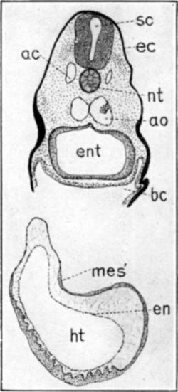
The first indication of the formation of the enteron is seen in the
very early embryo shown, from the dorsal aspect, in figure 1. The medullary folds and notochord are evident
at this stage, but no mesoblastic somites are to be seen.
A sagittal section of approximately this stage, shown in figure 1A, represents the
foregut, fg, as a shallow enclosure of the anterior region of the
entoderm, while the wide blastopore, blp, connects the region of
the hindgut with the exterior. No sign of a tail fold being present,
there is, of course, no real hindgut. The entoderm, which has the
appearance of being thickened because of the fact that the notochord has
not yet completely separated from it, is continuous, through the
blastopore, with the ectoderm. Posterior to the blastopore
2
the primitive streak, ps, is seen as a collection of scattered
cells between the ectoderm and the entoderm, apparently formed by
proliferation from the ventral side of the ectoderm.
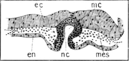
A slightly later stage is shown in figure 2,
a dorsal view of an embryo with five pairs of mesoblastic somites.
A sagittal section of this stage is shown in figure 2A. The foregut is
here more inclosed, and the notochord, nt, having separated from
the entoderm, en, is seen as a distinct layer of cells extending
from the foregut to the blastopore.
A transverse section through the headfold of this stage is shown in
figure 2B. The
foregut is seen as a wide cavity, ent, depressed dorsally,
apparently, by the formation of the medullary groove and the notochord;
it is wider laterally than in a dorso-ventral direction, and its walls
are made up of about three layers of closely arranged, irregular cells;
the wall is somewhat thinner on the dorsal side, just below the
notochord.
Figure 3 is a dorsal view of the next stage to
be described; about fifteen pairs of somites are present.
Figure 3A is
a transverse section through this embryo near the anterior end of the
enteron, ent, which cavity, cephalad to this region, is bluntly
pointed. As seen in the figure the enteron is here wide from side to
side, and is depressed dorso-ventrally except for a wide groove in the
ventral wall. This groove is lined with rather more closely arranged
cells, and marks the region where the mouth will break through at a
somewhat later stage. A short distance caudad to this region the
groove disappears and the pharynx is reduced to a shallow slit extending
almost to the superficial ectoderm on either side; then the slit-like
pharynx becomes suddenly reduced in a lateral and increased in a
dorso-ventral direction, to assume the outline shown in figures 3B and 3C. At a point about
one-third of the length of the embryo from the tip of the head, the
enteron opens to the yolk-sac, so that what now may be called the
foregut has this considerable extent. There is, however, not the
slightest indication of a tail-fold, so that there is no inclosed
hindgut at all. As is shown in figure 3D, the neurenteric canal, nc, still opens
ventrally, though the medullary canal, mc, has now no dorsal
opening to the exterior. The medullary canal continues for a short
distance (about fifteen sections of five microns thickness) posterior to
the opening of the neurenteric canal.
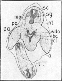
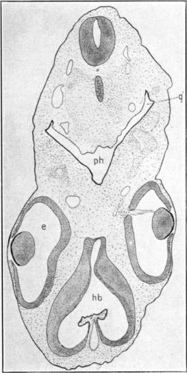
Figure 4 is a surface view of the next stage to
be described. There are here about twenty pairs of somites, though the
exact number cannot be determined. Although not visible externally in
the surface view shown, the gill clefts are beginning to form, and the
first one opens to the exterior as will be seen in sections of another
embryo
3
of this stage. The mouth has now broken through, putting the wide
pharynx into communication with the exterior; probably the mouth opening
is formed at about the time of the opening of the first gill cleft.
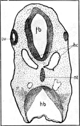
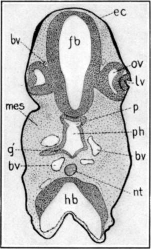
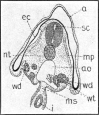
Figure 4A
represents a transverse section through the head of an embryo of the
approximate age of the one just described; it passes through both
forebrain, fb, and hindbrain, hb; through the extreme edge
of the optic vesicles, ov, and through the anterior end of the
notochord, nt. It is just cephalad to the anterior end of the
pharynx and to the hypophysis. The chief purpose in showing this section
is to represent the two large head-cavities, hc. The origin of
these cavities may be discussed at a later time. They are irregularly
oval in cross section, and extend in an antero-posterior direction for a
distance about equal to their long axis as seen in cross section. The
two cavities project towards each other in the middle line, and are
almost in contact with the notochord, in the region figured, but they do
not fuse at any point. These two head-cavities are the only ones to be
seen, in this animal, unless the small evaginations from their walls
represent other cavities fused with these. Their walls are thin but
distinct, and consist of a single layer of cells. These cells are
completely filled with their large, round nuclei, so that the wall has
the appearance, under higher magnification than is used in this figure,
of a band of closely strung, round beads.
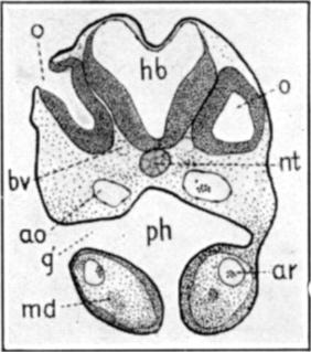
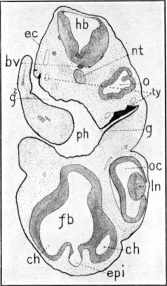
Figure 4B
represents the eighteenth section caudad to the one just described. It
passes through that region of the enteron, ph, which may be
called the pre-oral gut, since it lies cephalad to the now open mouth.
Owing to the plane of the section the upper angle of the first gill
cleft, g1, is seen on the left, although this would
not naturally have been expected in a section through the pre-oral gut.
The evagination to form the hypophysis, p, is seen against the
floor of the forebrain, fb. The wall of this region of the
enteron is comparatively thin, and consists of not more than two layers
of compactly arranged cells with round nuclei.
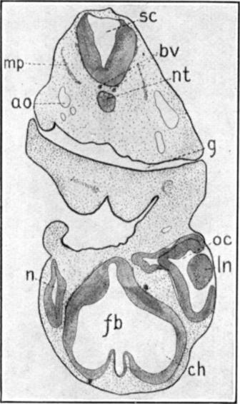
Figure 4C is
about forty sections caudad to the one just described. It passes through
the mouth, seen as a vertical opening between the two mandibular arches,
md. The hyomandibular cleft, g1, the only one
which opens to the exterior in this embryo, is very wide, and may be
traced through a number of sections; in this section the opening is seen
only on the left. The pharynx, ph, is very wide; as it is
followed caudad its ventral opening is gradually closed by the approach
of the two mandibular folds. The dorsal wall of this region of the
pharynx is very thin, consisting of a single layer of flat cells with
round nuclei; while the ventral wall, leading through
4
the mouth and lining the mandibular folds, is composed of two or three
layers of compactly arranged cells.
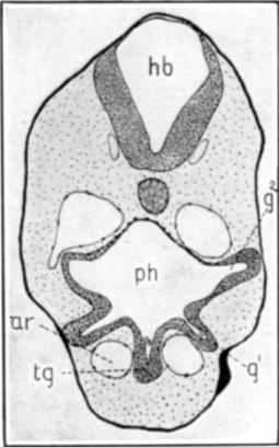
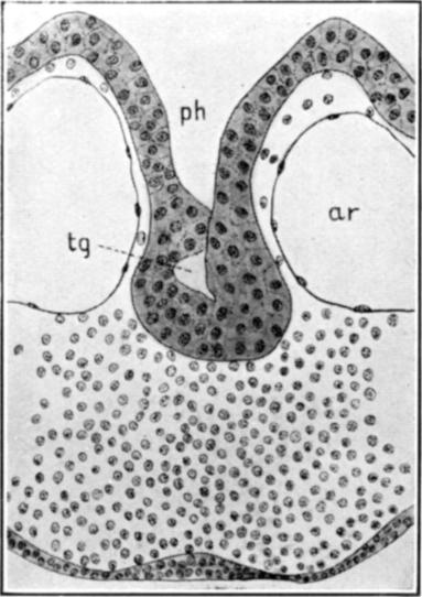
Figure 4D is
through a plane sixteen sections caudad to the last. In this region,
which is just caudad to the otic vesicles, the pharynx has still its
rectangular outline, and its walls are of the same character as in the
preceding figure. The posterior edges of the hyomandibular clefts are
seen projecting in a ventro-lateral direction, g1;
while dorsal to these are the wider, second pair of clefts,
g2. Where the mandibular folds come together posterior
to the mouth, they fuse first at their outer or ventral border, which
leaves a deep, narrow groove in the anterior floor of the mouth. As this
groove is followed caudad its ventral wall is seen to become much
thickened, tg, to form the anlage of the thyroid gland. In
the present section the walls of the groove are just fusing, to cut off
the cavity of the gland from the dorsal part of the groove. The next
section caudad to this shows the thyroid as a round, compact mass of
cells, with a very small lumen, still closely fused with the bottom of
the oral groove. The lumen may, in this embryo, be traced for only a few
sections, caudad to which the thyroid is seen as a small, solid mass of
cells unattached to the oral groove. Close to the sides of the thyroid
are seen two large blood vessels, ar, the mandibular arches,
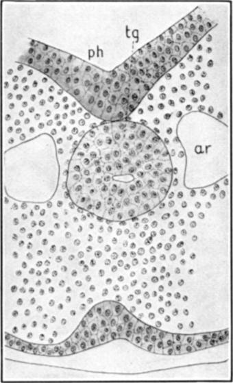
which unite into the single ventral aorta just caudad to the posterior
end of the thyroid. High power drawings of the thyroid just described
are shown in figures 4E and 4F.
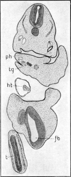
Figure 4G is
about fifty-five sections caudad to the preceding figure, and passes
through the middle region of the heart, ht. The enteron,
ent, is cut caudad to the last gill cleft, but it is nearly as
large as in the pharyngeal region described above; its walls are of a
more even thickness than in the more anterior sections, though there is
an area, just below the aorta, where the wall is still but one cell
thick. In the ventral wall of this part of the enteron, and, to some
extent, in the lateral walls, there seems to be a tendency for the
nuclei to become collected toward the side of the wall away from the
digestive cavity; this condition cannot be well seen in the figure owing
to the amount of reduction in reproduction.
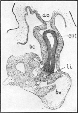
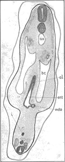
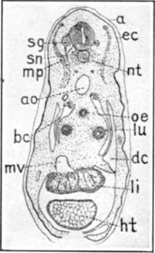
Figure 4H is
seventy-nine sections posterior to the last, and passes through the
foregut, ent, just cephalad to the anterior intestinal portal and
caudad to the heart. The outline of the enteron is here almost a
vertical slit, and the lining entoderm consists, in its dorsal and
lateral regions, of a single layer of columnar epithelium, while in its
ventral region, where it adjoins the liver trabeculae, it is made up of
several layers of cuboidal or irregular cells. The nuclei in the dorsal
and lateral regions of the entoderm are arranged in a very definite
layer
5
at the basal ends of the cells, though an occasional nucleus may be seen
near the center of the layer. The mesoderm that extends ventrad from the
mesentery, on each side of the entoderm just described, consists of a
thick layer of compactly arranged cells. The ventral end of the
entodermal wall is fused with the wall of a small cavity, li,
which may be traced several sections cephalad to this plane. This cavity
is a part of the system of hollow liver trabeculae seen as a group of
irregular masses of cells ventrad to the enteron at the opening of the
anterior intestinal portal. The large blood vessel, bv, is the
meatus venosus.
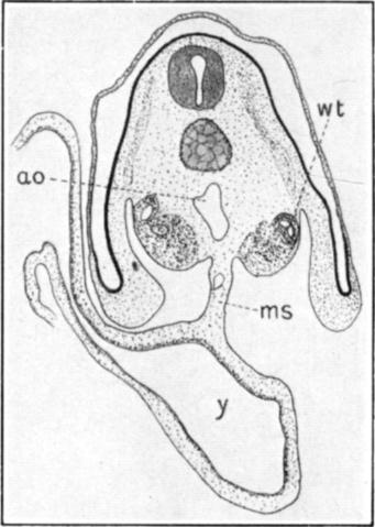
Figure 4I is
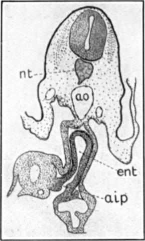
just four sections caudad to the preceding. It passes through the
anterior intestinal portal, aip. The medial liver trabecula into
which the enteron was seen to open, in the preceding figure, now opens
ventrally to the yolk-sac as the anterior intestinal portal. A few
liver trabeculae are to be seen on either side of the portal, but they
show no lumena, and may be traced through only a few sections. The
extent of this uninclosed region, the midgut, is very difficult to
determine with accuracy, but, at this stage, it comprises about one-half
of all the sections of the series. The difficulty is due partly to the
unavoidable tearing of the tissues in removing the embryo from the
yolk-sac, and partly to the indefiniteness of the posterior intestinal
portal, where the walls of the enteron are very thin. As seen in figure 4I the location
of the anterior intestinal portal is very distinct.
A short distance caudad to the anterior intestinal portal there is
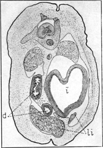
constricted off from the roof of the midgut a narrow diverticulum,
figure 4J,
i, the meaning of which is not apparent; it extends through only
ten to fifteen sections, tapering caudad till it disappears. The region
of the hindgut, at this stage, is about one-fifth of the entire length
of the embryo. Its anterior portion is wide and, as has been said,
rather indefinite in outline.
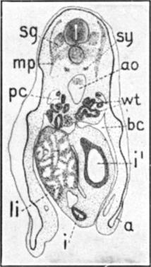
Figure 4K
represents a typical section through the midgut region of an embryo of
about the age of the one from which the preceding figures were drawn.
This and the following figures of this stage were drawn from an embryo
in which the posterior region was in better condition than in the embryo
from which the other figures of the stage were taken. The mesentery,
ms, is here of considerable length and continues around the yolk
in a layer of diminishing thickness. The epithelium of this region of
the enteron consists of a single layer of fairly regular cells, which
are columnar in the dorsal region, just beneath the mesentery, and
cuboidal or even flattened in regions more distant from the median
plane.
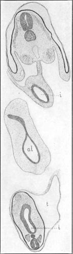
Figure 4L,
through the region of the hindgut, shows at i the completely
inclosed intestine; it is a comparatively narrow tube, lined
6
with columnar epithelium outside of which is a dense layer of mesoblast
continuous with the mesentery. In the center of the figure the
allantois, al, is seen as an irregular cavity, lined with a
single layer of columnar or cuboidal cells, and surrounded by a thick
mass of loosely arranged, stellate mesoblast cells. The allantois is
probably somewhat larger here than in the other embryos used for this
stage, in which it was torn away. The tail, t, of the embryo is
shown at the lower side of the figure, surrounded by the amnion; it is
cut in the region of a curve so that the caudal intestine, i, is
cut longitudinally and has the outline of an elongated ellipse. In this
embryo the caudal intestine could be followed to the end of the tail,
through several dozen sections; for some distance posterior to the
allantois it is extremely narrow, so that its lumen is almost
obliterated, and its walls are made up, in any one place, of not more
than a dozen cuboidal cells. Towards the posterior end of this region
the intestine is considerably enlarged as seen in figure 4L.
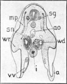
Figure 4M
passes through the region where both the allantois and the Wolffian
ducts open into the hindgut. The union of the allantois and the gut
accounts for the elongated outline of the enteron in this section. The
openings of the Wolffian ducts, wdo, are seen at the lower end of
the section of the enteron. The cells lining the Wolffian ducts are
smaller than those lining the enteron. In the lower side of the figure
are seen the structures of the tail, including the outline of the tiny
caudal intestine, i, mentioned above. No sign of a cloacal
invagination could be made out with certainty.
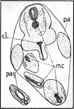
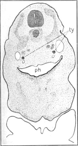
The next stage to be studied is shown in surface view in figure 5.
Figure 5A
represents a section through the head region of this embryo. Owing to
the obliquity of the plane of the section the figure is quite
asymmetrical. The pharynx, ph, is lined with a comparatively thin
epithelium and opens, on the left, at two places, one the mouth and the
other the second gill cleft, g2. In the dorsal wall of
this cleft, as well as in the corresponding wall of the opposite cleft,
is seen a thickening of the epithelium; these thickenings, ty,
are the rudiments of the thymus gland, whose development may be
described in detail in another paper. Compared to the size of the gill
clefts the cavity of the pharynx is, at this stage, comparatively
small.
Followed caudad the pharynx becomes depressed until, in the region
shown in figure 5B, it is a mere narrow slit, g, extending
transversely across the embryo and opening through the gill clefts to
the exterior on each side.
Figure 5C
passes through the posterior region of the pharynx, ph, the tip
of the forebrain, fb, the anterior edge of the heart, ht,
and the curve of the tail, t. The chief point of interest in this
section is
7
the thyroid gland, tg. It now lies deep in the tissue of the
floor of the pharynx, entirely separated from the pharyngeal epithelium.
It consists of a compact mass of cells, now showing a bilobed structure
in its anterior end, and extending through about twenty-five ten-micron
sections. It is solid throughout most of its extent, but, in the section
figured, which is near the anterior end, the lobe on the right side
shows a small but distinct cavity scarcely visible in the figure.
Caudad to the region just described the pharynx contracts suddenly to
form the oesophagus, a narrow, V-shaped slit, which soon divides
into an upper and a lower cylindrical tube, figure
5D, ent.
Followed caudad the lower of these tubes divides into the two
bronchial rudiments, figure 5E, lu, which, in the embryo here figured,
extend through nearly one hundred sections. In the region shown in
figure 5E the three tubes, oe
and lu, lie at the angles of an imaginary equilateral triangle,
while in the region of the liver, where the bronchial rudiments end, the
tubes lie in the same horizontal plane.
A short distance caudad to the ends of the bronchial rudiments the
oesophagus turns suddenly ventrad and becomes much enlarged to form the
stomach, figure 5F, i´, which may be traced through
twenty-five or thirty sections in this series. The epithelium of the
stomach is fairly thick, and consists of five or six layers of compact,
indistinctly outlined cells with spherical nuclei. Ventrad to the
stomach is seen, in figure 5F
a section of the duodenum, i, which extends, with gradually
diminishing caliber, for twenty-five or thirty sections caudad to the
posterior limit of the stomach, where it opens to the yolk-sac and is
lost.
The section that cut this embryo in the posterior region of the
stomach also passed through the hindgut in the region of the posterior
appendages, figure 5G. There the intestine, i, is a distinct,
cylindrical tube which extends, with not much variation in caliber, and
with little variation in position, from this point to the cloaca.
Followed cephalad, towards the posterior intestinal portal, it gradually
diminishes in caliber, as did the foregut on approaching the anterior
intestinal portal. The epithelium consists here of three or four layers
of compactly arranged cells, and has about the same appearance as in the
oesophagus and duodenum.
Figure 5H
represents a section through the cloacal region, cl, showing the
openings into the cloaca of the Wolffian ducts, wdo. Just
anterior to these openings the cloaca opens ventrally into a small,
anteriorly-projecting pouch, the rudiment of the allantois.
Caudad to the openings of the Wolffian ducts the cloaca extends
8
ventrad as a narrow, solid tongue of epithelium towards the exterior,
figure 5I, and
fuses with the superficial ectoderm at the caudal end of a prominent
ridge that lies in the mid-ventral line between the posterior
appendages. In this embryo the cloaca has no actual opening to the
exterior; the walls of the part that projects towards the exterior are
in close contact, except in the region of the openings of the Wolffian
ducts, as is shown in figure 5H.
Owing to the coiling of the end of the long tail the plane of the
section, as is seen in figure 5I, passes through the posterior end of the
embryo no less than four times. In the most posterior of these four
sections of the tail, beginning slightly caudad to the section here
shown, is seen a small cavity which may be called the post-anal gut,
pag. It has thick walls, and extends for about thirty-five
sections in the series under discussion. Its lumen is very large in its
caudal region, figure 5I, pag,
and tapers gradually cephalad until it disappears. Posteriorly the
post-anal gut ends quite abruptly not very far from the extreme tip of
the tail.
Figure 5J is
a composite drawing from reconstructions of the enterons of two embryos
of approximately this stage. One of these reconstructions was plotted on
paper from a series of transverse sections; the other was made in wax
from a series of sagittal sections. For the sake of simplicity the gill
clefts are not represented, and the pharynx, mouth, and liver are
represented in outline only. For the same reason the lung rudiment of
one side only is shown.
The relative size of the pharynx, ph, as seen in the figure,
is smaller than it is in reality because of the small dorso-ventral
diameter (the only one here shown) compared to the lateral diameter. The
end of the lung rudiment, lu, is slightly enlarged and lies in a
plane nearer to the observer than that of the oesophagus, oe,
though this is not well shown in the figure.
The oesophagus, oe, diminishes slightly in caliber for a short
distance caudad to the origin of the lungs, then gradually increases in
caliber until it suddenly bends to the side (towards the observer) and
merges into the wide stomach, i´. The stomach, which is
irregularly conical in shape, lies in a place slightly nearer the
observer than the end of the lung rudiment mentioned above.
Lying to one side of the stomach and duodenum, and extending cephalad
beyond the end of the lung rudiment is the liver, li, whose
outline is only roughly shown here by the broken line. The stomach opens
rather abruptly into the duodenum, d, which slopes back towards
the plane of the oesophagus (away from the observer).
The projection from the side of the duodenum, pan, not well
figured here, indicates the position of the pancreas, better shown in
9
the next reconstruction. The duodenum extends only a short distance
caudad to this point and then opens, aip, to the yolk-sac.
The yolk-stalk, or unclosed region of the enteron, is still of
considerable extent, though its exact boundaries are not easy to
determine. The distance between the anterior and posterior intestinal
portals is approximately shown in the figure under discussion.
The hindgut is cylindrical in cross section and of about the same
diameter throughout, except for a slight enlargement in the cloacal
region.
The post-anal gut is not shown here; it will be described in
connection with the next reconstruction where it is figured.
Figure 6 is a surface view in profile of an
embryo of the next stage to be studied. The manus and pes are here well
developed, and the general development of the embryo is in considerable
advancement over the last stage studied.
Figure 6A
represents a reconstruction, from a series of transverse sections, of
the enteron of an embryo of about the age of the one shown in
figure 6. The outlines of the entire embryo, of the eye, e,
and of the anterior, aa, and posterior, pa, appendages are
shown by broken lines. Its position being coincident with that of the
stomach, liver, and pancreas, the anterior appendage can scarcely be
seen. The enteron, including one lung only, for the sake of simplicity,
is shaded solid black, while the liver and pancreas, with their ducts,
are outlines with unbroken lines. As in the preceding reconstruction no
attempt is made to show the gill clefts, and only the dorso-ventral
profile of the enteron is shown. Caudad to the pharynx, the enteron
being more or less cylindrical in section, this profile gives a good
idea of its shape, but in the pharyngeal region, where the lateral
diameter is so much greater than the dorso-ventral, the reconstruction
gives but a poor idea of the size of that part of the enteron.
The widely-open mouth, m, leads, with no line of demarkation,
into the pharynx, ph, which is of irregular outline and, as has
been said, of much greater lateral than dorso-ventral diameter.
The pharynx becomes gradually constricted to form the oesophagus,
oe, a very long and slender structure, which, as will be
seen in cross section, is, at this stage, solid for the greater part of
its length. As in the case of the pharynx, the lateral diameter of the
oesophagus is generally greater than the dorso-ventral diameter.
From the floor of the caudal part of the pharynx is pushed out the
trachea, ta. In the reconstruction, especially in the anterior
end, the trachea appears several times the diameter of the oesophagus;
this is due to the great thickness and indefiniteness of its walls
rather than to a greater diameter of its lumen.
10
At about the position of the line ta the trachea divides into
the two bronchi (only one shown in the figure), which are somewhat
enlarged at the ends to form the lung rudiments, lu. While the
trachea and bronchi lie ventrad to the oesophagus, the lungs lie laterad
and even dorsad to the oesophagus and cardiac end of the stomach. Caudad
to the heart and in the region of the anterior appendages, aa,
the oesophagus suddenly enlarges to form the stomach, i´, which
has now quite the outline of the typical human stomach.
From the stomach the duodenum, d, extends, following a sort of
V-shaped course, towards the yolk-stalk, ys. In the region of the
yolk-stalk it is somewhat enlarged and ends in a blind sac like a
caecum. At the side of this sac is seen the opening of the enteron to
the yolk-stalk; the anterior and posterior intestinal portals are not
distinguishable from each other. From this point the hindgut, hg,
extends cephalad until it lies laterad to the middle region of the
duodenum, then bends through 180° and extends, in an almost straight
line, to the cloaca, cl, lying in the region of the posterior
appendage, pa.
The allantois, al, extends cephalad for some distance from the
floor of the cloaca. Some distance caudad to the cloaca, near the end of
the much coiled tail, is seen the post-anal gut, pag. This
structure as has been noted above, is quite distinct from the other
parts of the enteron. It is of elongated, pyriform outline, with the
pointed end extending cephalad.
In the narrow space between the stomach and the duodenum is the
elongated pancreas, pan, opening by two or more short ducts into
the duodenum.
The liver, li, in the figure under discussion, has about twice
the area of the stomach. It extends caudad and dorsal about the same
distance as the latter organ, but it extends ventrad and cephalad far
beyond the boundaries of the stomach.
Extending along the ventral border of the liver is a long narrow
duct, apparently the bile duct, bd. It connects, caudally, with
the anterior end of the pancreas, while at its other extremity, near the
antero-ventral corner of the liver; it ends blindly.
The transverse sections now to be described have been selected from
the series from which the reconstruction, just described, was made.
Figure 6B
represents a typical section through the pharynx. Its plane is
approximately shown by the line 400 of figure 6A though the plane apparently does not
cut the eye, e. The pharynx, ph, has here the outline of
an irregular V. Its walls, except at the outer
11
angles of the clefts, g1, are composed of but a single
layer of cells. In the dorsal wall these cells are flattened, while in
the ventral wall they are more rounded. This difference in the shape of
the cells accounts for the slightly greater thickness of the floor over
that of the roof of the pharynx. The gill clefts no longer communicate
with the exterior.
Figure 6C
represents the caudal half of the embryo in the plane 475 of figure
6A. The section of the pharynx,
ph, is here crescentic in outline, and the pharyngeal walls,
especially the floor, are somewhat thicker than in the more anterior
section just described. Lying a short distance dorsad to the pharynx are
seen two small, thick-walled openings, ty; these are the
rudiments of the thymus glands. They are here quite distinct from the
enteron, and may be traced through a large number of sections, being in
some regions solid and of a smaller diameter than in the present
section.
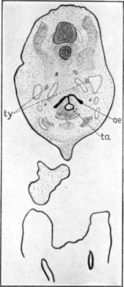
Figure 6D is
in the region of the line 500 in figure 6A. The thymus rudiments, ty, have about the
same appearance as in the preceding figure, except that they are
somewhat larger. The pharynx, ph, is much smaller than in the
last section, and though somewhat crescentic in outline, its convex side
is dorsal instead of ventral in position. The pharyngeal walls are here
thicker, and consist of two or three layers of cells, instead of the
single layer of more anterior sections.
In the median plane the floor of the pharynx is pushed down, as a
solid tongue of cells, gs, the anterior edge of the glottis.
Ventrad and laterad to the glottis a crescentic condensation of
mesoblast represents the beginning of the laryngeal cartilages,
la.
Two or three sections caudad to the one just described, the two
layers of which the tongue of cells from the floor of the pharynx is
composed separate slightly at the bottom to form a small cavity, the
trachea, ta; this condition is shown in figure
6E, which represents part of a
section through the plane 532 of figure 6A.
The oesophagus, oe, is here a solid, crescentic mass of cells,
the lumen being completely obliterated. The dorsal part of the tongue of
cells, mentioned above, connects the ventral side of the oesophagus with
the trachea, like a sort of mesentery. Above the oesophagus, on either
side, is the thymus rudiment, ty, in this section practically a
solid mass of cells instead of a tube. The epithelium of the trachea
here consists of three or four layers of compactly arranged cells; this
epithelium is surrounded by a dense mass of mesoblast which is
responsible for the greater thickness of the trachea as seen in figure 6A. As has been
said, the oesophagus here has no lumen, and when examined under high
magnification its walls are found to be completely
12
fused, not merely in close contact. The same is true of the tongue of
cells between the oesophagus and trachea. Two or three sections caudad
to the one under discussion this tongue of cells loses its connection
with the trachea, and the latter structure is entirely independent of
the oesophagus.
The solid condition of the oesophagus continues through about fifty
sections of this series, the horns of the crescent gradually shortening
until only the central part remains as the hollow cylinder seen in
figure 6F,
oe, which is a section through plane 650 of figure 6A. From about this point to its opening into the
stomach the oesophagus has essentially the same structure. Its
epithelium is of the simple columnar type, the cells being long, with
generally basally located nuclei.
In the section under discussion the trachea, ta, is of about
the same size as the oesophagus, but its epithelium is thicker and
consists of two or three layers of cells. The trachea extends, as a
separate and distinct structure, through about one hundred and fifteen
sections, and then, at a point four or five sections caudad to the
present section, it divides suddenly into the two bronchial tubes. Each
bronchus, like the trachea, is lined with an epithelium of three or four
layers of cells; but the epithelium is surrounded by a thin layer of
much condensed mesoblast. The bronchi continue caudad, with slightly
increasing caliber, through about fifty sections, when they suddenly
enlarge to form the lungs. As seen in figure 6A the
lungs are irregularly conical in outline and lie on either side of the
posterior end of the oesophagus.
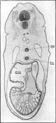
Figure 6G is
a section through the plane 750 of figure 6A. The oesophagus, oe, is seen as a small,
circular opening between two much larger openings, the lungs, lu.
The epithelium of the oesophagus is the same here as in the more
anterior regions described above; that of the lung rudiments is very
variable in thickness, even in different parts of the same section,
being in some places composed of a single layer of cuboidal or even
flattened cells, in other places consisting of four or five layers of
cells (not well shown in the figure). Surrounding the epithelium of the
lung rudiments is a thin layer of quite dense mesoblastic tissue.
A fairly well defined mesentery, ms, is now present in this
region.
Filling the greater part of the body cavity, below the oesophagus and
lung rudiments, is the liver, li; and ventrad to the liver the
section passes through a loop of the duodenum, d.
The epithelium of the duodenum consists of four or five layers of
compactly arranged cells, near the center of an oval mass of fairly
dense mesoblast. In a lateral projection of this mass of mesoblast
13
lies a small, circular opening, the bile duct, bd. Its epithelium
consists of a single layer of columnar cells. In more anterior sections
the bile duct is larger in cross section, being about one-half the
diameter of the oesophagus. As has been said it ends blindly at a point
a short distance anterior to the antero-ventral edge of the liver.
A few sections caudad to the one under discussion the bile duct
connects with the liver, figure 6A, bd´; and some distance caudad to this
the duct opens, bd´´, into the duodenum so close to the opening,
pan´, of the pancreas that it is difficult to determine whether
the latter organ has a separate opening into the duodenum or opens into
the bile duct.
At some distance ventrad to the structures just described the
intestine is cut, by the plane of the section, in two places, i.
The more dorsal of these is inclosed and has, under this magnification,
the same appearance as the duodenum, d; a higher
magnification, however, shows that its epithelium consists of a single
layer of tall, rather clear, columnar cells. The more ventral of the two
sections, above mentioned, which is continuous with the dorsal section a
very short distance caudad to this point, is in the region that opens to
the yolk—in fact a number of yolk-granules, y, may be seen
in the opening. The epithelium of this part of the intestine consists of
a single layer of clear, columnar cells, which, around the borders of
the opening, are thrown into numerous folds and are almost of goblet
form.
Figure 6H
represents a section through the plane 820 of figure 6A. The section is caudad to one lung and cuts the
extreme tip of the other, lu. The liver, li, and pancreas,
pan, are seen at the side of the stomach, i´, here cut
through its greatest transverse diameter. The epithelium of the stomach
varies somewhat in thickness and consists of two or three layers of
cells, the variation in thickness being due to a variation in the length
of the cells rather than to a variation in the number of layers.
Ventrad to the stomach the intestine, i, is cut in three
places, of which the most dorsal section is the largest. The epithelium
of these intestinal sections, especially the lower two, consists of
usually a single layer of columnar cells which are clearer than those of
the stomach. A fairly thin mesentery, ms, supports this
region of the intestine.
In the region of the posterior appendages, pa, the section
passes through the hindgut, hg, and allantois, al. The
former is of about the same size as the more anterior sections of the
intestine, but its epithelium is less clear and is composed of two or
more layers of cells. The allantois is cut near its opening into the
hindgut; its walls
14
are thin, the epithelium consisting of but a single layer of more or
less flattened cells.
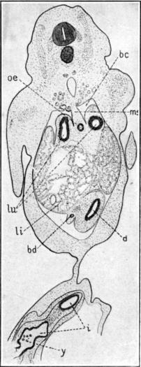
Figure 7 represents a reconstruction of the
enteron of an embryo of 42 mm. crown-rump length. Because of the body
flexure and large size of the embryo the head was amputated, in the
plane a-b, and cut sagitally, while the body was cut transversely
in the direction shown by the section planes. In the present figure the
outline of the embryo, including the eye, appendages, and umbilical
stalk, is shown by fine dotted lines; the outlines of the lungs and
liver are shown by heavier, broken lines; while the outlines of the
enteron proper and the trachea are shown in solid lines, filled in which
fine stippling. For the sake of simplicity only one lung and one
bronchus are shown.
Since the head has now quite a reptilian form, the oral cavity,
m, has more or less of the adult outline. A transverse
groove near the anterior end of the lower jaw marks off the tongue,
tn; and the rudiments of teeth are seen but not shown in the
figure because of the low magnification used.
The pharynx, ph, is a very extensive cavity that is sharply
separated from the mouth by a prominent transverse fold of skin, the
velum palitum, vp, just in front of the posterior nares,
pn, and by a less marked fold from the base of the tongue; it is
these two valves that enable the adult alligator to open its mouth under
the surface without getting water into the lungs. The mouth and pharynx
are lined at this stage with a thin, stratified epithelium, which
consists of a basal layer of rather tall columnar cells and one or two
superficial layers of flattened cells. The pharyngeal epithelium is
rather thicker than that of the oral cavity.
In the embryo from which this reconstruction was made the pharynx was
in direct communication with neither the oesophagus nor the trachea,
though the separation in each case was by a mere membrane. The trachea,
ta, opens, except for this membrane, into the pharynx a short
distance back of the transverse, dorsal and ventral folds mentioned
above, and almost directly ventrad to the posterior nares. The anterior
end of the oesophagus, oe, is in contact with the extreme
postero-ventral wall of the pharynx.
The trachea, which is already surrounded by distinct cartilaginous
rings, is long, and of about the same diameter throughout. In the region
of the anterior appendage, at the point marked X, it divides into the
two very short bronchi, which almost immediately open into the lungs,
lu. The lungs, whose structure will be shown in the sections of
this stage, are large, irregular bodies, extending about equal distances
cephalad and caudad to their openings into the
15
bronchi. The caudal ends of the lungs overlap the cephalic end of the
liver, li.
The oesophagus, oe, is large, and is laterally compressed so
that its dorso-ventral diameter, the one shown in the present figure, is
two or three times as great as its lateral diameter. This gives the
impression, in the reconstruction, that the oesophagus is nearly as
large as the stomach.
As has been said, the oesophagus does not open directly into the
pharynx, but is separated from it by a membrane which consists of the
flattened epithelial layers of both cavities separated by a thin layer
of mesoblast. This partition between the pharynx and the oesophagus is
not a mere fold of mucous membrane, but is a complete, though thin,
wall, easily seen in the series of sagittal sections from which this
region of the embryo was drawn. The anterior end of the oesophagus is
suddenly constricted so that the actual opening closed by this partition
is not large.
Followed caudad the dorso-ventral diameter of the oesophagus varies
somewhat, as does the lateral diameter, but it remains large throughout
and opens into the stomach with no sharp line of demarkation. The
character of the epithelium of the enteron caudad to the pharynx will be
discussed in connection with the sections to be described below.
The stomach, i´, is very different in outline from what was
seen in the last stage described, figure 6A. Instead of having approximately the form of
the typical mammalian stomach it is now so elongated that the opening
into the duodenum, the pylorus, py, seems to be nearer the
anterior than the posterior end. While the position of the pylorus is
very distinct it is difficult to distinguish the line of demarkation
between the stomach and the oesophagus.
The extreme caudal region of the stomach is enlarged to form a blind
sac, representing the gizzard, gz. A slight enlargement in
the region of the pylorus may represent the glandular region of the
adult stomach. The stomach opens, in a rather curious way, into the side
of the duodenum, d, the anterior end of the latter structure
having the appearance of a sort of caecum, to be seen in the next stage
of development.
The duodenum, d, makes a U-shaped bend at the side of the
stomach, and then, in the region of the caudal edge of the gizzard,
gz, dips suddenly ventrad and caudad towards the umbilical cord,
u, where it apparently ends blindly, though this appearance is
probably due to an artifact in the embryo from which the reconstruction
was made. It is likely that, in removing the embryo from the yolk, the
connection between the two loops, i, of the intestine was
broken.
16
The ascending intestinal loop is of slightly less caliber than the
descending loop above mentioned; it passes dorsal and cephalad to the
posterior border of the gizzard where its lumen is continuous, for a
short distance, with that of the descending loop above described. This
unusual condition is probably abnormal, but owing to lack of material
only one series of this stage was studied.
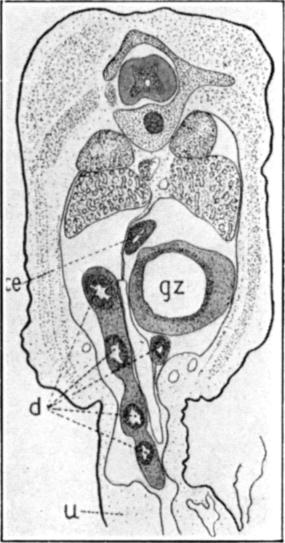
At the dorso-caudal angle of the gizzard the small intestine,
i, opens into the ventral side of a larger tube which may be
called the large intestine, il. The blind end of the large
intestine, cephalad to the opening of the small intestine, projects
forward, dorsal to the gizzard, as a sort of caecum, ce, though
this structure is generally stated to be wanting in the crocodilia, and
is not seen in the next stage.
From the caecum the large intestine passes in a ventro-caudal
direction, with gradually decreasing caliber, to the cloaca, from whose
anterior wall the intromittent organ, io, projects.
From the ventral wall of the large intestine, at a point about
one-third the distance from the cloaca to the caecum, projects ventrad
and cephalad the stalk of the allantois, al. Owing to its thin
walls and small lumen the allantois was traced only a short distance
into the umbilical stalk.
The profile of the liver, li, has, at this stage, about the
same area and even outline as that of the lung. It lies, of course, on
both sides of the enteron proper, and overlaps, anteriorly, as has been
said, the posterior end of the lung.
Figure 7A
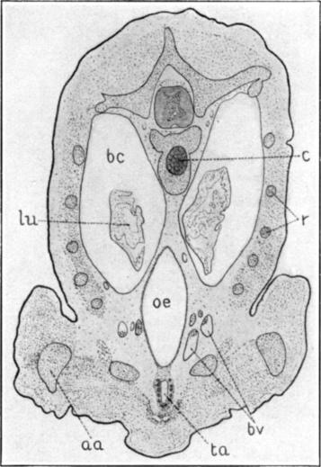
represents a section through the plane 305 of figure 7.
A considerable advance in the general development of the organs is
seen over the last stage studied. The spinal column is well outlined in
cartilage, and the ribs are cut at various places, r. In the body
wall a considerable differentiation of muscular tissue has taken place,
but it is only faintly shown in this series of figures. The scales,
especially along the mid-dorsal line, are shown as an area of less
closely dotted tissue.
The lungs, lu, cut here through their anterior ends, are
large, but do not nearly fill the cavities, bc, in which they
lie; they have the sacculated appearance characteristic of embryonic
lung tissue.
The oesophagus, oe, is cut through about its middle region,
where its caliber is greatest. As was said above, its dorso-ventral
diameter is more than twice its lateral diameter, caused partly by the
oblique angle at which it was cut. Its wall, figure
7H, is very thin and exhibits a
dense layer of mesoblastic tissue, in which circular and longitudinal
muscle layers are beginning to differentiate. It is lined by an
epithelium which here consists of a single layer of columnar
17
or cuboidal cells with large nuclei. On the ventral side, where the
oesophageal wall is in contact with that of the trachea the epithelium
is somewhat thickened by an increase in the number of cell layers. With
the low magnification used these details could not, of course, be
shown.
The trachea, ta, is of much smaller caliber than the
oesophagus, especially in its dorso-ventral diameter. While its
epithelial lining is not yet appreciably different from that of the
oesophagus, its connective tissue wall is much thicker and shows
numerous condensations, the rudiments of the cartilaginous rings. In the
region represented by this figure the connective tissue layers of the
trachea and oesophagus are continuous with each other, but cephalad and
caudad to this point they are distinct, though sometimes in contact.
Several large blood vessels, bv, on each side of the oesophagus
probably represent the carotids and jugulars, but they were not worked
out to determine with certainty which they were.
Eighty-five sections (figure 7, X)
caudad to the one under discussion the trachea divides into the two
bronchi. These bronchi gradually separate from each other until, at the
point at which they open into the lungs, about eighty sections caudad to
their point of separation, they lie on either side of the ventral third
of the oesophagus.
Figure 7B
represents a section through the plane 480 of figure 7. The section
is just cephalad to the heart, and passes through the caudal third of
the lungs, lu, which have the same appearance as in the preceding
figure; also through the extreme cephalic end of the liver, li.
The lungs here much more nearly fill the body cavity than in the
preceding figure. The section being caudad to their openings into the
lungs the bronchi do not, of course, show.
The oesophagus, oe, is here of much less diameter than in the
preceding figure, but is still laterally compressed. Its wall is
somewhat thicker than in the more cephalic region, the increase being
mainly due to the greater thickness of the connective tissue layer,
though the epithelium is also slightly thicker because of an increase in
the length of the lining cells. Instead of lying almost entirely ventrad
to the lungs, as in the preceding figure, the oesophagus here lies
directly between them.
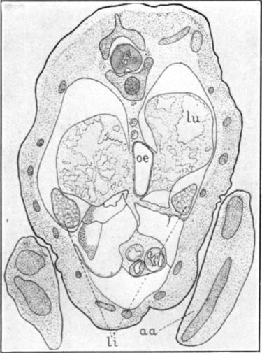
Figure 7C
represents a section through the plane 627 of figure 7. The plane
of the section passes through the opening of the stomach, i´,
into the duodenum, d. The cross section of the stomach is
somewhat larger than that of the oesophagus, but it differs from the
more anterior region mainly in the character of its walls. These are
much thicker than in the oesophagus; in the mesoblast which
18
forms the greater part of their thickness, muscle fibers are beginning
to differentiate. The epithelial layer also is thicker than in the
oesophagus; it consists of tall columnar cells that, at places, are
thrown into small folds, figure 7I. These folds, even under the low magnification
used, are more evident than is shown in the present figure. The pylorus,
py, is wide and, as has been noted in connection with
figure 7, is situated far cephalad to the caudal end of the
stomach. It opens into the side rather than into the end of the
duodenum, which projects cephalad as a short blind pouch, d. The
stomach and duodenum, in this section, are almost completely surrounded
by the liver, li.
Figure 7D
represents a section through the plane 680 of figure 7.
The stomach, i´, which is cut through its middle region, is
somewhat larger than in the preceding figures, though its walls have
about the same character. Its outer walls are continuous, to a
considerable extent, with the tissue of the surrounding body wall,
especially in the region just caudad to the plane of the present
section.
The duodenum, being cut through a double loop (see figure 7), is seen in two places, dorsally where it is
cut through the edge of one loop, and ventrally where it is cut square
across. In both sections the structure is the same, as might be
expected, figure 7J. The surrounding mesoblast is differentiated
into muscle fibers, figure 7J,
ml, which form a fairly distinct layer; inside of this layer is a
tall columnar epithelium, ep´, which is thrown into prominent
folds. A thin layer of mesoblast, probably the submucosa,
sl, lies beneath the epithelium and projects up into the folds.
About ten or twelve folds are seen in any one section; only the larger
ones are well seen in figure 7D.
Figure 7E
shows a section through the plane 770 of figure 7. It is in the
region of the umbilicus, u, and the extreme caudal end of the
stomach which has been called the gizzard, gz. The small size of
the gizzard is due to its being cut near its caudal margin. The enteron
is here cut in no less than seven places: the reason for this will be
evident on examination of the plane of the section as shown in figure 7. Dorsal to the gizzard the section cuts the
so-called caecum, ce, a little nearer its anterior end than
is shown in figure 7. The duodenum, d, is cut at five
points, and has about the same structure as in the preceding figure. The
character of the duodenal loops that causes the rather curious
appearance of the present figure will be readily understood by reference
to figure 7, though the reconstruction is not mathematically
accurate. The ventral projection of the lower loops of the duodenum into
the umbilicus is seen both in the present figure and in the
reconstruction. The loop of
19
the duodenum that, in the sections, is seen to lie directly ventrad to
the gizzard, in the reconstruction is shown too much to the side of the
latter organ. The descending loops of the duodenum are cut in such a way
that the surrounding mesoblast forms a continuous mass of tissue.
Figure 7F
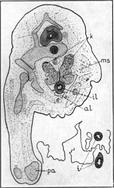
represents a section through the plane 901 of figure 7. The section
passes through the kidneys, k, the edge of one posterior
appendage, pa, the large intestine, il, and two regions of
the small intestine, i.
The large intestine is here a thick walled, cylindrical structure,
il, hanging from a thin mesentery, ms, in the much reduced
body cavity. The layers of its wall are much more fully differentiated
than in the more anterior regions of the enteron. The epithelium is here
stratified instead of simple columnar, and the folds into which it is
thrown are broader and less numerous than in the duodenum above
described.
Ventrad to the large intestine, and almost in contact with it, is
seen the allantois, al, whose general outline was noted in
connection with figure 7. It is an irregular structure, consisting
of a very thin outer layer of mesoderm, lined with a single layer of
flattened epithelial cells.
Lying at a considerable distance ventrad to the main body of the
section, are seen the two sections of the small intestine, i,
surrounded by irregular strands of tissue from the umbilicus. The
structure of these two intestinal loops is about the same as in the more
anterior region described above.
Figure 7G,
the last of this series, represents a section through the cloaca, caudad
to the urinary openings, in the plane 1060 of figure 7. The
epithelium of the cloaca is, of course, simply a continuation of that of
the surface of the body, somewhat thickened, perhaps, in the deeper
regions.
The intromittent organ, io, which projects cephalad from the
wall of the cloaca, is here seen as a three-pointed body of considerable
size, projecting ventrally from the body.
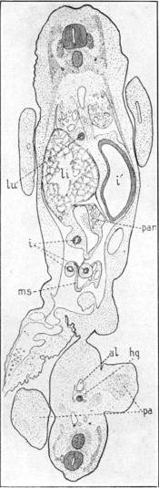
Figure 8 shows in outline the enteron, from the
ventral aspect, of an embryo of 20 cm. total length, or at about the
time of hatching. The drawing was made from a dissection and, for the
sake of simplicity, only the enteron, respiratory organs, heart, and
thymus are shown. The jaw is cut through on the left side and is turned
over to the right, thus bringing into view the roof of the mouth,
m, and the dorsal side of the tongue, tn. At the same time
the pharynx, ph, and the wide anterior end of the oesophagus,
oe, are cut open, exposing the glottis, gs, and vocal
cords, vc.
20
The lungs, lu, and trachea, ta, which are now fully
formed, are dissected loose and drawn over to the right side of the
animal, together with the heart, ht, and the thymus, ty;
only one side of the thymus is shown, the other half being hidden by the
trachea.
The mouth has reached nearly the outline of the adult. The lips are
formed and, in the anterior part of the lower jaw, four tooth rudiments,
to, are externally visible. The mucous membrane of the roof of
the mouth, m, is covered with rounded papillae, easily seen with
a lens but not shown in the figure. The tongue, tn, is fully
formed, and is free anteriorly and laterally to about the extent that is
seen in the adult; the papillae with which it is covered are not so
prominent as those seen on the roof of the mouth. At the base of the
tongue is the prominent transverse fold, noted in connection with
figure 7, that meets above the velum palitinum,
not shown here but shown in figure 7. Caudad to these folds is seen
the glottis, gs, a triangular opening with the vocal cords,
vc, at its base.
The mucosa of the inside of the pharynx and the anterior end of the
oesophagus, exposed by the dissection, is thrown into numerous
longitudinal folds, not shown in the figure; these well-marked folds
extend throughout the length of the oesophagus.
The oesophagus, oe, tapers gradually from the wide pharynx,
ph, and then continues as a cylindrical tube of uniform diameter
to the right side of the anterior end of the stomach, where it opens
into the latter organ. Its walls are thick, and its lumen is almost
obliterated by the longitudinal folds of the mucosa, mentioned
above.
The stomach, i´, is oval in outline, though somewhat flattened
laterally; it is depressed, dorso-ventrally, to a little more than half
the lateral diameter. As has been said, the oesophagus enters its right
anterior border; the pylorus is on the right side, 3 or 4 mm.
caudad to the oesophageal opening. The wall of the stomach is
comparatively thin except in the region of the oesophageal and pyloric
apertures, and at a point, opposite these apertures, on the left side.
At the latter point is an oval or disc-shaped area that is several times
as thick as the surrounding wall; it probably represents the gizzard
structure of the adult. The thickening mentioned in the region of the
two apertures seems to be mainly due to a wrinkling of the mucosa which,
in other parts of the stomach, is nearly smooth, so far as can be seen
with the naked eye. A sphincter thickening around the oesophageal
and, to some extent, around the pyloric aperture, causes each of these
structures to project into the stomach like an ileo-caecal valve.
The pylorus, py, opens into a small, pointed, thin-walled
diverticulum, di, and, at the same time, into the duodenum,
d. The diverticulum
21
noted, also, in connection with figure 7, has relatively thick,
wrinkled walls; its significance is not known to the writer. From this
diverticulum the duodenum, d, leads caudad and laterad for a
short distance as a narrow tube, then suddenly expands into the widest
part of the entire intestine. Into this wide part of the duodenum,
3 or 4 mm. from the pylorus, opens the bile duct, bd. The
bile sac, bs, is an elongated oval body with thin walls, lying to
the right of the pylorus, its connection with the liver was not
seen.
Lying between the anterior end of the duodenum and the posterior end
of the stomach, and extending caudad for 10 to 15 mm., in the median
plane of the animal is the pancreas, pan. It is a long narrow
body of a whitish color; its duct or ducts could not be determined by
dissection. The duodenum extends caudad, with gradually diminishing
caliber, from the enlarged region mentioned above. About 10 to 15 mm.
caudad to the stomach it makes a sort of double loop to the right,
a wide loop, lp, and a close one, lp´, nearer the
median plane. From the latter loop the intestine extends straight to the
left, for a distance of about 10 mm., where it makes a small loop
cephalad, lp2, and then opens to the yolk-sac,
y. The yolk-sac is shown here simply as an irregular piece of
tissue, the yolk having been removed.
The anterior intestinal portal, aip, and posterior intestinal
portal, pip, are in close proximity with each other.
From the posterior intestinal portal the intestine extends straight
cephalad to the posterior end of the stomach, dorsal to which it forms a
double loop, a wider one, lp3, and a narrow one,
lp4. From the latter loop, lp4, the
intestine extends straight caudad, parallel and near to the straight
region leading from the posterior intestinal portal, until it reaches
the region of the loop lp2, dorsal to which it forms a
small loop, lp5. From loop lp5 the
intestine, which is here of very small caliber, extends caudad for about
10 mm., where it forms another indistinctly double loop,
lp6.
From loop lp6 the large intestine, il,
extends, with gradually increasing caliber, to the cloaca, cl,
a distance of 10 to 15 mm.
Except in the enlarged region near the pylorus the lumen of the
intestine is almost obliterated by the folding of its thick walls, so
that little or nothing can be told of its lining with the naked eye.
A distinct mesentery holds the loops of the intestine in position and
binds the entire enteron close to the dorsal body wall. Because of the
lack of properly fixed tissue no sections of the enteron of this stage
were made.
REFERENCES
1. Bronn, H. G.: Klassen des
Thier-Reichs. (Vols. on reptiles.) 1890.
2. Chaffanjon, V.: Observations sur
Alligator mississippiensis (Tractus intestinalis und Mesenterium). Ann.
Soc. Linn. Lyon, vol. 28, p. 83 ff., 1881.
3. Eisler, P.: Zur Kentniss der
Histologie des Alligatormagens. Archiv f. Mikr. Anat., vol. 34, pp.
1-10, 1889.
4. Hertwig, O.: Comparative
Embryology of Vertebrates. Especially vol. 2, pp. 1-241, 1906.
5. Reese, A. M.: The Nasal Passages
of the Florida Alligator. Proc. Acad. Nat. Sc. Phila., 1901.
6.
Reese, A. M.: The Development of the
American Alligator. Smith. Misc. Coll., vol. 51, No. 1791, pp.
1-66, 1908.
23
DESCRIPTION OF FIGURES 1-8, PLATES 1-15
The word “Plate” refers to the physical pages on which the Figures were
printed. The word is not used as an illustration identifier. The
complete caption text from this section has been added to each Figure.
In the original, Figures were labeled only with number and letter. Based
on the author’s age at time of publication, “Miss C. M. Reese” is more
likely to have been his sister than his daughter.
The surface views were drawn, under the author’s direction, by Miss
C. M. Reese. The first two of these views were copied, by
permission, from S. F. Clarke; the others were drawn from the
specimens themselves.
All of the figures of any one stage are given the same number,
followed by distinguishing letters, so that it is possible to tell at a
glance what figures belong together.
All of the figures except those from Clarke were drawn under a camera
lucida.
Figure 1.
A surface view of an embryo, from the dorsal aspect, at the beginning of
the formation of the enteron.
Figure 1A.
A sagittal section of an embryo of approximately the age of the one
shown in figure 1. × 43.
Figure 2.
A dorsal view of an embryo with five pairs of mesoblastic somites.
Figure 2A.
A sagittal section of an embryo of the stage shown in figure 2.
× 43.
Figure 2B.
A transverse section through the headfold of an embryo of the stage
shown in figure 2. × 43.
Figure 3.
A dorsal view of an embryo with about fifteen pairs of somites.
× 20.
Figures 3A–3D. A series of transverse sections through an
embryo of the stage of the one shown in figure 3. × 43.
Figure 4.
A surface view of an embryo with about twenty pairs of somites.
× (about) 15.
Figures 4A–4D. A series of transverse sections through the
anterior end of an embryo of the approximate age of the one shown in
figure 4. × 20.
Figures 4E
and 4F. Two
transverse sections through the thyroid gland of this stage; more highly
magnified. × 102.
Figures 4G–4M. A series of transverse sections caudad to the
preceding. Figure 4H, × 43; other
figures, × 20.
Figure 5.
A surface view, in profile, of an embryo at the time of the origin of
the limbs. × (about) 5.
Figures 5A–5I. A series of transverse sections through an
embryo of the age shown in figure 5. × 7.
Figure 5J.
A composite drawing of reconstructions of the enterons of two embryos of
the age of the one shown in figure 5. One reconstruction was in
wax, from sagittal sections, the other was a plotted reconstruction from
transverse sections. × 14.
Figure 6.
A surface view, in profile, of an embryo with well developed manus and
pes. × (about) 5.
Figure 6A.
A reconstruction, plotted from transverse sections, of the enteron of an
embryo of about the age of the one shown in figure 6.
× 14.
Figures 6B–6H. Part of a series of transverse sections from
which the preceding reconstruction was made. × 7.
Figure 7.
A reconstruction of the enteron of an embryo of 42 mm. crown-rump
length.
Figures 7A–7G. A part of the series of transverse sections
from which the preceding reconstruction was made. × 7.
24
Figure 7H.
A high power drawing of a portion of the wall of the oesophagus in
the region of figure 7A. × 190.
Figure 7I.
A high power drawing of a portion of the wall of the stomach in the
region of figure 7C.
Figure 7J.
A high power drawing of a portion of the wall of the duodenum in the
region of figure 7D.
Figure 8.
An outline drawing, from the ventral aspect, of the enteron of an embryo
of 20 cm. length, at about the time of hatching; made from a dissection.
× 1.
Lettering for all Figures
a, head-fold of amnion. aa, anterior appendage. ac, anterior cardinal vein. aip, anterior intestinal portal. al, allantois. an, anterior nares. ao, aorta. ar, aortic arch. au, auricle. b, bulbus arteriosus. bc, body cavity. bd, bile duct. bd´, opening of bile duct to liver. bd´´, opening of bile duct to duodenum. blp, blastopore. bp, basilar plate. bs, bile-sac. bv, blood vessel. c, centrum of vertebra. ca, caudal artery. ce, caecum. ch, cerebral hemisphere. cl, cloaca. cm, circular muscle layer. cn, cranial nerve. cp, posterior choroid plexus. cv, cardinal vein. d, duodenum. dc, ductus Cuvieri. di, diverticulum of stomach. e, eye. ec, ectoderm. ec´, thickening of ectoderm. en, entoderm. en´, endocardium. ent, enteron. ep, epidermal layer of ectoderm. ep´, epithelium. epi, pineal body. es, embryonic shield. f, fronto-nasal process. fb, forebrain. fg, foregut. g1-5, gill clefts. gf1-6, gill folds. gl, glomerulus. h, head-fold. gs, glottis. gz, gizzard. hb, hindbrain. hc, head cavity. hg, hindgut. ht, heart. i, intestine. i´, stomach. il, large intestine. in, infundibulum. io, intromittent organ. ir, iris. it, iter. k, kidney. la, larynx. li, liver. lm, longitudinal muscle layer. ln, lens. lp, lp´, etc., loops of intestine. lu, lungs. lv, lens vesicle. m, mouth. ma, manus. mb, midbrain. me, medullary canal. me´, tip end of medullary canal. md., mandibular folds. | mes, mesoderm. mes´, myocardium. mf, medullary fold. mg, medullary groove. mk, Meckel’s cartilage. 25 ml, muscle layer. mp, muscle plate. ms, mesentery. mv, meatus venosus. mx, maxillary fold. myc, myocoel. n, nasal cavity. na, neural arch of vertebra. nc, neurenteric canal. nl, nervous layer of ectoderm. nt, notochord. o, ear vesicle. oc, optic cup. oe, oesophagus. on, optic nerve. os, optic stalk. ov, optic vesicle. p, pituitary body. pa, posterior appendage. pag, post-anal gut. pan, pancreas. pan´, opening of pancreas. pc, posterior cardinal vein. pe, pes. pg, primitive groove. ph, pharynx. pip, posterior intestinal portal. pl, pelvis. pn, posterior nares. pr, pericardial cavity. ps, primitive streak. pt, pecten. py, pylorus. r, rib. rt, retina. s, somites. sc, spinal cord. se, spenethmoid cartilage. sg, spinal ganglion. sl, submucosa. sm, splanchnic mesoblast. sn, spinal nerve. so, somatic mesoblast. st, stomodaeum. sy, sympathetic nervous system. t, tail. ta, trachea. tg, thyroid gland. tn, tongue. to, tooth anlage. tr, trunchus arteriosus. tv, tv´, third ventricle of brain. ty, thymus gland. u, umbilical stalk. v´, v´´, v´´´, first, second, and third cerebral va, vascular area. vc, vocal cords. vm, vitelline membrane. vn, ventricle of heart. vp, velum palitum. vv, vitelline blood vessels. wd, Wolffian duct. wdo, opening of Wolffian duct. wr, Wolffian ridge. wt, Wolffian tubule. x, point of origin of bronchi. y, yolk. ys, yolk-stalk. |
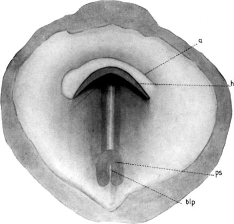
Fig. 1.
A surface view of an embryo, from the dorsal aspect, at the beginning of
the formation of the enteron.
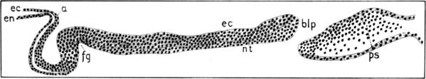
Fig. 1a.
A sagittal section of an embryo of approximately the age of the one
shown in figure 1. × 43
2
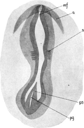
Fig. 2.
A dorsal view of an embryo with five pairs of mesoblastic somites.
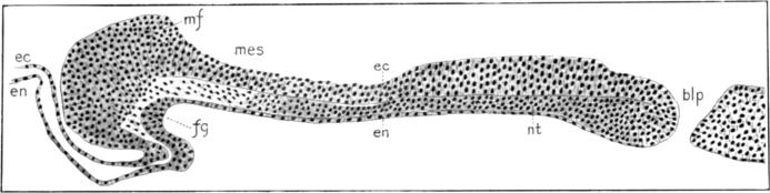
Fig. 2a.
A sagittal section of an embryo of the stage shown in figure 2.
× 43.
Fig. 2b.
A transverse section through the headfold of an embryo
of the stage shown in figure 2. × 43.
3
Fig. 3.
A dorsal view of an embryo with about fifteen pairs of somites.
× 20.
 |
 |
| Fig. 3a. | Fig. 3b. |
4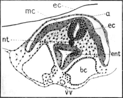 |
 |
| Fig. 3c. | Fig. 3d. |
|
A series of transverse sections through an embryo of the stage of the one shown in figure 3. × 43. | |
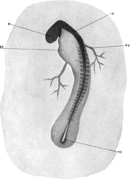
Fig. 4.
A surface view of an embryo
with about twenty pairs of somites. × (about) 15.
5
 |
 |
| Fig. 4a. | Fig. 4b. |
 |
 |
| Fig. 4c. | Fig. 4d. |
|
A series of transverse sections through the anterior end of an embryo of the approximate age of the one shown in figure 4. × 20. | |
 |
 |
| Fig. 4e. | Fig. 4f. |
|
Two transverse sections through the thyroid gland of this stage; more highly magnified. × 102. | |
6
 |
 |
 |
| Fig. 4g. | Fig. 4h. | Fig. 4k. |
 |
 |
 |
| Fig. 4i. | ||
 | ||
| Fig. 4j. | Fig. 4l. | Fig. 4m. |
|
A series of transverse sections caudad to the preceding. Figure 4H, × 43; other figures, × 20. | ||
7
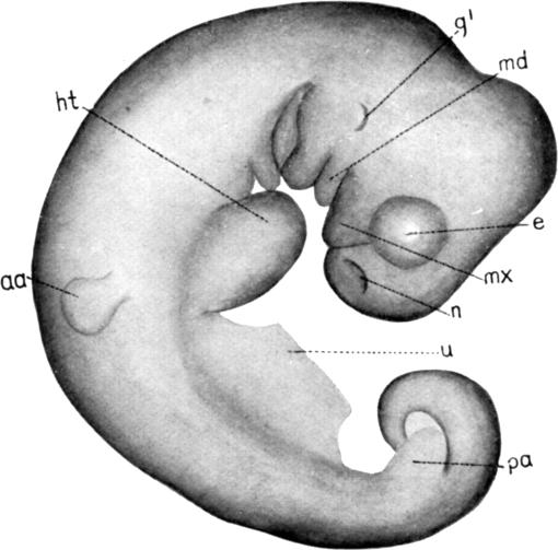
Fig. 5.
A surface view, in profile, of an embryo
at the time of the origin of the limbs. × (about) 5.
 |
 |
 |
| Fig. 5a. | Fig. 5b. | Fig. 5c. |
8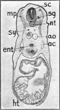 |
 |
 |
| Fig. 5d. | Fig. 5e. | Fig. 5f. |
 |
 |
 |
| Fig. 5g. | Fig. 5h. | Fig. 5i. |
|
A series of transverse sections through an embryo of the age shown in figure 5. × 7. | ||
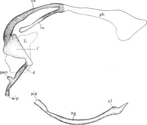
Fig. 5j.
A composite drawing of reconstructions of the enterons of two embryos of
the age of the one shown in figure 5. One reconstruction was in
wax, from sagittal sections, the other was a plotted reconstruction from
transverse sections. × 14.
9
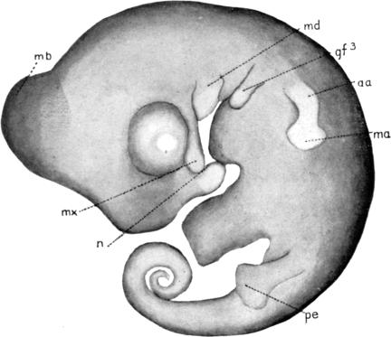
Fig. 6.
A surface view, in profile, of an embryo
with well developed manus and pes. × (about) 5.
10
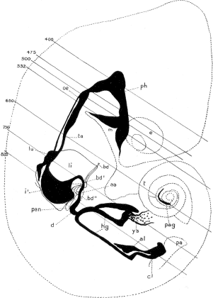
Fig. 6a.
A reconstruction, plotted from transverse sections, of the enteron of an
embryo of about the age of the one shown in figure 6.
× 14.
9b
 |
 | ||
| Fig. 6b. | Fig. 6c. | ||
11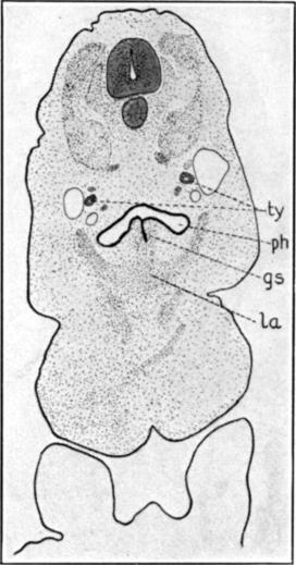 |
 |
 | |
| Fig. 6d. | Fig. 6e. | Fig. 6f. | |
 |
 | ||
| Fig. 6g. | Fig. 6h. | ||
|
Part of a series of transverse sections from which the preceding reconstruction was made. × 7. | |||
12
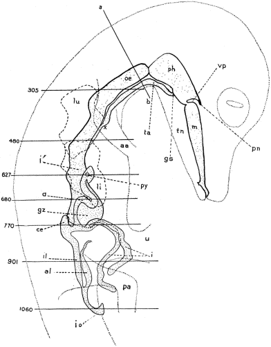
Fig. 7.
A reconstruction of the enteron of an embryo of 42 mm. crown-rump
length.
 |
 |
| Fig. 7a. | Fig. 7b. |
13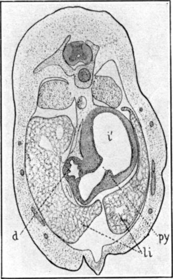 |
 |
| Fig. 7c. | Fig. 7d. |
 |
 |
| Fig. 7e. | Fig. 7f. |
14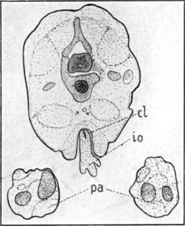 | |
|
Fig. 7g. A part of the series of transverse sections from which the preceding reconstruction was made. × 7. | |
 |
 |
|
Fig. 7h. A high power drawing of a portion of the wall of the oesophagus in the region of figure 7A. × 190. |
Fig. 7i. A high power drawing of a portion of the wall of the stomach in the region of figure 7C. |
Fig. 7j.
A high power drawing of a portion of the wall
of the duodenum in the region of figure 7D.
15
Fig. 8.
An outline drawing, from the ventral aspect, of the enteron of an embryo
of 20 cm. length, at about the time of hatching; made from a dissection.
× 1.